O
RIGINALA
RTICLE Revista Brasileira de FisioterapiaEffects of cryotherapy, transcutaneous electrical
stimulation and their combination on femoral
nerve electrical activity in rats
Efeitos da crioterapia, estimulação elétrica transcutânea e da sua associação na
atividade elétrica do nervo femoral em ratos
Santuzzi CH1,2, Gonçalves WLS1, Rocha SS2, Castro MEC1,3, Gouvea SA2, Abreu GR1
Abstract
Background: Clinical reports suggest that the therapeutic association between cryotherapy (CRYO) and transcutaneous electrical
stimulation (TENS) favors local analgesia. Objective: To evaluate the electrical activity of the femoral nerve (FNA), at rest and during
single and combined application of TENS and CRYO, in rats. Methods: Nine adult Wistar rats weighting ±300g were used in this study.
After inducing anesthesia (Urethane, 1mg/g i.p.), the right femoral nerve was isolated in order to record the FNA at baseline and during the therapeutic modalities. After attaching the electrodes to the lower third of the right thigh, TENS (50Hz, 10mÅ) was applied for five minutes, and CRYO and the combined therapy (CT) for ten minutes. The FNA was recorded continuously by means of an action potential amplifier and the recordings from the first, fifth and tenth minutes were subsequently evaluated using arbitrary units (aU). One-way analysis of variance (ANOVA) was used, with Dunnett’s test as post-hoc analysis. The values were expressed as the mean ±SEM and differences were established at p<0.05. Results: The femoral nerve activity increased (p<0.01) after TENS (0.358±0.09aU) and CT
(0.230±0.07aU) and was unchanged after CRYO (0.063±0.003aU), in relation to the baseline (0.009±0.0003aU). In the fifth minute, we observed significant (p<0.05) attenuation of FNA in the CT (0.144±0.027aU) in relation to TENS alone (0.324±0.089aU). Conclusions:
The association between CRYO and TENS noninvasive analgesia significantly attenuates the effects produced by TENS alone on the FNA of anesthetized rats.
Key words: TENS; nerve activity; cryotherapy; Physical Therapy; analgesia.
Resumo
Contextualização: Relatos clínicos sugerem que a associação terapêutica entre crioterapia (CRIO) e estimulação elétrica transcutânea
(TENS) favorece analgesia local. Objetivo: Avaliar a atividade elétrica do nervo femoral (ANF), em repouso e durante a aplicação
isolada, e associada de TENS e CRIO em ratos. Métodos: Foram utilizados nove ratos (Wistar) adultos com peso de ±300g. Após
anestesia (Uretana, 1mg/g i.p.), o nervo femoral direito foi isolado para registro da ANF basal e durante as modalidades analgésicas. Depois da fixação dos eletrodos no terço inferior da coxa direita, foram aplicadas TENS (50Hz, 10mÅ) por cinco minutos, CRIO isolada e terapia associada (TA) por dez minutos. Os registros contínuos da ANF foram realizados por meio de um amplificador de potenciais de ação, avaliados posteriormente no primeiro, quinto e décimo minuto em unidades arbitrárias (Ua). Utilizaram-se a análise de variância (ANOVA) uma via e o teste de Dunnett como post-hoc. Valores expressos como média ±EPM e as diferenças
fixadas emp<0,05. Resultados: A atividade do nervo femoral aumentou (p<0,01) na TENS (0,358±0,09Ua) e na TA (0,230±0,07Ua)
e ficou inalterada após CRIO (0,063±0,003Ua), em relação ao basal inicial (0,009±0,0003Ua). No quinto minuto, observou-se uma significante (p<0,05) atenuação da ANF na modalidade TA (0,144±0,027Ua) versus TENS isolada (0,324±0,089Ua).Conclusões:A
associação entre as modalidades analgésicas não-invasivas CRIO e TENS atenua significativamente os efeitos produzidos pela TENS isoladamente sobre a ANF de ratos anestesiados.
Palavras-chave: TENS; atividade nervosa; crioterapia; Fisioterapia; analgesia.
Received: 04/09/2007 – Revised: 05/02/2008 – Accepted: 04/08/2008
1 Postgraduate Program in Physiological Sciences, Health Sciences Center, Universidade Federal do Espírito Santo (Ufes) – Vitória (ES), Brazil 2 Escola de Ciências da Saúde da Santa Casa de Misericórdia de Vitória (Emescam) – Vitória (ES), Brazil
3 Physical Therapy Department, Faculdade Novo Milênio – Vila Velha (ES), Brazil
Correspondence to: Glaucia Rodrigues de Abreu, Programa de Pós-graduação em Ciências Fisiológicas, Centro de Ciências da Saúde da Universidade Federal do Espírito Santo (Ufes), Avenida Marechal Campos, 1.468, Maruípe, CEP 29040-577, Vitória (ES), Brazil, e-mail: [email protected]
Introduction
Transcutaneous Electrical Nerve Stimulation (TENS) is an analgesic technique used in a variety of frequencies, intensities and pulse duration, classiied as high frequency (>50Hz), low frequency (<10Hz) and burst (alternate high and low frequencies)1-3. Conventional TENS is a
continu-ous high-frequency (50 and 150Hz), low-intensity stimula-tion of the fast conducstimula-tion nervous ibers. he intensity of TENS should not cause muscle contractions, but only a not-unpleasant feeling of paresthesia, adjusted according to individual sensibility4. Studies show that intensities between
ten and 30 milliamperes (mÅ) are more comfortable and do not cause signiicant fasciculation in the pulse time which varies from 40 to 75µs. In this type of stimulation, analgesia occurs immediately or ten minutes after the application. his efect can last from 20 or 30 minutes up to two hours, which explains why this method is preferably used to treat acute pain1-5.
TENS promotes analgesia predominantly through the mechanism of gate control theory of pain, proposed by Melzack and Wall5. According to this theory, analgesia is provoked by the
selective activation of the large-diameter tactile ibers (A-beta ibers), without activation of the small-diameter nociceptive ibers (A-delta and C ibers). he activity generated in A-beta ibers inhibits the current activity of the nociceptive neurons located in the dorsal horn of the spinal cord5. Additionally, the
analgesic mechanism of TENS also seems to be related to the activation of endogenous opioid receptors in the spinal cord6.
Recent studies demonstrate that low-frequency TENS specii-cally activates µ-opioid receptors, serotonin receptors and spi-nal muscarinic receptors. Conversely, the aspi-nalgesia produced by high-frequency TENS activates delta-opioid and muscarinic receptors in the dorsal horn of the spinal cord and the supraspi-nal delta-opioid receptors1,6-9.
Studies show that therapeutic cryotherapy (CRYO) appli-cation gradually reduces impulse transmission in the sensitive nerves because of the decreased nerve conduction velocity10,11.
However, after a prolonged cold compress application, the du-ration of the action potentials of sensorial nerves can increase due to longer refractory periods. Clinically, CRYO is commonly applied in the management of acute injuries, muscle spasms and inlammatory processes11-13.
Studies prove that CRYO and TENS can reduce pain in patients with several conditions such as in the post-operative period14. Clinical reports that promote analgesia
currently recommend the combination of the two thera-peutic modalities (CT) for a better response in pain control. However, the electrophysiological mechanism that involves the nervous conduction and, therefore, the analgesic efects
of the combination of both techniques have not been suf-iciently explained in the literature15-17. CT creates a paradox
among the physiological mechanisms of the two therapeutic modalities: while one reduces the nerve conduction velocity, the other stimulates the nervous ibers. he aim of this study was to evaluate the efects of TENS and CRYO, isolated or combined, on the frequency of the action potentials of the femoral nerve.
Methods
he investigation was conducted in accordance with the established norms of Guide care and use of laboratory18 and
approved by the Animal Testing Ethics Committee of Uni-versidade Federal do Espírito Santo (Ufes), protocol number 026/2007.
Animals and experimental design
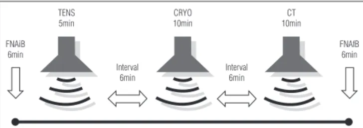
Nine Wistar rats with body mass between 300 and 350 grams were used in a single group. he animals were main-tained in the Research Vivarium of the Postgraduate Pro-gram in Physiological Sciences of Ufes. All the procedures were conducted according to the biomedical research guide for use of laboratory animals, as determined by the Fed-eration of Experimental Biology Societies. Rats were main-tained in individual cages, on a 12-hour light/dark cycle at a controlled temperature of 22ºC, under artiicial lighting and with ad libitum food and water. he evaluated parameter in this study was the femoral nerve electrical activity (FNA) of anesthetized rats, without painful stimuli, before and during the application of two modalities (isolated and combined) which originate three types of intervention: TENS, CRYO and CT.
Figure 1. Recording protocol for femoral nerve activity (FNA) in anesthetized rats during application of the noninvasive analgesic modalities.
FNAiB 6min
TENS 5min
Interval 6min
CRYO 10min
Interval 6min
CT 10min
FNAfB 6min
TENS=transcutaneous electrical nerve stimulation; CRYO=cryotherapy (compress); CT=combined therapy; FNA (Ua)=continuous line; FNAiB=initial baseline;
FNAfB=final baseline; =intervals.
Surgical procedures and femoral nerve electrical
activity records
Rats were initially anesthetized with a single intraperi-toneal (i.p.) dose of Urethane (1mg.g-1 of body mass). his
anesthetic was chosen because it is widely used in experi-ments which involve electrical activity, and therefore recom-mended for small changes in nerve activity19,20. he animals
were placed in the supine position, and the femoral nerve of the right limb was exposed through a 1.5 to 2cm longitudinal rectilinear skin incision on the inner thigh (pelvic area). he space between the hip abductor and the femoral muscles was dissected, and the femoral vascular nerve plexus was identiied. A microscope was used (M900, DF Vasconcelos®,
São Paulo, Brazil) for the femoral nerve selection and subse-quent accommodation in silver electrodes to record nervous activity. Mineral oil and petroleum jelly were placed on the incision to avoid nerve lesion, to lubricate and to maintain the integrity of the femoral nerve. After this procedure, the extracellular action potentials at baseline were recorded in an ampliier (NL 104, Neurolog®, Digitimer, Welwyn Garden,
UK). he signs were iltered (NL 126, Neurolog®, Digitimer,
Welwyn Garden, UK) and connected to an audio ampliier (NL 120, Neurolog®, Digitimer, Welwyn Garden, UK) and
linked to an oscilloscope (Tektronix 2205, General Electric®,
NJ, USA). After that, the signs were processed in a Spike trigger action potential discriminator (NL 200, Neurolog®,
Digitimer, Welwyn Garden, UK) and in a pulse integrator (NL 601, Neurolog®, Digitimer, Welwyn Garden, UK). hose signs
were simultaneously converted by the software Acknowledge for Windows (Biopac System®, Santa Barbara, CA, USA) for
subsequent analysis.
Isolated transcutaneous electrical stimulation
TENS (EMPI® Eclipse Inc., Minneapolis, MN, USA) was
ap-plied for ive minutes at 50Hz and inal sensorial intensity of 10mÅ. he electrodes (0.5cm diameter) were attached with adhesive tape to the medial and the lateral area of the right knee joint, before the beginning of the experiment in order to avoid possible changes in the FNA recording. he sensorial intensity was determined by increasing the parameters up to levels in which muscle contractions were not evident. his therapeutic modality characterizes an application of conven-tional TENS and those parameters remained constant1,6-9.
Im-mediately after the TENS application (at the end of the ifth minute), the equipment was switched of, and six minutes were allowed so that the nerve reached stability at the action potential frequency (Figure 1). Studies conducted in the same
laboratory as the present study demonstrate that TENS stim-ulates the nerve quickly, and its response is the same after ive or ten minutes. herefore, the shortest stimulation period ( ive minutes) was chosen to minimize possible lesions dur-ing nerve exposure.
Isolated cryotherapy and combined therapies
After the frequency was stabilized, CRYO application began with an ice compress over the right knee joint of the back limb for ten minutes. During this period, the femoral nerve action potentials were recorded. At the end of the procedure, the ice was removed and six minutes were allowed once again. After this, CT was applied for ten minutes (Figure 1). he ten-minute period for CRYO application was also based on pilot studies which showed this to be the longest time needed to afect nerve activity. At the end of the study, the animals were euthanized with a lethal dose of anesthetic.
Statistical analyses
he results were analyzed using statistics software (Graph-Pad® Prism4). One-way analysis of variance (ANOVA), followed
by the Dunnett test, was used to compare the variables. Values (aU) were expressed as mean ±SEM. he α level considered for analyses was set at 0.05.
Results
As demonstrated in Figure 2A, in the irst minute after the start of the analgesic modalities, FNA increased signiicantly (p<0.01) in TENS (0.358±0.094aU) and CT (0.230±0.074aU), and it did not change in CRYO (0.063±0.037aU). In the ifth minute, the analgesic modalities TENS (0.324±0.089aU) and CRYO (0.035±0.015aU) maintained the FNA means observed
Initial Baseline
TENS CRYO CT
End TENS
End CRYO and CT
Final baseline 0,7
0,6 0,5 0,4 0,3 0,2 0,1 0,0
0 5 10 15
Minutes
FNA (Arbitrar
y Units)
FNA (Arbitrar
y Units)
0,55 0,45 0,35 0,25 0,15
0,02 0,01 0,00
Groups 0,03
Baseline TENS CRYO CT **
**
*
**
*
Figure 2: A. Temporal evaluation of the effects of the isolated (TENS or CRYO) and combined (CT) analgesic modalities on femoral nerve activity (FNA) in anesthetized rats; B. Definition of baseline FNA before and after application of the noninvasive analgesic modalities. Values expressed as mean ±SEM; one-way ANOVA and Dunnett test for multiple comparisons.
A
B
*p<0.05 **p<0.01 versus baseline; n=9.
in the irst minute, however CT (0.144±0.027aU) attenuated the FNA levels compared to the isolated applications. In the tenth minute, there were no signiicant diferences among the three analgesic modalities, emphasizing that TENS was interrupted in the ifth minute. Figure 2B demonstrates that the mean of FNA at baseline in the anesthetized rats was 0.009±0.0003aU. his value was similar to those obtained in the intervals of the application of TENS (0.004±0.002aU), CRYO (0.001±0.0006aU) and CT (0.007±0.001aU).
Discussion
he present study demonstrated the efects of isolated and combined TENS and CRYO on the frequency of femoral nerve
action potentials. he data we obtained showed that isolated TENS in anesthetized rats increases FNA, which was signii-cantly attenuated when CT was used. However, the results also showed that isolated CRYO did not change the FNA of the ex-perimental animals.
These data demonstrated that TENS induces this an-algesic effect by promoting peripheral nerve stimulation, specifically in the tactile proprioceptive fibers1-6. Conversely,
CRYO attenuated FNA possibly because it increases refrac-tory periods and reduces nerve conduction velocity11-15. The
literature has demonstrated that the use of isolated TENS or CRYO produces significant analgesic effects in acute and chronic inflammatory processes1,6,10,11,16. These studies
report that CRYO produces analgesia by two main local mechanisms, the neural and the vascular mechanisms. In the neural mechanism, the topical ice application reduces the local temperature which lowers the activation thresh-olds of tissue nociceptors and, consequently, the transmis-sion signs of pain. With regard to the vascular effects of CRYO, the analgesia is associated with a decrease in blood flow, caused by cold-induced vasoconstriction as well as reduced neural metabolism11-15. The means by which these
effects occur have yet to be fully explained11-15.
Traditionally, TENS promotes analgesia through the selec-tive activation of A-beta tactile ibers, which inhibit the cur-rent activity of nociceptive neurons in the dorsal horn of the spinal cord. Moreover, several studies on the presence of pain demonstrate that the analgesic action mechanism of TENS is also related to the activation of opioid receptors in the spinal supraspinal cordl1-5,21-23 and not only through the gate control
theory.
In fact, CT has been routinely observed in clinical practice with the purpose of increasing the analgesic efects of these therapies and possibly producing hyperalgesia. In spite of the common (and conventional) simultaneous use of these tech-niques in physical therapy practice, the literature has little evidence of the beneits of CT.
his study demonstrated that the combined therapy pro-motes attenuation in FNA which was previously elevated by isolated TENS. his is explained by the reduction in nerve conduction velocity promoted by the ice10; cold decelerates
the axoplasmatic transport, i.e. substance low along the axon; ATP and creatine phosphate concentrations do not change, therefore, it appears that the blockade is caused by a metabolic reduction in the ATP use by nerves due to reduced enzymatic activity10-15. hus, it is possible to speculate that the use of
CT not only opposes the stimulating efect and reduces the therapeutic actions of isolated TENS, but also reduces tactile sensibility10,11 making it more diicult to identify the ideal
However, the sequential use of TENS and CRYO is jus-tified in the presence of pain and other factors, such as osteoarthritis with edema, and because of the arthrogenic muscle disinhibiting effect promoted by CRYO. This occurs because the edemas that accompany the pain of these joint injuries sensitize the capsular mechanoreceptors. These receptors inhibit the spinal alpha motoneurons and, con-sequently, the signs transmitted to the muscle10,11. Although
the present study was not conducted during a painful or in-flammatory process, the electroneurographic data obtained indicate that the sequential use of TENS and CRYO (in that order) can improve the analgesic pattern6-8 and allow better
joint manipulation, without lowering the pain threshold, respectively. In clinical practice, this fact is incorrectly in-terpreted as hyperalgesia10,11.
New studies involving painful processes are necessary for a better understanding of the action mechanisms involved in neural activity during the use of isolated and combined analgesic techniques. Finally, caution is recommended when applying CRYO and TENS simultaneously in physi-cal therapy cliniphysi-cal practice as this combination produced a new pattern of FNA response in laboratory animals, i.e. FNA was increased by TENS and attenuated by the CT in anesthetized rats.
Acknowledgments
To the Conselho Nacional de Desenvolvimento Cientíico e Tecnológico (CNPq) for their inancial support.
445
References
1. Sluka KA, Walsh D. Transcutaneous electrical nerve stimulation: basic science mechanisms and clinical effectiveness. J Pain. 2003; 4(3):109-21.
2. Lampl C, Kreczi T, Klingler D. Transcutaneous electrical nerve stimulation in treatment of chronic pain: predictive factors and evaluation of the methods. Clin J Pain. 1998;14(2):134-42.
3. Jensen H, Zesler R, Christensen T. Transcutaneous electrical nerve stimulation (TNS) for painful osteoarthrosis of the knee. Int J Rehabil Res. 1991;14(4):356-8.
4. Katz J, Melzack R. Auricular transcutaneous electrical nerve stimulation (TENS) reduces phantom limb pain. J Pain Symptom Manage. 1991;6(2): 73-83.
5. Melzack R, Wall PD. Pain mechanisms: a new theory. Science. 1965;150(699):971-8.
6. King EW, Sluka KA. The effect of varying frequency and intensity of transcutaneous electrical nerve stimulation on secondary mechanical hyperalgesia in an animal model of inflammation. J Pain. 2001;2(2):128-33.
7. Maeda Y, Lisi TL, Vance CG, Sluka KA. Release of GABA and activation of GABA(A) in the spinal cord mediates the effects of TENS in rats. Brain Res. 2007;1136(1):43-50.
8. Somers DL, Clemente FR. Transcutaneous electrical nerve stimulation for the management of neuropathic pain: the effects of frequency and electrode position on prevention of allodynia in a rat model of complex regional pain syndrome type II. Phys Ther. 2006;86(5):698-709.
9. Bolfe VJ, Ribas SI, Montebelo MIL, Guirro RRJ. Comportamento da impedância elétrica dos tecidos biológicos durante estimulação elétrica transcutânea. Rev Bras Fisioter. 2007;11(2):153-9.
10. Hopkins J, Ingersoll CD, Edwards J, Klootwyk TE. Cryotherapy and Transcutaneous Electric Neuromuscular Stimulation Decrease Arthrogenic Muscle Inhibition of the Vastus Medialis After Knee Joint Effusion. J Athl Train. 2002;37(1):25-31.
11. Nadler SF, Weingand K, Kruse RJ. The physiological basis and clinical application of cryotherapy and thermotherapy for the pain practitioner. Pain Physician. 2004;7(3):395-9.
12. Algafly AA, George KP. The effect of cryotherapy on nerve conduction velocity, pain threshold and pain tolerance. Br J Sports Med. 2007;41(6):365-9.
13. Tanaka M, Owens NC, Nagashima K, Kanosue K, McAllen RM. Reflex activation of rat fusimotor neurons by body surface cooling, and its dependence on the medullary raphe. J Physiol. 2006;572(Pt 2):569-83.
14. Kanlayanaphotporn R, Janwantanakul P. Comparison of skin surface temperature during the application of various cryotherapy modalities. Arch Phys Med Rehabil. 2005;86(7):1411-5.
15. Bleakley C, McDonough S, MacAuley D. The use of ice in the treatment of acute soft-tissue injury: a systematic review of randomized controlled trials. Am J Sports Med. 2004;32(1):251-61.
16. Rooney SM, Jain S, McCormack P, Bains MS, Martini N, Goldiner PL. A comparison of pulmonary function tests for postthoracotomy pain using cryoanalgesia and transcutaneous nerve stimulation. Ann Thorac Surg. 1986;41(2):204-7.
17. Lanham RH Jr, Powell S, Hendrix BE. Efficacy of hypothermia and transcutaneous electrical nerve stimulation in podiatric surgery. J Foot Surg. 1984;23(2):152-8.
19. Abreu GR, Futuro Neto HA, Cabral AM, Vasquez EC. Ouabain produces diverse excitatory effects on afferent baroreceptor nerve activity in SHR and WKY animals. Clin Exp Hypertens. 1998;20(1):85-94.
20. Abreu GR, Futuro-Neto HA, Cabral AM, Vasquez EC. L-arginine restores the effects of ouabain on baroreceptor activity and hypertension. Hypertension. 1999;34(4 Pt 2):729-32.
21. Sluka KA, Vance CG, Lisi TL. High-frequency, but not low-frequency, transcutaneous electrical nerve stimulation reduces aspartate and glutamate release in the spinal cord dorsal horn. J Neurochem. 2005;95(6):1794-801.
22. Ainsworth L, Budelier K, Clinesmith M, Fiedler A, Landstrom R, Leeper BJ, et al. Transcutaneous electrical nerve stimulation (TENS) reduces chronic hyperalgesia induced by muscle inflamation. Pain. 2006;120(1-2):182-7.