159 Umbilical cord cyst and fetal anomalies
Radiol Bras. 2008 Mai/Jun;41(3):159–162 Original Article • Artigo Original
Correlation between isolated sonographic finding
of umbilical cord cyst and fetal anomalies*
Correlação entre o achado ultra-sonográfico isolado de cisto de cordão umbilical e anomalias fetais
Sérgio Kobayashi1, Juliana Ferreira Lobo dos Santos2, Vladimir Monteiro Fernandes2, Maria Cristina Chammas3, Giovanni Guido Cerri4
OBJECTIVE: To correlate the isolated sonographic finding of umbilical cord cyst with fetal anomalies such as chromosomopathies and structural changes. According to the medical literature, the clinical implications of the sonographic finding of umbilical cord cyst in the second and third trimesters of pregnancy are well established; however, the meaning of this finding in the first trimester still remains controversial. MATERIALS AND METHODS: A retrospective study was developed with consecutive, pregnant women with single living fetuses presenting with umbilical cord cyst as an isolated finding, over a 10-year period (1996–2006). Ultrasound studies were performed in all cases for screening of fetal anomalies after the diagnosis of umbilical cord cyst. Neonates and umbilical cords were evaluated after delivery for the presence of abnormalities. RESULTS: Nine cases presenting umbilical cord cyst as a sole finding with no other sonographic marker for fetal abnormality were evaluated. Two cases were detected in the first pregnancy trimester and seven cases in the second and third trimesters. Fetal cytogenetic study was done by means of amniocentesis in two cases. No newborn presented with structural anomalies or aneuploidy. CONCLUSION: Isolated sonographic finding of umbilical cord cyst did not imply increased risk for fetal structural anomalies or aneuploidies. Keywords: Fetus; Umbilical cord; Cysts; Ultrasonography; Chromosome aberrations.
OBJETIVO: Correlacionar o achado ultra-sonográfico isolado de cisto de cordão umbilical com anomalias fetais, como cromossomopatias e alterações estruturais. Segundo a literatura médica, as implicações clíni-cas do achado ultra-sonográfico de cisto de cordão nos segundo e terceiro trimestres de gestação estão bem estabelecidas, entretanto, quando no primeiro trimestre, o significado ainda permanece controverso. MATERIAIS E MÉTODOS: Foi realizado estudo retrospectivo de gestantes da população geral, consecutivas, com fetos únicos e vivos, que apresentavam apenas o achado de cisto de cordão umbilical, num período de dez anos (1996–2006). Em todos os casos foram realizados exames ultra-sonográficos para o rastreamento de anomalias fetais após o diagnóstico de cisto de cordão. Os recém-nascidos e o cordão umbilical foram examinados após o parto para se verificar a presença de anomalias. RESULTADOS: Foram estudados nove casos que apresentavam cisto de cordão umbilical como único achado, sem outros marcadores ultra-sono-gráficos de anomalias fetais. Detectaram-se dois casos no primeiro trimestre de gestação e sete nos segundo e terceiro trimestres. Dois casos foram submetidos a estudo citogenético fetal, por meio de amniocentese. Nenhum recém-nascido apresentou anomalias estruturais ou aneuploidia. CONCLUSÃO: O achado ultra-sonográfico isolado de cisto de cordão umbilical não significou aumento de risco para anomalias estruturais ou aneuploidias.
Unitermos: Feto; Cordão umbilical; Cistos; Ultra-sonografia; Aberrações cromossômicas.
Abstract
Resumo
* Study developed at Centro de Diagnóstico por Imagem do Hospital Sírio-Libanês, São Paulo, SP, and Instituto de Radiolo-gia do Hospital das Clínicas da Faculdade de Medicina da Uni-versidade de São Paulo (InRad/HC-FMUSP), São Paulo, SP, Brazil.
1. PhD, Physician Assistant at Instituto de Radiologia do Hos-pital das Clínicas da Faculdade de Medicina da Universidade de São Paulo (InRad/HC-FMUSP), São Paulo, SP, Brazil.
2. MD, Residents in Radiology and Imaging Diagnosis at Hos-pital Sírio Libanês, São Paulo, SP, Brazil.
3. PhD, Director for the Unit of Ultrasonography, Instituto de Radiologia do Hospital das Clínicas da Faculdade de Medicina da Universidade de São Paulo (InRad/HC-FMUSP), São Paulo, SP, Brazil.
4. Full Professor, Department of Radiology and Imaging Diag-nosis at Faculdade de Medicina da Universidade de São Paulo (FMUSP), São Paulo, SP, Brazil.
Mailing address: Dr. Sérgio Kobayashi. Avenida Angélica,
utilized for detecting the presence of preg-nancy, fetal vitatility and singleton or twin gestation, obstetric ultrasonography cur-rently is useful in the diagnosis of small anatomical abnormalities or even to sug-gest the presence of chromosomal anoma-lies. Therefore, this method is a useful tool in pregnancy prediction and management, considering that the majority of pregnant women with malformed fetuses do not present any risk factor(1).
Kobayashi S, Santos JFL, Fernandes VM, Chammas MC, Cerri GG. Correlation between isolated sonographic finding of umbilical cord cyst and fetal anomalies. Radiol Bras. 2008;41(3):159–162.
2395, ap. 112, Higienópolis. São Paulo, SP, Brazil, 01227-907. E-mail: [email protected]
Received July 21, 2007. Accepted after revision October 5, 2007.
INTRODUCTION
Ultrasonography has been acknowl-edged as the modality of choice in routine prenatal examination, and has been utilized for more than three decades for this pur-pose in the obstetric practice. Previously
160
Kobayashi S et al.
Radiol Bras. 2008 Mai/Jun;41(3):159–162 Among sonographic malformation
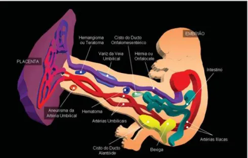
markers, umbilical cord anomalies involv-ing vascular, structural findinvolv-ings, cysts and masses may be found.
The umbilical cord develops around the seventh gestational week, and can be sonographically visualized at the eighth week(1,2).
Umbilical cord cysts are divided into true and pseudo-cysts, the first ones pre-senting an epithelial lining and subdivided into omphalomesenteric duct cysts and al-lantoic duct cysts.
Generally, true umbilical cord cysts de-velop in the fetal end of the umbilical cord, and reports in the literature describe their association with gastrointestinal and geniturinary tracts abnormalities (omphalo-cele, persistence of the urachus and ob-structive uropathy)(1,3,4).
On the other hand, pseudo-cysts do not present an epithelial lining, and originate from a focal edema of the Wharton’s jelly or from its absence because of degenera-tive alterations. Pseudo-cysts are more fre-quent than the true ones and have been found in cases of omphalocele and 18 tri-somy(5).
The exact differentiation between true and pseudo-cysts cannot be achieved by means of ultrasonography; this can be done only by means of a histopathologic study. Differential diagnosis of umbilical cord cysts is umbilical cord masses including
tumors, hematomas, varices and aneurysms (Figure 1).
The present study is focused on umbili-cal cord cyst which has been associated to fetal anomalies and aneuploidies since 1988 in a study developed by Jauniaux et al.(6). Correlation between umbilical cord cysts and anomalies was found in up to 50% of cases, particularly in findings oc-curred at the second and third trimesters of gestation. It is believed that the finding of umbilical cord cysts in such conditions constitute an indication for a karyotype study during the pregnancy.
However, other studies(7–14) have evalu-ated the presence of cord cysts at the first gestational trimester, observing the transi-tory nature of this finding and birth of nor-mal children in most of cases, so fetal karyotyping would not be justified.
In the literature, the prevalence of um-bilical cord cysts sonographically found at different gestational ages ranges between 0.4% and 3.4%(7,8).
The present study was aimed at corre-lating the isolated sonographic finding of umbilical cord cyst and fetal anomalies such as chromosomopathies and structural alterations.
MATERIALS AND METHODS
During the period from June 1996 to June 2006, nine cases of prenatal
sono-graphic studies whose sole finding had been the presence of umbilical cord cyst. These cases were collected and recorded in two units of obstetric ultrasonography uti-lizing equipment Logic 500, Voluson 730 and Logic 9 (GE Medical Systems; Wis-consin, EUA) with convex multifrequency (3–5 MHz) and transvaginal (5–8 MHz) transducers.
The cases identified involved non-se-lected, consecutive, pregnant women from the general population, with single living fetuses.
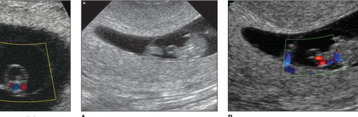
Umbilical cord cyst has been defined as an anechoic, thin-walled structure occur-ring along the umbilical cord and within the amniotic cavity, separated from the fetal pole and physiological herniation. Cord cyst is distinguished from the vitelline sac that has more echogenic walls and extra-amniotic location. Color Doppler was uti-lized for excluding differential diagnoses (Figure 2). The parameters considered in the present study are the same adopted in previous studies in the literature, and are aimed at reducing the incidence of diagnos-tic failure, as well as optimizing the ultra-sonographic method(9,10).
Some of the cases were offered a cyto-genetic study by means of amniocentesis conducted by an obstetrician, with no in-terference from the investigator. Addition-ally, data regarding postnatal physical health outcomes for the neonates and pres-ence, or not, of structural anomalies were collected by means of a review of the pa-tients’ records and contact with involved obstetricians and pediatricians.
RESULTS
In the present study, nine prenatal ultrasonographic images presenting um-bilical cord cyst as the sole abnormality finding with no other marker for fetal ab-normality were evaluated, two of them at the first gestational trimester (Figure 3) and seven at the second and third gestational trimesters (Figure 4). Table 1 demonstrates the results of this evaluation.
It is important to note that in all of the cases the cord cyst was medially located in relation to the length of the umbilical cord, and eccentrically located (para-axial) in re-lation to its diameter.
161 Umbilical cord cyst and fetal anomalies
Radiol Bras. 2008 Mai/Jun;41(3):159–162
Figure 4. Case 4. A: Sonographic image of a thin-walled cyst proximal to the placenta and the fetus. It is possible to observe that the cystic content is anechogenic, even in relation to the amniotic fluid that presents fine débris. B: The utilization of Power Doppler demonstrates the umbilical cord vessels.
Postnatal outcome was normal in all of the cases. Karyotyping by amniocentesis also resulted normal.
DISCUSSION
Umbilical cord cyst etiology is still not completely known, but it is believed to be
related to an increase in the hydrostatic pressure inside the umbilical cord vessels and extravasation of fluid content into the involved region(10,11). At the first gesta-tional trimester, the increase in the hydro-static pressure may be associated with the presence of a physiological herniation of the primitive bowel, whose presence and
disappearance are compatible with reports of transitory umbilical cord cysts in this gestational period. On the other hand, at the second and third gestational trimesters, the increase in the hydrostatic pressure could be a result from fetal growth restriction, omphalocele and chromosomopathies(10). As previously mentioned, the preva-lence of umbilical cord cysts ranged be-tween 0.4% and 3.4%(8,9). The high preva-lence reported by Ross et al.(9) can be attrib-uted to the high-risk population involved in the evaluation. In other studies, the prevalence, in general, were lower(11). Ad-ditionally, the mentioned study has demon-strated that, amongst the first-trimester cysts with spontaneous resolution, i.e., the transitory cases, association with fetal ab-normalities has occurred in 13% of cases, achieving 26% including cases of the cyst persistence up to the third gestational tri-mester. An increase was observed in the risk for fetal abnormality in cases of cord cysts located near the cord placental or fe-Figure 3. Case 1. A: Cystic image at the first gestational trimester, within the amniotic membrane. B:
Utilization of Color Doppler allows the correlation between the image and the umbilical cord.
Figure 2. Case 3. Transverse, cystic image eccen-trically located in the umbilical cord, within the amniotic membrane. The vitelline vesicle is shown at left.
Table 1 Description of cases.
Case
1 2 3 4 5 6 7 8 9
Cyst localization
Median* / Eccentric†
Median / Eccentric Median / Eccentric Median / Eccentric Median / Eccentric Median / Eccentric Median / Eccentric Median / Eccentric Median / Eccentric
Gestational age (DLM)
8 weeks and 3 days 27 weeks and 7 days 9 weeks
22 weeks and 4 days 30 weeks and 2 days 23 weeks
21 weeks and 1 day 22 weeks and 3 days 26 weeks and 4 days
Indication for the study
Routine Routine Routine Routine Routine Routine Routine Routine Routine
Cyst diameter (mm)
2.5 30.7
3.0 32.0 17.2 27.3 25.7 13.8 30.0
Postnatal outcome
Normal Normal Normal Normal‡
Normal Normal Normal‡
Normal Normal
* Location of the cyst in relation to the cord length; † Location of the cyst in relation to the cord circumference; ‡ Cases with normal karyotyping. DLM, date of the last
menstruation.
A B
162
Kobayashi S et al.
Radiol Bras. 2008 Mai/Jun;41(3):159–162 tal insertion, eccentric location in relation
to the longitudinal axis of the cord, and in cases of cysts with larger diameter.
Other two studies(8,10) have evaluated a total of 18 cases of umbilical cord cysts at the first gestational trimester, with normal postnatal outcomes in all of them. A study developed by Sepulveda et al.(10) has con-cluded that the incidental detection of umbilical cord cysts at the first gestational trimester is not associated with a unfavor-able prognosis for the gestation, as far as a low-risk population is concerned. On the other hand, Skibo et al.(8) indicate the first-trimester umbilical cord cyst as a new, many times transitory, sonographic finding, not associated with fetal abnormalities, and that may be clinically non-significant.
The mentioned authors(8–10) consider that the persistence of the finding exceed-ing the first trimester should be differently approached and even indicate fetal karyo-typing, considering the higher risk for aneuploidies and malformations.
So far, the approach suggested by stud-ies in the literature is that a fetal karyotyp-ing is performed in cases of umbilical cord cyst persistence at the second and third ges-tational trimesters, and detailed ultrasono-graphic studies are performed by an expe-rienced sonographist for evaluating the fetal anatomy looking for other markers for
fetal abnormalities in cases of umbilical cord cysts at the first gestational trimester. It is important to note that, unlike in the present study, in the majority of above mentioned studies the finding of umbilical cord cyst was not isolated.
Therefore, despite the small casuistic in the present study, the isolated sonographic finding of umbilical cord cyst did not rep-resented an increase in the risk for struc-tural or aneuploidic anomalies. Based on these findings, there is no indication for fetal karyotyping in cases of isolated sonographic finding of umbilical cord cyst at any gestational age.
Acknowledgements
The authors thank for the collaboration of Rita Ortega (Library of the Institute of Teaching and Research of Hospital Sírio Libanês), and Fabio Augusto dos Santos for the formatting of the present paper.
REFERENCES
1. Harris RD, Alexander RD. Ultra-sonografia da placenta e cordão umbilical. In: Callen PW, edi-tor. Ultra-sonografia em obstetrícia e ginecologia. 4ª ed. Rio de Janeiro: Guanabara Koogan; 2002. p. 563–90.
2. Moore KL, Persaud TVN. Formação do embrião humano. In: Moore KL, Persaud TVN, editores. Embriologia clínica. 5ª ed. Rio de Janeiro: Gua-nabara Koogan; 1994. p. 51–66.
3. Stella A, Babbo GL. Omphalocele and umbilical
cord cyst. Minerva Ginecol. 2000;52:213–6.
4. Smith GN, Walker M, Johnston S, et al. The sonographic finding of persistent umbilical cord cystic masses is associated with lethal aneuploidy and/or congenital anomalies. Prenat Diagn. 1996; 16:1141–7.
5. Kiran H, Kiran G, Kanber Y. Pseudocyst of the umbilical cord with mucoid degeneration of Wharton’s jelly. Eur J Obstet Gynecol Reprod Biol. 2003;111:91–3.
6. Jauniaux E, Donner C, Thomas C, et al. Umbili-cal cord pseudocyst in trisomy 18. Prenat Diagn. 1988;8:557–63.
7. Rempen A. Sonographic first-trimester diagnosis of umbilical cordcyst. J Clin Ultrasound. 1989; 17:53–5.
8. Skibo LK, Lyons EA, Levi CS. First-trimester um-bilical cord cysts. Radiology. 1992;182:719–22. 9. Ross JA, Jurkovic D, Zosmer N, et al. Umbilical cord cysts in early pregnancy. Obstet Gynecol. 1997;89:442–5.
10. Sepulveda W, Leible S, Ulloa A, et al. Clinical significance of first trimester umbilical cord cysts. J Ultrasound Med. 1999;18:95–9.
11. Sepulveda W. Beware of the umbilical cord ‘cyst’ [comment]. Ultrasound Obstet Gynecol. 2003;21: 213–4.
12. Ghezzi F, Raio L, Di Naro E, et al. Single and multiple umbilical cord cysts in early gestation: two different entities. Ultrasound Obstet Gynecol. 2003;21:215–9.
13. Emura T, Kanamori Y, Ito M, et al. Omphalocele associated with a large multilobular umbilical cord pseudocyst. Pediatr Surg Int. 2004;20:636– 9.