www.jcol.org.br
Journal of
Coloproctology
☆
Study conducted at Service of Pathology, Hospital Federal de Ipanema, Ministério da Saúde, Rio de Janeiro, RJ, Brazil. * Corresponding author.
E-mail: [email protected] (L.P.d. Barcellos).
2237-9363/$ - see front matter. © 2014 Sociedade Brasileira de Coloproctologia. Published by Elsevier Editora Ltda. All rights reserved. http://dx.doi.org/10.1016/j.jcol.2013.11.002
Original article
Value of conventional cytology in the presence of
macroscopic lesions of the anal canal
☆Lêda Pereira de Barcellos
a,*
, Fábio Russomano
b, José Ricardo Hildebrandt Coutinho
a,ca Hospital Federal de Ipanema M.S, Rio de Janeiro, RJ, Brazil
b Instituto Nacional de Saúde da Mulher, da Criança e do Adolescente Fernandes Figueira (IFF/FIOCRUZ), Rio de Janeiro, RJ, Brazil
c Instituto de Pesquisa Clínica Evandro Chagas (IPEC/FIOCRUZ), Rio de Janeiro, RJ, Brazil
a r t i c l e i n f o
Article history:
Received 27 September 2013 Accepted 25 November 2013
Keywords: HPV
Prevention of anal neoplasia Conventional cytology
a b s t r a c t
Objectives: To verify the value of conventional cytology for the diagnosis of macroscopic le-sions of the anal canal and to describe the limitations of the samples.
Method: We evaluated 395 conventional cytology samples obtained by brushing the anal canal of patients (predominantly male, HIV-positive) and compared them to the presence of macroscopic lesions of the anal canal observed under anorectal examination.
Results: Of the total, 91.6% of samples were classii ed as adequate. Cellular elements repre-sentative of the anal transformation zone were observed in 63.5% of samples. Sensitivity in the presence or absence of cellularity was 80% and 31%, respectively.
Conclusion: The study demonstrates the feasibility of using conventional anal cytology in outpatients.
© 2014 Sociedade Brasileira de Coloproctologia. Published by Elsevier Editora Ltda. All rights reserved.
Valor da citologia convencional na presença de lesões macroscópicas do canal anal
Palavras-chave: HPV
Prevenção de neoplasia anal Citologia convencional
r e s u m o
Objetivo: verii car o valor da citologia convencional no diagnóstico de lesões macroscópicas do canal anal e descrever as limitações das amostras obtidas.
Método: avaliamos 395 exames citológicos convencionais obtidos por escovado do canal anal de pacientes predominantemente do sexo masculino, soropositivos para HIV, e com-paramos com a presença de lesões macroscópicas do canal anal constatadas ao exame proctológico.
Conclusão: O estudo demonstra a possibilidade de utilização da citologia anal convencional no rastreio de lesões macroscópicas do canal anal em pacientes ambulatoriais.
© 2014 Sociedade Brasileira de Coloproctologia. Publicado por Elsevier Editora Ltda. Todos os direitos reservados.
Introduction
The incidence of anal squamous cell carcinoma and its pre-cursor lesions increased in recent decades, and it has been widely proven the participation of HPV (human papillomavi-rus) types 16 and 18 in its pathogenesis.¹
Although still a relatively rare cancer, its incidence has been rising alarmingly in younger patients, especially men who practice receptive anal sex with men (MSM, so-called by its acronym in English), independent of HIV infection, but with greater involvement of those infected with this virus. Women with a history of multicentric squamous intraepithe-lial neoplasia, heterosexual men and women HIV-positive or immunosuppressed for other reasons also have contributed to the increased incidence of this neoplasia.²
In Brazil we do not have data on the incidence of anal can-cer, because they are included in the colon and rectum to-pography. In these topographies 14,180 and 15,960 new cancer cases in men and in women, respectively, are expected, cor-responding to an estimated risk of 15 new cases per 100,000 men and 16 new cases per 100,000 women.3 Regarding the
situation of AIDS, there has been an increase in the number of cases of the disease in young people included in the MSM exposure category.4
The highly active antiretroviral therapy (HAART) seems not to modify the pathophysiology of HPV anal lesion in HIV-infected patients, suggesting that a progression to serious in-jury should occur in patients with prolonged survival.5
The similarities between cervical and anal cancers, for in-stance, the association with HPV, the greater occurrence in the existing epithelial transformation zone in both topogra-phies and the ability to diagnose early lesions susceptible to less aggressive treatments, led to the use of exfoliative cy-tology as a diagnostic test for more than a decade, and this procedure can be performed by the conventional method or in liquid medium, with similar results. Still, the current inci-dence of anal cancer is similar to that of cervical cancer be-fore the establishment of prevention programs, bringing great expectations in the use of anal cytology.¹,²
Several studies show that cytology has a good sensitivity but low speciicity. A meta-analysis reported variations in sen-sitivity between 42 and 98% and of speciicity between 16 and 96%.¹ An important factor for this variation may be due to lack of standardization of how the cytobrush should be introduced into the anal canal6 and to the greater dificulty in the
obtain-ment and preparation of anal canal samples, compared with the cervical collection.7 The evaluation of the screening
effec-tiveness for long-term prevention of anal as well as cervical cancer still remains to be done,¹ A contributing factor to this performance variation is the little experience of cytopatholo-gists with the sampling procedure in the anal canal. Aiming at
the improvement of this procedure, the College of American Pathologists (CAP) recommends the ampliation of its use.²
In this study we report the performance of this test in the presence of macroscopic lesions in the anal canal in outpa-tients seen at the Hospital Federal de Ipanema/MS/Rio de Janeiro, using the conventional method, a technology widely available in our country. Should it prove appropriate, it will contribute for the prevention of anal canal cancer in outpa-tient visits.
Material and method
From April 2005 to December 2011, 395 cytopathology exams of anal canal were compared with the clinical diagnosis of presence of macroscopic lesions. The information was ex-tracted from cytologic requisitions and medical records of the patients examined.
The study was approved by the Ethics Committee on Hu-man Research of the Hospital Federal dos Servidores do Es-tado (CEP_HFSE - 000474 protocol).
Patients whose samples were included in this trial were mostly men attended at our proctology outpatient clinic. The reasons of the visits were anal sex practice, some anal claim arising out of sexually transmitted disease or otherwise, pres-ence of warts, or seropositivity for HIV. Samples of three pa-tients from the gynaecology outpatient clinic were also includ-ed. One of them had perianal condylomata and the other two a diagnosis of vulvar intraepithelial neoplasia (VIN II and VIN III). In most cases, the sampling was done in two slides at the irst consultation, because no prior preparation is necessary. Patients who reported having receptive anal sexual relation-ship or made use of suppositories, creams or enema the day before the consultation were told to return later, observing the necessary precautions before collection.
The cytobrush containing the collected material was then rotated with a uniform motion on the central part of each slide, resulting in thin smears arranged longitudinally that, within 10 seconds and still wet, were totally immersed in hydrated ethyl alcohol for i xation of the samples (Fig. 1).
After collection, an inspection with conventional touch and anoscopy was performed. In patients older than 50 years a rec-tosigmodoscopy was also performed. Then, the samples were sent for the Pathology Service, along with the application form containing clinical data; there, the slides were stained by Papa-nicolaou. The reports were issued by an experienced cytopa-thologist reporting the representativeness of epithelia present in the sample and the diagnostics, based on cytomorphological criteria for diagnostic elaboration according to The Bethesda System for Reporting Cervical Cytology8 (Fig. 2). Considering
that the conventional method was used, the criteria for the ad-equacy of the sample were based on the 1991 version of The Bethesda System, which dei nes the sample as “paucicellular”
when it covers less than 10% of the surface of the slide, and “prejudiced” when i xation artefacts, overlapping, blood stains and other contaminants affect 50-75% of the sample.
Samples composed exclusively of keratinized squamous cells; paucicellular samples; or those with artefacts of i xation, overlapping, exudate, erythrocytes and faecal material that prevented the diagnosis were judged unsatisfactory.
Data were stored in spreadsheets and analyzed using SPSS v. 21.
Results
The sample consists of 395 cytological exams of patients pre-dominantly male, and, among these, of HIV-seropositive ones (59.7%). Most patients (61.2%) presented lesions at the time of collection. The injury to the anal canal, visualized by conven-tional anoscopy, was present in 31.7% of men and in 22.5% women (Table 1).
At the moment of the collection, the clinical inspection of the perianal region and a conventional anoscopy showed pre-dominance of macroscopic lesions in HIV-positive patients (62.5%) (data not shown).
Most of the samples were judged satisfactory by evalua-tion according to the criteria dei ned by Bethesda (Table 2). The most frequent cause of limitation of samples was
pauci-Table 1 – Characteristics of patients from whom the specimens for cytopathological exam of anal canal (HFI/ MS, 2005-2011) were obtained (cytobrushing).
Characteristics Male Female Total
Mean age in years (range) 36.8 (16-84) 41.9 (21-80) 37.9 (16-84) HIV situation
HIV+ 188 (59.7%) 15 (18.7%) 203
HIV - 64 (20.3%) 29 (36.2%) 93
HIV unknown 63 (20%) 36 (45%) 99
Presence of macroscopic lesion
Perianal 102 (32.3%) 22 (27.5%) 124
Anal canal 45 (14.3%) 15 (18.7%) 60
Both 55 (17.6%) 3 (3.8%) 58
Absence of perianal or anal canal lesion
113 (35.8%) 40 (50%) 153
Total 315 80 395
Fig. 1 – Appearance of adequate Pap smears after staining.
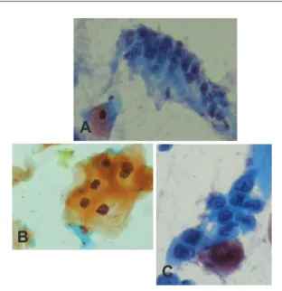
Fig. 2 – A, Columnar cells from the anal transformation zone. B, Low-grade anal intraepithelial lesion. C, High-grade anal intraepithelial lesion. (Conventional cytology stained by Papanicolaou method ×400.)
Table 2 – Classifi cation of the adequacy of smears obtained by anal cytobrushing (HFI/MS, 2005-2011).
Smear characteristics
Male Female Total
Satisfactory samplesa 292 (92.6%) 70 (87%) 362 (91.6%)
Unsatisfactory samplesb
6 (60%) 4 (40%) 10 (2.5%)
Limited samplesc 17 (73.9%) 6 (26%) 23 (5.8%)
Total 315 80 395
a Representative and preserved cellularity, composed of keratinized
and non-keratinized squamous cells and/or transitional and/ or columnar cells. bUnrepresentative cellularity, consisting
exclusively of keratinized squamous cells. c Limited cellularity, with
poor i xation, presence of blood, exudate or faecal material.
A
B
cellularity (43.4%) followed by exudate, bacteria and hematic background. The artefacts of ixation (13%) and faecal mate-rial presence (4.3%) had low signiicance.
The analysis of the frequency of unsatisfactory samples over time demonstrates a downward trend, but the same was not observed with respect to the limited samples. There was a increase in this category in 2011, due to paucicellularity and exudate. The downward trend in the proportion of inadequate samples was statistically signiicant (linear regression, p = 0.048821) (Table 3).
Representative elements of the anal transformation zone in 63.5% of samples were found, with no signiicant difference between genders (data not shown).
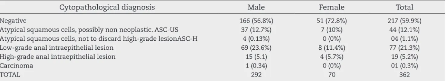
Among the tests considered satisfactory and altered, a predominance of low-grade lesions was perceived, especially in men; but it is worth mentioning a signiicant percentage of diagnoses of major relevance (Table 4).
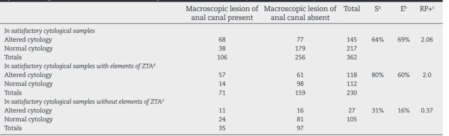
The analysis of the diagnostic performance of anal cytol-ogy for detection of macroscopic lesions of the anal canal showed signiicant increase in sensitivity when the collection brought representative elements of the anal transformation zone (Table 5).
Discussion
The prevention of anal cancer in Brazil is critical. Informa-tion campaigns are essential to reach a populaInforma-tion exceed-ing 1.5 million MSM, accordexceed-ing to a survey conducted on attitudes and practices in the Brazilian population in 2008 (PCAP-BR)9 and women with a history of genital
intraepithe-lial neoplasia.10
The methodology of liquid medium is still incipient among us and the biased and critical view of the conventional meth-od, which is widely available, has resulted in few services per-forming exfoliative cytology of the anal canal.
The use of this method in services that do not have an anoscope, including those providing care for HIV+ patients, should be encouraged to evaluate the presence of injury and to referral of patients with abnormal tests for centres with adequate equipment.11 Protocols and diagnostic algorithms
based on cytologic changes are already suggested, in order to reduce the incidence of anal cancer in high-risk populations.12
The intent of this study was to evaluate the performance of conventional exfoliative cytology in the diagnosis of mac-roscopic lesions in the anal canal. We also analyzed the procedure as a function of sample quality. The lack of in-formation about subclinical lesions prevents a more accu-rate analysis of this performance, but many patients remain continue to be followed-up, having been submitted to high-resolution anoscopy and biopsies, both liable to assessment in a next step.
The reduced percentage of unsatisfactory samples in this study is similar to the 6% reported by Mathews WC et al.,13
while studying 2,947 conventional smears of the anal canal. We believe that the training given to proctologists, coupled with the fact that the same physician collected most samples (78%), has contributed to our results, being an essential step in implementing the use of this practice in a health care facil-ity. On the other hand, Sherman et al.,14 after months of
train-ing, considered most of conventional samples unsatisfactory, attributing the failure to the fact that the professionals in-volved in the collection, both medical and nonmedical, had no prior experience in collecting this type of sample.
Table 3 – Evolution of the percentage of inadequate samples in smears obtained from anal cytobrushing over the years of study (HFI/MS, 2005-2011).
2005 2006 2007 2008 2009 2010 2011 Total
Number of exams
14 22 68 57 52 81 101 395
Unsatisfactory samples
3 1 5 0 0 1 0 10
Limited samples
0 1 9 4 3 1 5 23
Total of inadequate samples*
3 2 14 4 3 2 5 33
% 21.4% 9.0% 20.5% 14.0% 5.7% 2.46% 4.95%
* Result of the sum of unsatisfactory and limited samples.
Table 4 – Cytopathological diagnoses of smears considered as satisfactory, obtained by anal cytobrushing (HFI/MS, 2005-2011).
Cytopathological diagnosis Male Female Total
Negative 166 (56.8%) 51 (72.8%) 217 (59.9%)
Atypical squamous cells, possibly non neoplastic. ASC-US 37 (12.7%) 7 (10%) 44 (12.1%)
Atypical squamous cells, not to discard high-grade lesionASC-H 4 (0.13%) 0 (0%) 04 (1.1%)
Low-grade anal intraepithelial lesion 69 (23.6%) 8 (11.4%) 77 (21.3%)
High-grade anal intraepithelial lesion 15 (5.1) 4 (5.7%) 19 (5.2%)
Carcinoma 1 (0.34) 0 (0%) 01 (0.3%)
Our experience with anal canal cytology began in 2005 with the examination of smears obtained from patients in-cluded in this study, when several studies had already de-scribed the differences between cytological samples of cer-vical and anal canal.7,14-16 One of our concerns was to meet
the criteria contained in The ABCs of anal-rectal cytology CAP May 2004,17 limiting the inconclusive diagnoses of
atyp-ical cells of undetermined signiicance (ASC-US and ASC-H). The works de Ruiter et al.18 and Palefsky et al.19 correlated
the representativeness of the anal transformation zone in the conventional sample with diagnostic performance and presence of lesions in the anal canal. In the irst trial, 154 conventional cytological samples versus microanoscopy-guided histological diagnoses of anal biopsy were evaluated, and the presence of anal transformation zone cells was con-sidered as a criterion of adequacy. These authors found a sensitivity of 87.5% and a speciicity of 16.3%.
Palefsky et al.19 evaluated 658 MSM, and most remained
in follow-up for two years. These authors correlated the conventional cytopathological exam with high-resolution anoscopy and histopathology. The results were grouped ac-cording HIV status and representativeness of the anal trans-formation zone in the sample. The HIV-positive participants had sensitivity of 69% and speciicity of 59%; and the sero-negative participants, sensitivity of 47% and speciicity of 92%. Palefsky et al. concluded that the absence of columnar cells did not affect the diagnosis.
The study showed discrepant results when compar-ing the samples with and without components of the anal transformation zone, in disagreement with the opinion of some authors, who consider the presence of cells of the anal transformation zone as a qualitative data, without im-pact on the result. However, recent review articles point out that there are few publications addressing the minimum standard of adequacy of anorectal samples, and how the cellularity inluences the sensitivity and speciicity of the method.1,2
Anal cancer is related to a natural body oriice, and there-fore its diagnosis should be easy and established early. How-ever, the fact that is the anus, with all its burden of stigma and prejudice, coupled with the fact that its symptoms are indistinguishable from symptoms of the most common anal
Table 5 – Diagnostic performance of cytopathologic exam of smears considered satisfactory, obtained by anal cytobrushing, in the detection of macroscopic lesions of anal canal (HFI/MS, 2005-2011).
Macroscopic lesion of anal canal present
Macroscopic lesion of anal canal absent
Total Sa Eb RP+c
In satisfactory cytological samples
Altered cytology 68 77 145 64% 69% 2.06
Normal cytology 38 179 217
Totals 106 256 362
In satisfactory cytological samples with elements of ZTAd
Altered cytology 57 61 118 80% 60% 2.0
Normal cytology 14 98 112
Totals 71 159 230
In satisfactory cytological samples without elements of ZTAd
Altered cytology 11 16 27 31% 16% 0.37
Normal cytology 24 81 105
Totals 35 97
aSensibility; bspeciicity; codds ratio positive; danal transformation zone.
diseases, contributes for diagnoses often obtained at more advanced clinical stages and not always of easy solution.20
The recognition of the risk of developing anal cancer is still undervalued by physicians and even by those social groups whose patients of the so-called risk group have visibility.2
Conclusion
For the authors, this study demonstrated that the use of the conventional cytology method, widely available in our coun-try, is a viable technology, provided that all the steps of col-lecting and ixing the samples be performed carefully, includ-ing allowinclud-ing their transport when the samples are taken at distant locations from the laboratory.
Conlicts of interest
The authors declare no conlicts of interest.
Acknowledgements
I would like to acknowledge the cooperation of all adminis-trative and technical employees of the Service of Pathology, Hospital Federal de Ipanema, Marcos Costa, Marcos Duque and my daughter Cristina for her invaluable help.
R E F E R E N C E S
1. Bean SM, Chhieng DC. Anal-rectal cytology: a review. Diagn Cytopathol 2010; 38: 538–46.
2. Darragh TM, Winkler B. Anal cancer and cervical cancer screening: key differences. Cancer 2011; 119: 5–19. 3. MINISTÉRIO DA SAÚDE. Instituto Nacional de Câncer José
Alencar Gomes da Silva (INCA). 2ª edição revista e atualizada. Rio de Janeiro, RJ. 2012 .
4. Monitoramento e Avaliação em DST/AIDS. 1/26. Programa Municipal de Boletim Epidemiológico 4º Trimestre de 2008. Sítio de Excelência em DST/ Aids.
6. Nadal SR, Horta SH, Calore EE, Nadal LR, Manzione CR. [Quanto a escova deve ser introduzida no canal anal para avaliação citológica mais eicaz?] Rev Assoc Med Bras 2009; 55: 749–51. 7. Darragh TM, Jay N, Tupkelewicz BA, Hogeboom CJ, Holly EA,
Palefsky JM. Comparison of conventional cytologic smears and Thin Prep preparations from the anal canal. Acta Cytol 1997; 41: 1167–70.
8. Darragh TM, Birdsong GG, Luff RD, Davey DD: Anal-rectal cytology. In: Solomon D, Nayar R, eds. The Bethesda System for Reporting Cervical Cytology: Deinitions, Criteria and Explanatory Notes. 2nd ed. New York: Springer; 2004:169-175.Solomon D, Nayar R, editors. The Bethesda System for reporting cervical cytology. New York: Springer; 2004. 9. Pesquisa de Conhecimentos, Atitudes e Práticas Relacionadas
às DST e Aids da População Brasileira de 15 a 64 anos de idade – MS, 2008.
10. Jacyntho C, Giraldo P. A importância do exame cito-anuscópico sob visão ampliada para o diagnóstico das neoplasias intra-epiteliais anais em pacientes com neoplasias intra-intra-epiteliais genitais. Revista Brasileira de Ginecologia e Obstetrícia 2005; 27, 44-45.
11. Nadal SR, Calore EE, Nadal LR, Horta SH, Manzione CR. Citologia anal para rastreamento de lesões pré- neoplásicas . Rev Assoc Med Bras 2007; 53: 147–51.
12. Park IU, Palefsky JM. Evaluation and management of anal intraepithelial neoplasia in HIV-negative and HIV-positive men who have sex with men. Curr Infect Dis Rep. 2010;12:126-133.
13. Mathews WC, Sitapati A, Caperna JC, Baber RE, Tugend A, Go U. Measurement characteristics of aAnal cytology, histopathology, and high-resolution anoscopic visual impression in an anal dysplasia screening program. J Acquir Immune Deic Synd 2004;37:1610-15.
14. Sherman ME, Friedman HB, Busseniers AE, Kelly WF, Carner TC, Saah AJ. Cytologic diagnosis of anal intraepithelial neoplasia using smears and cytyc thin-preps. Mod Pathol 1995; 8: 270–4.
15. Friedlander MA, Stier E, Lin O. Anorectal cytology as a screening tool for anal squamous lesions: cytologic,
anoscopic, and histologic correlation. Cancer 2004; 102: 19–26. 16. Arain S, Walts AE, Thomas P, Bose S. The anal Pap smear:
cytomorphology of squamous intraepithelial lesions. Cytojournal 2005; 2: 4.
17. Darragh TM, Winkler B. The ABCs of anal-rectal cytology. CAP TODAY. May 2004:42–50.
18. de Ruiter A, Carter P, Katz DR, Kocjan G, Whatrup C, Northover J, et al. A comparison between cytology and histology to detect anal intraepithelial neoplasia. Genitourin Med. 1994 Feb;70(1):22–25.
19. Palefsky JM, Holly EA, Hogeboom CJ, et al. Anal cytology as a screening tool for anal squamous intraepithelial lesions. J Acquir Immune Deic Syndr Hum Retrovirol. 1997; 14: 415-22. 20. Coutinho, JRH. Rastreamento de lesões pré-neoplásicas do