www.jcol.org.br
Journal of
Coloproctology
* Corresponding author.
E-mail: [email protected] (J.A.D.d. Cunha e Silva).
2237-9363/$ - see front matter. © 2014 Sociedade Brasileira de Coloproctologia. Published by Elsevier Editora Ltda. All rights reserved. http://dx.doi.org/10.1016/j.jcol.2013.12.005
Original article
Anal melanoma: a rare, but catastrophic tumor
Andréa da Costa Veloso, Jaime Coelho Carlos Magno, José Antonio Dias da Cunha e Silva*
Colorectal Division of Hospital Naval Marcílio Dias, Rio de Janeiro, RJ, Brazila r t i c l e i n f o
Article history:
Received 11 September 2013 Accepted 5 December 2013
Keywords:
Anal Rectal Melanoma
a b s t r a c t
Introduction: Malignant melanoma of the anal canal is a rare and aggressive disease, which early diagnosis is difi cult. Its presentation with no specii c symptoms leads to a late diag-nosis at an advanced stage. The progdiag-nosis of anorectal malignant melanoma is poor and frequently related to distant metastasis and absence of response of chemoradiotherapy. Surgery remains the mainstay of therapy; otherwise, the best approach is controversial. Considering no survival benei ts for APR, wide local excision should be considered as the treatment of choice.
Methods: This report collects nine cases of anorectal melanoma treated at our division from 1977 to 2006, as well as a review of the literature.
Results: There were eight females and one male, of medium age 69 years (range: 41-85 years). Most frequent presentation was bleeding. Wide Local Excision (WLE) was performed in seven of them. Mean survival was 24 months, and six of them died on account of meta-static disease.
Conclusion: Anorectal melanoma remains challenging. Efforts should be taken to early diag-nosis, and wide local excision with negative margins is the preferred treatment. Abdomi-noperineal resection (APR) is a reasonable option for bulky tumors or when the sphincter is invaded.
© 2014 Sociedade Brasileira de Coloproctologia. Published by Elsevier Editora Ltda. All rights reserved.
Palavras-chave:
Anal Reto Melanoma
r e s u m o
Melanoma anal: tumor raro, mas catastrói co
Introdução: O melanoma maligno do canal anal é uma doença rara e agressiva, em que o diagnóstico precoce se torna difícil. Apresenta-se sem sintomas especíi cos, levando ao diagnóstico tardio e em fase avançada. O prognóstico é ruim e frequentemente relacionado a metástases a distância, bem como à ausência de resposta à rádio e à quimioterapia. A cirurgia permanece como terapia de escolha, no entanto a melhor abordagem ainda é con-troversa. Considerando não haver benefício na sobrevida da amputação abdômino-aerineal do aeto (AAPR), a excisão local ampla deve ser considerada o tratamento de escolha.
realizada em sete pacientes. A sobrevida média foi de 24 meses.
Conclusão: O melanoma anorretal continua desaiante. Todos os esforços devem ser feitos para o diagnóstico precoce, tornando assim possível realizar a excisão local com margens negativas. A AAPR ainda é uma opção factível para tumores avançados ou quando o esfínc-ter anal está comprometido.
© 2014 Sociedade Brasileira de Coloproctologia. Publicado por Elsevier Editora Ltda. Todos os direitos reservados.
Introduction
Anal malignant melanoma is a rare and aggressive disease with no speciic clinical symptoms and a poor outcome. It is often diagnosed at an advanced stage, presenting with hematoge-nous metastasis. The anorectal region is the most common site for development of primary melanoma within the alimentary tract. The majority of the lesions arises from the dentate line of the anal canal and tends to spread submucosally.
Historically, anal melanoma was treated with abdomino-perineal resection (APR), but recently some studies demon-strated no difference of the survival rates comparing to wide local excision (WLE). It appears that the only beneit of radical surgery is to obtain clear margins in tumor that are particularly bulky or invading the sphincter, and here APR should be con-sidered. So, WLE offers the advantage of avoiding a permanent colostomy and is the preferred treatment when negative mar-gins can be achieved.1-4
Methods
The data of nine patients with anorectal melanoma were col-lected and retrospectively analyzed. From 1977 to 2006 we had a total of nine cases, including one male and eight fe-males. Patients complaint, tumor characteristics, type of sur-gery, distant metastasis and overall survival were analyzed.
Due to the small number of patients, statistics analysis was not carried out.
Results
There were one male and eight females, of median age of 69 (41-85) years. Most frequent complaint was rectal bleeding. Two patients had amelanotic melanomas. Among nine pa-tients, none presented with distant metastasis when the time of diagnostic. Six were Caucasian.
Seven patients underwent WLE, and one patient whose bi-opsy revealed melanoma refused any further treatment. This patient returned two years later with a bulky bleeding tumor, and a palliative sigmoidostomy was performed combined with embolization due to severe bleeding. One patient, at the time of diagnosis, presented as frozen pelvis, and had not enough time to a surgical approach. The median overall sur-vival was 24 months. Among patients who underwent WLE we missed the follow up of one patient, one patient presented
a stroke and died ive years after the procedure, and one pa-tient is still alive (Figs. 1-6) (Table 1).
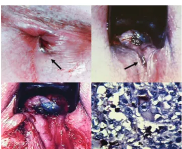
Fig. 2 – In clock hour: Dark pigmentation arising in anal verge extending to polypoid lesion in distal rectum (arrows). Wide local excision performed preserving the esincter muscle. Melanotic pigment in the cytoplasm of neoplasic cells (case two).
Discussion
Anorectal malignant melanoma is a rare mucosal disease with a particularly aggressive biology compared with cutaneous melanoma of equal stage, accounting for approximately 1-4% of anorectal malignancies,2,5 but it is the third most common site
after skin and eye and represents 0.6-1.6% of all melanomas.6,7
The majority of patients are Caucasian, with the highest incidence during sixth and seventh decade. There is a slight female preponderance. The anorectal melanoma arises from melanocytes present in the transitional zone of the surgical anal canal and tends to spread submucosally.8,9 Related to its
no speciic symptoms, most of them, presenting with bleed-ing, prolapsed mass and anorectal pain, almost 60% of patients have already disseminated disease at initial diagnosis.
Melano-mas are commonly mistaken for hemorrhoidal disease, and the inal diagnosis can be conirmed by positive excisional biopsy.1,2
The lesion is not pigmented in about 30% of cases and may re-semble a villous carcinoma, and carries a worse prognosis by its invasive nature.2,7,9
Immunohistochemical studies should always be done for establishing the diagnosis of melanoma. It is mostly positive for protein S-100 with a reported rate of 100%, melanoma anti-gen HMB-45 and vimentin.8,10 Others tests, as endoluminal
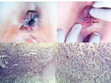
ul-trasound and computed tomography, should be performed to address the extension and presence of metastatic disease as its Fig. 3 – In clock hour: Typical dark nodular lesion in anal
margin with black pigmentation (arrows). Hystopathologic
indings suitable of malignant melanoma (case four). Fig. 5 – In clock hour: Fungiform dark mass in distal rectum. After neoadjuvant radiotherapy, wide local
excision with loop ileostomy. Brain methastasis one month after surgery. Hystopathologic indings show the lesion beginning at anal canal and extending to distal rectum (case seven).
Fig. 6 – In clock hour: Nodular lesion with dark pigmentation at the basis in anal verge (arrows). Local excision with sphincter preservation and marsupialization of the surgical incision. Complete healing at 180 days (case eight).
diagnosis. The computed tomography can reveals intraluminal masses in the distal rectum, with perirectal iniltration and of-ten enlarged lymph nodes. Endorectal ultrasound is helpful in the evaluation of the tumor thickness and its nodal status.2,8
Various factors, including duration of symptoms, inguinal lymph node involvement, tumor stage, the presence of amela-notic melanoma on histology, tumor necrosis, perineural inva-sion and tumor thickness, have been suggested to be negative prognostic factors. However, female sex has been suggested to be a positive prognostic factor for patients with cutaneous mel-anoma, and with an improved survival after radical resection for regional stage of anal melanoma. This reason is unclear.8,9,11
The optimal treatment remains controversial, and surgery is based on two operative options: wide local excision (WLE) and abdominoperineal resection (APR). In the past, APR was advo-cated for the nonmetastatic disease, but as the prognosis was poor regardless of surgical approach, the goal of the surgical procedure should be based on obtaining negative margins and maintaining sphincter function. Most studies shows APR to pro-vide a better local control, but no clear improvement in survival, and further consideration must be given to quality of life issues when making decisions between these two options.2,5,8,9,12,13,15
Tumor thickness is a strong predictor factor for the risk of local recurrence and is used to plan therapeutic proce-dures. Tumor thickness below 1mm can be performed by local sphincter-saving excision with a 1cm safety margin. Tumor thickness between 1-4 cm should be excised by sphincter-saving excision and 2 cm safety margin, and tumor with thickness above 4 cm or with the involvement of sphincter should be treated with APR.8,9,11,13,14,18
Stoidis et al. hypothesized that systemic dissemination is an early event in tumorigenesis, and by the time the lesion is clinically apparent micrometastases are well established. Me-tastases occur via lymphatic and hematogenous routes. Lym-phatic spread to mesenteric nodes is more common than to inguinal nodes, while lungs, liver and bones are the most fre-quent sites of distant metastases.8
Prophylactic lymph node resection has no value, and the therapeutic lymph node resection should be performed only in the presence of positive inguinal nodes. Sentinel lymph node mapping (SLNM) has inluenced the extension of surgi-cal resection and seems to be helpful in preventing understat-ing patients, who are pathologically node-positive but clini-cally node-negative and can allow early beneicial completion lymphadenectomy.5
The role of adjuvant therapy remains unknown. The re-sponse of anorectal melanoma to radio and/or chemotherapy continues to be poor. No systemic therapy regimen for meta-static anal melanoma is considered standard of care. Treat-ment is based on drugs developed for advanced cutaneous melanoma, although the clinical, biologic, and molecular seems to be different.8 Kim et al. used a combination of
temo-zolomide, cisplatin and liposomal doxorubicin for metastatic anal melanoma with encouraging results.4,17
Regardless of surgical approach, melanoma remains a highly lethal malignancy with overall 5-years survival rate less than 20%. The median survival is 34 months for patients with local disease and 10 months for those with metastatic disease.8,9
The early diagnosis is the key to improved survival rate for patients with anal melanoma, due to the fact that the stage of the disease is the most important determinant in anorec-tal melanoma. A standard approach has not been established because of the limited number of patients of all anal mela-noma reports. Further studies of the molecular mechanisms and tumor progression are needed to develop new treatment paradigms and improve survival.
Conclusion
Considering that anal melanoma is an aggressive tumor often diagnosed in advanced stages, a wide local excision should be considered as a therapeutic approach, despite controversial. CASES Year Age Gender Local Complaint Tumor
appearance Surgery Adjuvant therapy Metastasis or recurrence (months) Survival
1 1977 71 Female Anal Bleeding Hemorrhoidal thrombosis
Local excision Imuno + QT
Liver (24m) 70m
2 1977 69 Male Anorectal Bleeding Anal pigmented nodule
Local excision QT Rectum (6m) 14m
3 1993 58 Female Anorectal Anal discharge
Rectal ulcerated mass
Local excision QT + interferon
Lung (18m) 23m
4 1995 85 Female Anal Bleeding Anal pigmented nodule
Local excision No Groin (2m) 29m
5 1996 71 Female Anorectal Bleeding Rectal polyp Local excision No Rectum lymphnode (8m)
12m
6 1996 42 Female Anorectal Bleeding Anal polyp Sigmoidostomy + embolization
No Liver (35m) 40m
7 2004 41 Female Anorectal Bleeding Rectal pigmented nodule
Local excision + ileostomy
RT Brain (1m) 8m
8 2006 81 Female Anal Nodule Anal pigmented nodule
Local excision No No alive
9 2006 58 Female Anorectal Bleeding Rectal pigmented nodule and ulcerated mass
There´s no signiicant impact in general survival with APR. So, WLE has its beneits in quality of life.13,16
Conlicts of interest
The authors declare no conlicts of interest.
R E F E R E N C E S
1. Zhou HT, Zhou ZX, Zhang HZ, Bi JJ, Zhao P. Wide local excision could be considered as the initial treatment of primary anorectal malignant melanoma. Chin Med J (Engl). 2010 Mar;123(5):585-8.
2. Trzcinski R, Kujawski R, Mik M, Sygut A, Dziki L, Dziki A. Malignant melanoma of the anorectum-a rare entity. Langenbecks Arch Surg. 2010 Jan 12.
3. Khaled A, Hammami H, Fazaa B, Kourda N, Kamoun MR, Ben Jilany S, Zoghlami A. Primary amelanotic anorectal melanoma: an uncommon neoplasia with poor prognosis. Pathologica. 2009 Jun;101(3):126-9.
4. Kim KB, Sanguino AM, Hodges C, Papadopoulos NE, Eton O, Camacho LH, et al. Biochemotherapy in patients with metastatic anorectal mucosal melanoma. Cancer 2004, 100:1478-1483.
5. Iddings DM, Fleisig AJ, Chen SL, Faries MB, Morton DL. Practice patterns and outcomes for anorectal melanoma in the USA, reviewing three decades of treatment: is more extensive surgical resection beneicial in all patients? Ann Surg Oncol2010;17:40-44.
6. Nivatvongs S. Perianal and analcanal neoplasms. In: Gordon PH, Nivatvongs S, eds. Colon, Rectum and Anus. 3rd ed. New York: Informa Healthcare; 2007:369-390.
7. Garrett K, Kalady MF. Anal neoplasms. Surg Clin North Am 2010;90:147-161, Table of Contents.
8. Stoidis CN, Spyropoulos BG, Misiakos EP, Fountzilas CK, Paraskeva PP, Fotiadis CI. Diffuse anorectal melanoma: review of the current diagnostic and treatment aspects based on a case report. World J Surg Oncol. 2009 Aug 11;7:64.
9. Sayari S, Moussi A, Bel Haj Salah R, Gherib SB, Haouet K, Zaouche A. Primary anorectal melanoma: a case report. Tunis Med. 2010 Jun;88(6):430-2.
10. Seya T, Tanaka N, Shinji S, Shinji E, Yokoi K, Horiba K, et al.2007. A case of rectal malignant melanoma showing immunohistochemial variability in a tumor. J Nippon Med Sch 74:377–381.
11. Kiran RP, Rottoli M, Pokala N, Fazio VW. Long-term outcomes after local excision and radical surgery for anal melanoma: data from a population database. Dis Colon Rectum. 2010 Apr;53(4):402-8.
12. Bullard KM, Tuttle TM, Rothenberger DA, et al. Surgical therapy for anorectal melanoma. Journal of the American College of Surgeons2003;196:206-211.
13. Thibault C, Sagar P, Nivatvongs S, Ilstrup DM, Wolff BG. Anorectal melanoma — an incurable disease? Dis Colon Rectum1997;40:661-668.
14. Weyandt GH, Eggert AO, Houf M, Raulf F, Brocker EB, Becker JC. Anorectal melanoma: surgical management guidelines according to tumour thickness. Br J Cancer 2003;89:2019-2022. 15. Yeh JJ, Shia J, Hwu WJ, et al. The role of abdominoperineal
resection as surgical therapy for anorectal melanoma. Ann Surg2006;244:1012-1017.
16. Martínez-Hernández-Magro P, Villanueva-Sáenz E, Chávez-Colunga L. Anal malignant melanoma. Case report and literature review. Rev Gastroenterol Mex. 2009 Jan-Mar;74(1):39-44.
17. Hay A, Liong J, Kumar D, Glees J. A striking response of anorectal melanoma to radiotherapy (locoregional disease conined to perineum and anal canal). Ann R Coll Surg Engl. 2010 Jan;92(1):W10-2.