www.jcol.org.br
Journal of
Coloproctology
* Corresponding author.
E-mail: [email protected] (R.O. Perez).
2237-9363/$ - see front matter. © 2014 Sociedade Brasileira de Coloproctologia. Published by Elsevier Editora Ltda. All rights reserved. http://dx.doi.org/10.1016/j.jcol.2013.12.007
Original article
Total mesorectal excision and sphincter preservation —
the early steps of rectal cancer surgery
Rodrigo O. Perez
a,*, Guilherme P. São Julião
b, Igor Proscurshim
b, Charles Sabbagh
b,
Esteban Grzona
b, Patricio B. Lynn
b, Joaqim Gama-Rodrigues
b,ca Colorectal Surgery Division, Faculdade de Medicina, Universidade de São Paulo, São Paulo, SP, Brazil
b Angelita & Joaquim Gama Institute, São Paulo, SP, Brazil
c Faculdade de Medicina, Universidade de São Paulo, SP, Brazil
a r t i c l e i n f o
Article history:
Received 15 October 2013 Accepted 11 December 2013
Keywords: Rectal cancer
Sphincter preservation Total mesorectal excision Intersphicteric resection
a b s t r a c t
The treatment of rectal cancer has evolved signii cantly over the last 100 years. Standardiza-tion of total mesorectal excision and the development of techniques for sphincter preserva-tion have resulted in signii cant improvements in the management of this disease. Still, local disease control and functional outcomes of sphincter preserving procedures remain a rel-evant issue. In this historical paper, the oncological and functional outcomes of patients with rectal cancer treated between 1960 and 1971 by a pioneer woman surgeon using a sphincter preserving approach and a technique resembling total mesorectal excision performed at that time are reported. The results rel ect one of the earliest steps of partial intersphincteric resec-tion and total mesorectal excision with good oncological outcomes (2% local recurrence) and acceptable functional outcomes in a highly selected group of patients.
© 2014 Sociedade Brasileira de Coloproctologia. Published by Elsevier Editora Ltda. All rights reserved.
Excisão total do mesorreto e esfíncter — os primeiros passos de uma cirurgia de câncer retal
Palavras-chave: Câncer retal
Preservação do esfíncter Excisão total do mesorreto Ressecção interesi nctérica
r e s u m o
J C O L O P R O C T O L . 2 0 1 4 ;3 4 ( 1 ): 4 1 – 4 7
42
Introduction
Rectal cancer management has evolved signiicantly over the past 100 years.1 In fact, many contributions in the ields of
surgery, radiology, pathology, and medical and radiation on-cology have led to improved oncological outcomes. In surgery, perhaps the two most signiicant contributions have been standardization of total mesorectal excision and sphincter preserving operations. A total mesorectal excision with pre-cise, sharp dissection of the mesorectum through an avas-cular plane, initially described by Heald in the early 1980’s, led to a signiicant decrease in local recurrence rates.2 Many
surgeons have claimed they routinely performed total meso-rectal excision prior to its standardized description in medi-cal journals and long before computers and internet were available. Sphincter preservation however was initially con-sidered possible only for the most upper rectal cancers, com-monly referred to the distal part of the pelvic colon, as early as 1950.3,4 Up until the early 1970’s, middle and distal rectal
cancers were most commonly treated by proctectomy with-out primary anastomosis (Miles’ operation).5
There was a common belief that primary restoration of bowel continuity for patients with mid/distal rectal cancers was unsuitable and would inevitably lead to unacceptable functional results. Fortunately, a few surgeons were already “thinking outside the box”. In 1972, a most improbable sci-entiic work was presented suggesting successful outcomes following primary anterior resection with primary colorectal anastomosis for mid/distal rectal cancers. In the male world of surgery, a woman surgeon (irst in her country) was able to break through and treat a number of patients undergoing restorative proctectomy with a technique whose detailed de-scription matches that of a total mesorectal excision. In ad-dition, circular staplers were not avaliable at the time and hand-sewn low colorectal and coloanal anastomoses were performed through the anal canal, similar to how inter-sphincteric resections are performed today.
In this historical paper, the oncological and functional out-comes of patients with mid/distal rectal cancers treated by radical proctectomy, total mesorectal excision and delayed coloanal anastomosis between 1960 and 1971 are document-ed. The methods, results and illustrations described here were taken from the original thesis entitled “Indicações e re-sultados da retocolectomia abdominoendoanal no tratamen-to do câncer do retratamen-to/ Indications and results of abdominal-endoanal rectocolectomy in the treatment of rectal cancer” presented to the University of São Paulo School of Medicine by Angelita Habr-Gama in 1972 to obtain the title of Associate Professor of Surgery.6 Not only the scientiic data is of
inter-est but also the historical setting in which a woman surgeon, clearly ahead of her time, challenged the surgical community in a call for a change in the management of rectal cancer. This thesis describes one of the predecessor techniques of the
par-tial intersphincteric resection with delayed coloanal anasto-mosis following previous descriptions by Bacon and Black on techniques for proctectomies with sphincter preservation.7,8
Methods
Between 1960 and 1971, consecutive patients with resect-able extraperitoneal rectal cancer were managed (handled/ treated) by a single surgeon at the Hospital das Clínicas of the University of São Paulo School of Medicine. Initial assessment included a physical and digital rectal examination, rigid proc-toscopy, barium enema and chest radiograph
Patients received no preoperative radiation or chemothera-py treatments. Patients with hepatic and/or peritoneal metas-tases detected intraoperatively or with unresectable disease were excluded from the study. Prior to surgery all patients received full bowel preparation including low-residue diet, laxatives and enemas for three days. All patients received oral antibiotic prophylaxis with Neomycin and Sulfamycin.
Surgery
The original description of the surgery included a two-staged procedure, the primary resection and at a later date, delayed endocoloanal anastomosis:
FIRST STAGE
Abdominal phase. The patient was positioned in supine posi-tion with legs apart and lexed at a 45-degree angle. A mid-line incision was performed between the pubis and xiphoid. Systematic examination of the abdominal cavity in search of liver metastases and peritoneal implants was followed by the placement of a Gosset’s abdominal wall retractor. The small bowel was retracted upward and to the right by the irst as-sistant and protected with sterile towels.
Incision of the mesosigmoid was performed laterally to identify the left ureter and gonadal vessels and continued an-teriorly to reach the level of the base of the bladder or vagina. At this point, the surgeon decided on the resectability of the tumor.
Once the tumor was considered resectable, the left colon was completely mobilized including the splenic lexure in all cases. The proximal colon above the tumor was occluded us-ing gauze to avoid possible dissemination of cancerous cells. The left ureter and gonadal vessels were again identiied, now in this more cranial position, and the mesocolon was incised proximal to the ligament of Treitz. The inferior mesenteric vein (IMV) was identiied and ligated close to the inferior bor-der of the pancreas. The inferior mesenteric artery (IMA) was ligated close to its origin in the aorta and prior to the exit of the left colic branch (Fig. 1).
The dissection of the rectum began by its posterior aspect, releasing adhesions to the presacral fascia in an avascular e a excisão total do mesorreto com bons resultados oncológicos (2% de recidiva local), e os resultados funcionais aceitáveis em um grupo altamente selecionado de pacientes.
Fig. 1 – Illustration showing high-ligation of the inferior mesenteric vein and artery.
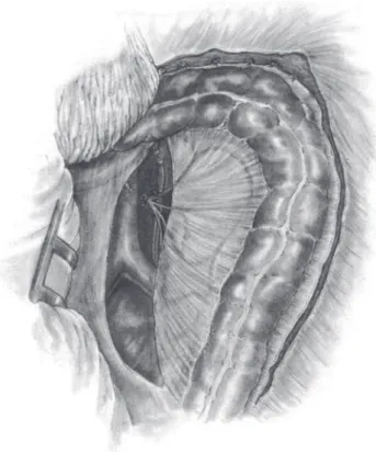
Fig. 3 – Illustration of the circumferential incision of the rectal wall allowing access to the intersphincteric plane. Fig. 2 – Illustration showing complete mobilization of the rectum through an avascular plane suggesting total mesorectal excision (there are no vessel ligatures). plane down to the level of the levator muscles. The anterior
wall was dissected, preferably behind Denonvilier’s fascia: in male patients down to the level of the prostate gland pre-serving the seminal vesicles and deferens ducts; and in fe-male patients, posterior dissection of the uterine wall to the vaginal dome. The lateral ligaments of the rectum were taken near the pelvic wall. Dissection of the rectum was then com-pleted to the level of levator muscles and its distal end, was tied with gauze (Fig. 2).
Once the proximal colon was completely mobilized and the rectum completely dissected, the selection of the de-scending colon segment to be lowered to the anal canal was performed taking into consideration its reach over the pubic bone without any signii cant tension. Adequacy of the arte-rial blood l ow at this point was routinely evaluated prior to resection.
The surgeon moved to the perineum while the assistant remained in the abdomen to help with the i nal passage of the descending colon through the pelvis.
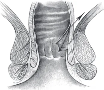
Perineal phase. Following standard antiseptic rinsing of the perineum, gentle digital dilatation of the anus was performed and Allis’ clamps were placed at the cardinal points of the anus to allow good exposure of the anorectal region. A cir-cumferential incision 0.5 cm above the dentate line was made starting at the posterior wall, followed by submucosal dissec-tion along with the identii cadissec-tion of the internal sphincter muscle (Fig. 3). An incision was made through the muscular layer of the rectal wall (with partial resection of the cranial portion of the internal sphincter) circumferentially and distal to the previously made occlusion of the rectum (tied gauze),
thus communicating with the abdominal dissection (Fig. 4). The rectal stump was then closed with interrupted sutures and the specimen extracted through the abdomen.
J C O L O P R O C T O L . 2 0 1 4 ;3 4 ( 1 ): 4 1 – 4 7
44
colostomy. Cardinal sutures were placed between the dentate line and the descending colon (Fig. 5). Special care was taken in order to maintain the anastomosis tension free and prop-erly vascularized. A tubular drain was placed in the presacral space and the abdominal wall was closed.
SECOND STAGE (Resection of the perineal colon stump) Ideally, the second stage of the operation was planned to be performed within 10 to 20 days from the original procedure. Under spinal anesthesia, the patient was positioned in gyne-cological position with the aid of leg stirrups. A circular inci-sion of the colon at the level of coalescence to the dentate line was made and the marginal arcade was ligated. Reinforce-ments to the previously sutured coloanal anastomoses were performed with additional interrupted sutures.
Postoperative complications were considered immediate when occurring up to 30 days from the initial surgical proce-dure and late when occurring after that period.
None of the patients underwent postoperative chemo-therapy. Follow-up included visits to the colorectal surgeon one month after surgery and then every three months during the i rst year and every 6 months thereafter. Recurrence was considered local in the presence of endoluminal coni rmation of adenocarcinoma. Systemic recurrences were considered in the presence of clinical or radiological evidence (with or with-out pathological coni rmation) of distant metastases.
All patients underwent assessment for urological, sexual and anorectal function. Urological assessment included a questionnaire of urological symptoms (dysuria, urinary in-continence) and cystometry 2 months after surgery. Sexual function was assessed with a questionnaire of postoperative sexual function (retrograde ejaculation, orgasm and erection). Fecal continence was assessed with a questionnaire of post-operative events specii cally addressing gas, liquids or sol-ids incontinence every 6 months until 2 years of follow-up completion.. In addition, patients underwent a radiographic enema and manommetry. All questionnaires included objec-tive questions to the presence or absence for each of the men-tioned symptoms.
Results
Fifty patients with resectable rectal cancer were included. Pa-tients’ demographics are described in Table 1.
Median hospital stay was 18 days and the median interval for resection of the perineal colostomy was 20 days. Immedi-ate postoperative mortality was 8% (4 patients). Postopera-tive complications, included necrosis (mucosal or transmu-ral) of the large bowel used for anastomosis, were detected in 5 patients (10%) (Table 2). Postoperative complications and difi culties in hospital admittance (a public University Hospital) for resection of the perineal colostomy led to a signii cant increase in the interval time between the stages of the procedure. Long term complications were mainly re-lated to stricture of the anastomosis occurring in 7 patients (14%). Nearly half of these strictures were short in extent (3
Fig. 5 – Perineal colostomy created through exteriorization of a segment of well-vascularized and tension- free proximal colon.
Fig. 4 – Illustration of the intersphincteric dissection showing a partial resection of the internal anal sphincter (arrow).
Table 1 – Patient’s demographics.
N 50 (100%)
Age (yrs) 55.7 (28-81)
Gender (male/female) 31 – 19 (62%)
Mean distance from anal verge (cm) 9.6 (5-15)
Previous surgery (diverting colostomy) 4 (8%)
patients) while the remaining developed long tubular stric-tures (4 patients) (Table 2).
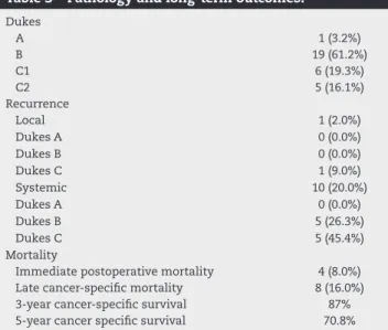
Pathology was available for 31 patients and classiied using Dukes’ staging system. Dukes A (pT1-2N0) tumor was present in one patient, Dukes B (pT3-4N0) in 19 patients and Dukes C in 11 (5 with C1/pT1-2N+ and 6 with C2/pT3-4N+) (Table 3).
Median follow-up was 60 months. There was one local recurrence (2%) and 10 systemic recurrences (20%). Eight pa-tients died due to disease progression during the study pe-riod. Three year and 5 year cancer speciic survival was 87% and 71% respectively.
Only 6.5% of patients had any urinary symptoms during the postoperative period (> 30 days). All were transitory and all pa-tients became asymptomatic at 2 months of follow-up. Cysto-metric values were normal 2 months after surgery in all cases. Five out of 12 male patients that completed the postop-erative questionnaire complained of sexual dysfunction after surgery (48%). There were 2 patients with exclusively retro-grade ejaculation and 3 with impaired erection and orgasm.
Tables 4 and 5 provide information on bowel function and episodes of gas, liquids and solids incontinence. Bowel func-tional outcomes were obtained from 40 patients. In general, 50% of patients had bowel function perceived as normal (no diarrhea or constipation) after 1 year from the primary surgi-cal procedure. Complete continence to solid stool increased from 30% in the immediate postoperative period to 92% after 2 years of follow-up. Continence to gas increased from 9% to 75% during the same period. Finally, among the 20 patients that underwent radiographic enema, all were able to com-pletely retain the contrast during the study.
Discussion
Even though the retrospective observations of the present study may not seem a breakthrough in the management of rectal cancer today, it is clearly an outstanding accomplish-ment when the data and the results are put into historical perspective. After all, more than 50 years ago and 50 years after Miles original description of the abdominal perineal ex-cision of the rectum, preservation of the anus with coloanal anastomosis for tumors as low as 5cm from the anal verge was unthinkable by most surgeons, perhaps considered mad-ness by many. Still, the idea that sphincter preservation for these patients was considered a merely pleasant possibility and never a necessarily required outcome is clearly empha-sized in the thesis.6,9
Apart from the obvious differences in terms of hospital stay (18 days!), immediate postoperative mortality and ab-sence of stapling devices, this paper draws attention for the similarity to “modern” rectal cancer management. First, the fact that an operation resembling total mesorectal excision was used during proctectomy stands out immediately, even though not as clearly described as Heald did years later.2 The
description of the surgical procedure speciically mentioning the dissection through an avascular plane close to the presa-cral fascia, also indicated by the drawing, clearly illustrates an embryonary total mesorectal excision with no vessel liga-tion and what appears to be an intact mesorectal plane. In addition to the actual description and illustration of the tech-nique, the 2% local recurrence rate strongly suggests that an operation very close to TME had been performed and contrib-uted to the excellent local disease control. This by no means Table 2 – Surgical outcomes.
Mean hospital stay (days) 18.6
Mean interval for perineal colostomy removal (days) 20 (11-60)
Overall Morbidity 24 (48%)
Early complications (<30 days)
Urinary retention 3 (6%)
Urinary retention with infection 4 (8%)
Renal failure 1 (2%)
Heart failure 1 (2%)
Stroke 1 (2%)
Pseudomembranous Enteritis 2 (4%)
Wound infection 4 (8%)
Evisceration 2 (4%)
Partial colonic necrosis (mucosal ischemia) 2 (4%)
Complete colonic necrosis (transmural ischemia) 3 (6%)
Pelvic sepsis 5 (10%)
Late complications (>30 days)
Anastomotic dehiscence 3 (6%)
Short anastomotic stricture 3 (6%)
Long anastomotic stricture 4 (8%)
Table 3 – Pathology and long-term outcomes.
Dukes
A 1 (3.2%)
B 19 (61.2%)
C1 6 (19.3%)
C2 5 (16.1%)
Recurrence
Local 1 (2.0%)
Dukes A 0 (0.0%)
Dukes B 0 (0.0%)
Dukes C 1 (9.0%)
Systemic 10 (20.0%)
Dukes A 0 (0.0%)
Dukes B 5 (26.3%)
Dukes C 5 (45.4%)
Mortality
Immediate postoperative mortality 4 (8.0%)
Late cancer-speciic mortality 8 (16.0%)
3-year cancer-speciic survival 87%
5-year cancer speciic survival 70.8%
Table 4 – Functional outcomes - bowel function.
Bowel function Normal Diarrhea
After 3 months 6 (13.9%) 32 (74.4%)
After 6 months 12 (30.0%) 20 (50.0%)
After 1 year 20 (50.0%) 10 (25.0%)
Table 5 – Functional outcomes - continence.
Continence Solid stools Liquid stools and gas
After 6 months 13 (32.2%) 4 (9.3%)
After 12 months 35 (87.5%) 20 (50.0%)
J C O L O P R O C T O L . 2 0 1 4 ;3 4 ( 1 ): 4 1 – 4 7
46
diminishes the relevance of Heald’s proper and meticulous description of the technique, or conirmation of its relevance in terms of local disease control done some years later in properly designed studies.10-12 The efforts and works of Prof.
Heald allowed widespread education and training of surgeons worldwide with a signiicant impact in the practice and out-comes for rectal cancer surgery.10,11 The early use of TME
pre-sented in this study merely reinforces that total mesorectal excision is so appropriate that to some surgeons it was nearly instinctive. Still, one should take into account that strict se-lection of patients may also have accounted for this excellent result in local disease control since many more advanced or aggressive cases could have been managed by alternative ab-dominal perineal resection during the same study period.
Second, the concern of the colorectal surgeon with the urinary, sexual and fecal continence consequences after rec-tal surgery with sphincter preservation is rather remarkable. The observation that fecal incontinence improved over time, conirmed much later in randomized controlled trials, was already observed after straight colorectal or coloanal anas-tomosis.12-14 None of the currently used fecal incontinence
scores were available at the time (most proponents were not even born yet!) and the author used a simple assessment of gas, liquids and solids incontinence for dysfunction evalua-tion.15 The same applies to the attempt of assessing sexual
and urinary functions.16,17
Third, is the description of what we know today as a par-tial intersphincteric resection (ISR). Instead of a complete re-moval of the internal anal sphincter, partial intersphincteric resections include the most cranial portion of the internal sphincter as described by Yamada et al.18 In fact, sphincter
preservation with coloanal anastomoses had already been clearly described at that time, particularly by works from Babcock and Bacon in the late 40’s.7,19 Also known as
“ab-domino-anal pull-through”, these authors described a tech-nique with removal of the lining of the anal canal and bring-ing the mobilized colon through the canal leavbring-ing a segment of nearly 5cm protruding beyond the anal verge. By this tech-nique, the anal sphincters were divided and then sutured to the protruding colon. It was Black, nearly 10 years later that modiied the technique to preserve the anal canal lining and anal sphincters (by dilation instead of sectioning).8 The
for-mer description is the closest to the technique employed in the present manuscript that had also been described for the treatment of Chagasic megacolon in 1961. 20
Still, at that time, a 5cm distal free margin was the rule for curative rectal cancer surgery. Therefore, sphincter pres-ervation would rarely be considered for distal tumors (< 5cm) from the anal verge. Progressively, the 5cm has been replaced by 2cm, 1cm and now distal margins even less than 1cm are currently considered appropriate for most patients with rec-tal cancer, particularly for tumors undergoing neoadjuvant therapies and without direct invasion of the sphincters.21
This incision of the rectum at (or closely higher to) the den-tate line with delayed coloanal anastomosis is clearly de-scribed in the manuscript and illustrated by the drawings of the time. In fact, in a time where surgical staplers were still unavailable, this was the only technical option for the preservation of large bowel continuity to the anus. Curiously the same year,,1972, Sir Alan Parks published the technique
for transanal coloanal anastomosis with primary suturing.22
Years later, intersphincteric resection for rectal cancer be-came increasingly popular.23,24 The only difference between
today’s partial intersphincteric resection and the technique described in the 1972 manuscript is that delayed anastomo-sis with perineal colostomy is now rarely used. This has cur-rently been replaced by temporary stomas (most frequently ileostomies).25 Still, considering that none of the patients
required stomas in the author’s series and that the mean in-terval between the inal resection of the perineal stoma and deinitive colonanal anastomosis was 20 days, leads to a re-lection whether this approach should be revisited and con-sidered in speciic situations by the experienced colorectal surgeon. This is particularly relevant considering the mor-bidity and mortality directly associated with stoma creation and closure.26,27
Finally, if all of this was not already a signiicant leap in rectal cancer management between 1960’s and 1970’s, the fact that all patients had been exclusively intervened by a single woman surgeon is signiicant. At that time, there were very few women in surgery and did not usually play key roles in the advancement of our specialty. In order to introduce so many new concepts in rectal cancer management, one needed to literally invade an almost exclusively men’s world.
Sphincter preservation and functional outcomes fol-lowing rectal cancer surgery has been a matter of interest and concern for many years. However, there is still contro-versy in the use of sphincter preservation and ISR for the management of distal rectal cancer. Particularly, appropri-ate comparison of oncological, functional and quality of life outcomes to abdominal perineal excision in the setting of proper cylindrical (also known as extra-levator APE) is war-ranted.28 While technical advancements seem to have been
overcome, it is our task to take the next step to provide deinitive answers for its use. Otherwise, we will continue teaching new dogs old tricks instead of teaching old dogs new tricks.
Conlicts of interest
The authors declare no conlicts of interest.
R E F E R E N C E S
1. Campos FG, Habr-Gama A, Nahas SC, Perez RO (2012) Abdominoperineal excision: evolution of a centenary operation. Dis Colon Rectum 55l:844-853. doi:10.1097/ DCR.0b013e31825ab0f7
2. Heald RJ, Husband EM, Ryall RD (1982) The mesorectum in rectal cancer surgery--the clue to pelvic recurrence? Br J Surg 69l:613-616
3. Goligher JC, Dukes CE, Bussey HJ (1951) Local recurrences after sphincter saving excisions for carcinoma of the rectum and rectosigmoid. Br J Surg 39l:199-211
4. Dixon CF (1948) Anterior Resection for Malignant Lesions of the Upper Part of the Rectum and Lower Part of the Sigmoid. Ann Surg 128l:425-442
terminal portion of the pelvic colon (1908). CA Cancer J Clin 21l:361-364
6. Habr-Gama A (1972) Indicações e Resultados da
Retocolectomia Abdominoendoanal no Tratamento do Câncer de Reto. University of Sao Paulo, Sao Paulo
7. Bacon HE, Giambalvo GP (1948) Cancer of the rectum; its surgical management without colostomy and with preservation of the internal and external sphincter. J Int Coll Surg 11l:452-463
8. Black BM (1952) Combined abdominoendorectal resection; technical aspects and indications. AMA Arch Surg 65l:406-416 9. d AF, Morgan CN, Lloyd-Davies OV (1950) Discussion on
conservative resection in carcinoma of the rectum. Proc R Soc Med 43l:697-710
10. Kapiteijn E, Putter H, van de Velde CJ, Cooperative investigators of the Dutch ColoRectal Cancer G (2002) Impact of the introduction and training of total mesorectal excision on recurrence and survival in rectal cancer in The Netherlands. Br J Surg 89l:1142-1149. doi:10.1046/j.1365-2168.2002.02196.x 11. Martling A, Holm T, Rutqvist LE, Johansson H, Moran BJ,
Heald RJ, Cedermark B (2005) Impa ct of a surgical training programme on rectal cancer outcomes in Stockholm. Br J Surg 92l:225-229. doi:10.1002/bjs.4834
12. Fazio VW, Zutshi M, Remzi FH, Parc Y, Ruppert R, Furst A, Celebrezze J, Jr., Galanduik S, Orangio G, Hyman N, Bokey L, Tiret E, Kirchdorfer B, Medich D, Tietze M, Hull T, Hammel J (2007) A randomized multicenter trial to compare long-term functional outcome, quality of life, and complications of surgical procedures for low rectal cancers. Ann Surg 246l:481-488; discussion 488-490. doi:10.1097/SLA.0b013e3181485617 13. Heriot AG, Tekkis PP, Constantinides V, Paraskevas P, Nicholls
RJ, Darzi A, Fazio VW (2006) Meta - analysis of colonic reservoirs versus straight coloanal anastomosis after anterior resection. Br J Surg 93l:19 - 32. doi:10.1002/bjs.5188
14. van Duijvendijk P, Slors F, Taat CW, Heisterkamp SH, Obertop H, Boeckxstaens GE (2003) A prospective evaluation of anorectal function after total mesorectal excision in patients with a rectal carcinoma. Surgery 133l:56-65. doi:10.1067/msy.2003.3 15. Baxter NN, Rothenberger DA, Lowry AC (2003) Measuring fecal
incontinence. Dis Colon Rectum46l:1591-1605. doi:10.1097/01. DCR.0000098906.61097.1C
16. Lange MM, van de Velde CJ (2011) Urinary and sexual dysfunction after rectal cancer treatment. Nat Rev Urol 8l:51-57. doi:10.1038/nrurol.2010.206
17. Ho VP, Lee Y, Stein SL, Temple LK (2011) Sexual function after treatment for rectal cancer: a review. Dis Colon Rectum 54l:113-125. doi:10.1007/DCR.0b013e3181fb7b82 18. Yamada K, Ogata S, Saiki Y, Fukunaga M, Tsuji Y, Takano
M (2009) Long-term results of intersphincteric resection for low rectal cancer. Dis Colon Rectum 52l:1065-1071. doi:10.1007/DCR.0b013e31819f5fa2
19. Babcock WW (1947) Radical single stage extirpation for cancer of the large bowel, with retained functional anus. Surg Gynecol Obstet 85l:1-7
20. Haddad J, Simonsen O, Raia A, Netto AC (1961)
[Complications of abdominoperineal rectosigmoidectomy in the treatment of acquired megacolon]. Rev Paul Med 59l:1-8
21. Fitzgerald TL, Brinkley J, Zervos EE (2011) Pushing the envelope beyond a centimeter in rectal cancer: oncologic implications of close, but negative margins. J Am Coll Surg 213l:589-595. doi:10.1016/j.jamcollsurg.2011.07.020 22. Parks AG (1972) Transanal technique in low rectal
anastomosis. Proc R Soc Med 65l:975-976
23. Lyttle JA, Parks AG (1977) Intersphincteric excision of the rectum. Br J Surg 64l:413-416
24. Schiessel R, Karner-Hanusch J, Herbst F, Teleky B, Wunderlich M (1994) Intersphincteric resection for low rectal tumours. Br J Surg 81l:1376-137825.
25. Matthiessen P, Hallbook O, Rutegard J, Simert G, Sjodahl R (2007) Defunctioning stoma reduces symptomatic anastomotic leakage after low anterior resection of the rectum for cancer: a randomized multicenter trial. Ann Surg 246l:207-214. doi:10.1097/SLA.0b013e3180603024 26. Perez RO, Habr-Gama A, Seid VE, Proscurshim I, Sousa AH,
Jr., Kiss DR, Linhares M, Sapucahy M, Gama-Rodrigues J (2006) Loop ileostomy morbidity: timing of closure matters. Dis Colon Rectum 49l:1539 - 1545. doi:10.1007/s10350-006-0645-8
27. Chow A, Tilney HS, Paraskeva P, Jeyarajah S, Zacharakis E, Purkayastha S (2009) The morbidity surrounding reversal of defunctioning ileostomies: a systematic review of 48 studies including 6,107 cases. Int J Colorectal Dis 24l:711-723. doi:10.1007/s00384-009-0660-z