Cop
yright
© ABE&M t
odos os dir
eit
os r
eser
vados
.
Molecular markers in the
diagnosis of thyroid nodules
Marcadores moleculares no diagnóstico de nódulos tireoidianos
Laura S. Ward1, Richard T. Kloos2
SUMMARY
An indeterminate thyroid nodule cytology result occurs about every sixth ine-needle aspiration. These indeterminate nodules harbor a 24% risk of malignancy (ROM); too high to ignore, but driving surgery where most nodules are benign. Molecular diagnostics have emerged to ideally avoid surgery when appropriate, and to trigger the correct therapeutic surgery when indicated, as opposed to an incomplete diagnostic surgery. No current molecular test offers both high sen-sitivity and high speciicity. A molecular diagnostic test with high sensen-sitivity (e.g. Airma Gene Expression Classiier sensitivity 90%) offers a high Negative Predictive Value when the ROM is relatively low, such as < 30%. Only such tests can “rule-out” cancer. In this setting, a molecularly benign result suggests the same ROM as that of operated cytologically benign nodules (~6%). Thus, clinical observation can replace diagnostic surgery; increasing quality of life and decrea-sing medical costs. However, its low speciicity cannot “rule-in” cancer as a suspicious result has a Positive Predictive Value (PPV) of ~40%, perhaps too low to routinely relex to deinitive cancer surgery. Conversely, high speciicity tests (BRAF, RAS, PPAR/PAX-8, RET/PTC, PTEN) offer high PPV results, and only these tests can “rule-in” cancer. Here a positive molecular result war-rants deinitive therapeutic surgery. However, their low sensitivity cannot “rule-out” cancer and a negative molecular result cannot dissuade diagnostic surgery; limiting their cost-effectiveness. Whether or not there is a useful and cost-effective role to sequentially combine these approaches, or to modify existing approaches, is under investigation. Arq Bras Endocrinol Metab. 2013;57(2):89-97
Keywords
Biopsy, ine-needle; DNA mutational analysis; gene expression; genomics; molecular diagnostic techniques
SUMáRio
Resultados indeterminados na citologia de um nódulo tireoidiano ocorrem em cerca de um a cada seis punções aspirativas por agulha ina. Esses nódulos indeterminados apresentam risco de malig-nidade (RM) de cerca de 24%, um valor alto demais para ser ignorado e que leva à cirurgia em casos em que a maioria dos nódulos é benigna. O diagnóstico molecular é uma forma ideal de se evitar a cirurgia quando apropriado e de se levar ao correto procedimento cirúrgico terapêutico quando in-dicado, em oposição à cirurgia diagnóstica incompleta. Atualmente, não existem testes moleculares com alta sensibilidade e especiicidade. Um teste molecular de alta sensibilidade (por exemplo, a sensibilidade do teste Airma Gene Expression Classiier é de 90%) tem um alto Valor Preditivo Nega-tivo quando o RM é relativamente baixo, por exemplo, < 30%. Apenas esses testes podem “excluir” o câncer. Nesse contexto, um resultado molecular benigno sugere o mesmo RM de nódulos com re-sultado benigno na citologia e operados (~6%). Assim, a observação clínica pode substituir a cirurgia diagnóstica, aumentando a qualidade de vida e diminuindo os custos médicos. Entretanto, a baixa especiicidade não pode “incluir” o câncer como um resultado suspeito quando esse resultado tem um Valor Preditivo Positivo (VPP) ~40%, que é talvez baixo demais para levar, rotineiramente, à cirur-gia deinitiva para o câncer. Por outro lado, testes com alta especiicidade (BRAF, RAS, PPAR/PAX-8, RET/PTC, PTEN) têm alto VPP, e apenas esses testes podem “incluir” o câncer. Nesse caso, um resul-tado molecular positivo leva à recomendação de cirurgia terapêutica deinitiva. Entretanto, sua baixa sensibilidade não pode “excluir” o câncer, e um resultado molecular negativo não pode dissuadir o médico de executar a cirurgia diagnóstica, limitando seu custo-benefício. Ainda se investiga se existe ou não um modo útil e com alto custo-benefício de se combinar essas abordagens sequencialmente, ou de se modiicar as abordagens existentes. Arq Bras Endocrinol Metab. 2013;57(2):89-97
Descritores
Biópsia, agulha ina; análise de mutações no DNA; expressão gênica; estudo do genoma; técnicas de diagnóstico molecular
1 Laboratory of Cancer Molecular Genetics, Faculty of Medical Sciences, University of Campinas (FCM- Unicamp), Campinas, SP, Brazil 2 Veracyte, Inc., South San Francisco, CA, United States
Correspondence to:
Laura S. Ward
Laboratory of Cancer Molecular Genetics,
Faculty of Medical Sciences, PO Box 6111
Rua Tessália Vieira de Camargo, 126 13083-887 – Campinas, SP, Brazil [email protected]
Cop
yright
© ABE&M t
odos os dir
eit
os r
eser
vados
.
iNTRoDUCTioN
P
rior to the advent of thyroid nodule ine-needle aspiration (FNA) biopsy, these nodules were rou-tinely referred for diagnostic surgery because of their 5%-15% risk of malignancy (ROM) (1). FNA decreased diagnostic thyroidectomies by one-half as most FNAs are conclusively diagnosed as benign cytologically (2). Still, 15%-30% of thyroid FNAs are cytologically inde-terminate, i.e. not clearly benign nor malignant (1,3). When cytologically indeterminate thyroid nodules undergo diagnostic surgery, three-quarters prove to be benign (4,5). Therefore, a signiicant opportunity exists to improve the care of patients with cytologically indeterminate nodules using novel genomic diagnostic technology. Current opportunities include the accurate pre-operative identiication of (i) benign nodules to sa-fely avoid diagnostic surgery, (ii) malignant nodules to direct an optimal initial treatment (e.g. total thyroidec-tomy instead of a diagnostic hemithyroidecthyroidec-tomy with a subsequent second surgery for completion thyroidec-tomy), and (iii) rare neoplasms requiring evaluations and/or treatments that differ from typical differentia-ted thyroid cancer (e.g. medullary thyroid cancer, me-tastases to the thyroid).Cost, morbidity, and risk of mortality from surgery
The costs of diagnostic surgery include direct costs of the procedure (and its complications), indirect costs of time lost from work and responsibilities of daily living, and impaired quality of life (the fear of potentially ha-ving cancer and the anxiety of undergoing surgery, and the post-operative pain and suffering from surgery, and the potentially impaired quality of life from iatrogenic hypothyroidism with (6), or without (7), a normal se-rum TSH and its consequences).
Thyroid surgeries carry a perioperative mortality rate of 0.1%-0.2%, with rates as high as 0.5% (8-10). Se-rious or permanent non-lethal complications of thyroi-dectomy include hypocalcemia, recurrent laryngeal nerve damage, re-bleeding and wound infection (11). These complications are more frequent than commonly appreciated, and are strongly related to surgeon expe-rience (volume) and expertise. In Brazil, complications occurred in 34.7% of the thyroidectomies performed in a University Hospital, including hypoparathyroidism in 8.8% (12). Complication rates of thyroid surgeries in a US population-based study were found to be 10.1% for surgeons who did between one and nine cases per year,
and 5.9% for surgeons who did more than one hun-dred cases per year (13). In fact, over 50% of thyroid surgeries in the US are performed by surgeons who do ive or fewer cases per year (14), placing many patients at a higher risk of complications. In Spain, a study of surgeons performing less than 5 thyroid surgeries per year demonstrated hypocalcemia rates of 50% and 19% at 6 and 12 months, respectively (15). Figures in Brazil are likely similar.
Current management of cytologically benign thyroid FNA biopsies
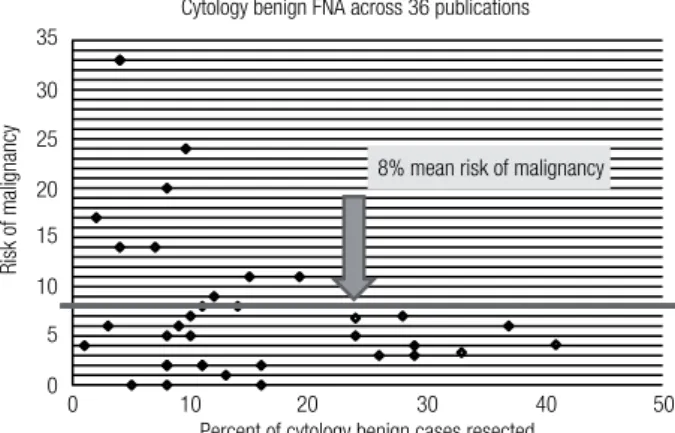
The gold-standard to determine benign versus malig-nant status for all thyroid nodules, including those with benign FNAs, is surgical histology. Current ATA and AACE guidelines recommend close clinical and sono-graphic follow-up of cytologically benign nodules six to eighteen months following the FNA biopsy, despite an average ROM [1- negative predictive value (NPV)] for operated cytologically benign nodules of 6%-8% across multiple studies (4,16-21) (Figure 1), and ranging as high as 33% (17). Thus, the average NPV, or accuracy of a benign diagnosis for thyroid FNA is 92%-94% (4,17,18). The NPV of the Airma Gene Expression Classiier (GEC) applied to cytologically indetermina-te thyroid nodules (Bethesda caindetermina-tegories III and IV) is therefore similar to that of a benign cytological diag-nosis (Figure 2) (20). It follows that these two thyroid nodule scenarios should be treated similarly. Watchful waiting in lieu of diagnostic thyroidectomy is conside-red safe for cytologically benign thyroid nodules be-cause thyroid cancer is relatively indolent, except for anaplastic thyroid cancer whose malignant nature is usually both clinically and cytologically obvious. Thus,
Figure 1. Risk of malignancy among cytologically benign thyroid FNAs across 36 publications (17).
Percent of cytology benign cases resected Cytology benign FNA across 36 publications
8% mean risk of malignancy
0 10
35
30
25
20
15
10
5
0
20
Risk of malignancy
Cop
yright
© ABE&M t
odos os dir
eit
os r
eser
vados
.
even when the benign cytological diagnosis is wrong, if the patient undergoes surgery within twelve months their ive-year risk of mortality and local recurrence is unchanged (1). In fact, papillary thyroid carcinoma (PTC) of any size that is conined to the thyroid gland (no extraglandular extension or lymph node metastases at presentation) has a favorable outcome whether or not they are treated in the irst year after diagnosis (22). Indeed, the indolent nature of PTC has prompted ob-servational trials in microPTC (23).
lar lesion of undetermined signiicance (AUS/FLUS, category III), follicular and Hürthle cell neoplasm or suspicious for same (FN/SFN, category IV), and suspi-cious for malignancy (category V) (26). Unfortunately, it appears that this expansion created less reproducible categorization (27), and has not solved the problem.
The Bethesda system postulated that the FN/SFN category would require diagnostic surgery, while the ROM in the AUS/FLUS category would be signiican-tly lower and best served by a repeat FNA that may resolve the majority of cases. Currently, this differential approach is being questioned. The literature strongly suggests that the ROM for AUS/FLUS lesions is sig-niicantly higher than postulated by the Bethesda Clas-siication creators (20,24,28-30). Additionally, some literature suggests that the ROM in AUS/FLUS may not be signiicantly lower than FN/SFN (21,29). Bon-giovanni and cols. reported a 22% ROM (29), a inding consistent with a recent large multicenter prospective study where the ROM for AUS/FLUS was 24% whi-le the ROM for FN/SFN was 25% (20). Thus, if the ROM for AUS/FLUS lesions is higher than originally projected, or if the ROM is similar to that of the FN/ SFN category, then is it optimal to surgically resection FN/SFN nodules while repeating the FNA for AUS/ FLUS nodules (28)?
In addition, some studies (28,31) [but not all (30,32)] suggest that the ROM in a benign FNA after an initial AUS/FLUS biopsy is of intermediate risk be-tween the two categories (i.e. the second benign FNA cannot make the irst AUS/FLUS FNA go away). The-se studies suggest that the remaining ROM in a follow--up benign FNA remains high enough that diagnostic surgery may not be avoidable.
AppliCATioN oF iMMUNohiSToCheMiSTRY To
FNA CYTologY
A number of immunohistochemical approaches have been applied to indeterminate cytology specimens in an effort to reclassify these samples, including galec-tin-3, HBME-1, and CK19 (33,34). This approach is best at identifying classic PTC, but sensitivity progres-sively declines when one encounters fvPTC, FTC, and less common malignant variants. Further, speciicity is impaired as some immunohistochemical approaches are positive in benign tissues, including follicular adeno-mas, hyperplastic nodules, and normal thyroid tissue. Most importantly, blinded, prospective, large-scale,
Figure 2. Airma Gene Expression Classiier (GEC) reclassiies cytologically indeterminate thyroid nodules with a low risk of malignancy (ROM) to GEC Benign (20). ROM is 1-negative predictive value (NPV).
Cytopathology diagnosis (N) 6%
24%
5%
25%
6%
62%
15%
Benign (47) Atypia/FLUS (129) Follicular or hürthle cell Neoplasm (81)
Suspicious for malignancy (55) 70%
60%
50%
40%
30%
20%
10%
0%
Pre-test risk of malignancy (ROM) Pre-test Airma GEC Benign result (ROM)
Risk of malignancy (1 – NPV)
Cytologically indeterminate FNAs are problematic, so can they be reclassiied?
Physicians recognize that indeterminate FNA results do not resolve the clinical question proposed: is the no-dule benign or malignant? One solution would be to label fewer FNAs as cytologically indeterminate. Cyto-logically indeterminate samples comprise 10%-26% of all samples, averaging 17% (4). Some cytopathology experts claim that they can safely render indetermi-nate diagnoses much less often, but considering the potentially high number of false negative results (mis-sed cancers) in operated cytologically benign nodules (4,17,18), there may be signiicant risk with very low cytology indeterminate rates, assuming the rate is lo-wered by moving many of these samples to the benign cytology category (24).
follicu-Cop
yright
© ABE&M t
odos os dir
eit
os r
eser
vados
.
multicenter clinical trials investigating immunohisto-chemical markers are lacking (34). Thus, most cytopa-thologists view immunohistochemistry as having a very limited role in thyroid cytology (29).
MicroRNA analyses
MicroRNAs are small RNA sequences (19-25 nucleoti-des) that function to regulate the expression of genes. Amid mixed results, several studies have suggested that aberrant miR expression proiles may separate thyroid cancers from benign thyroid lesions and normal thyroid tissue (34-36). However, this approach has yet to be tested on indeterminate FNAs in a large, prospective, blinded, multicenter study (34,35).
DNA mutation/rearrangement testing
Malignant thyroid nodules with indeterminate Bethes-da categories III and IV cytology have a low incidence of BRAF mutation (37,38). This is not unexpected as these thyroid nodule categories include malignant tu-mors that less commonly (or never) harbor BRAF mu-tations such as follicular thyroid cancer, Hürthle cell thyroid cancer, follicular variant PTC, and medullary thyroid cancer. However, the low incidence is also ex-plained by the inding that BRAF mutated PTCs have cytological features that cytopathologists recognize and classify as suspicious for malignancy or malignant (Bethesda categories V and VI); cytological categories typically treated with total thyroidectomy regardless of BRAF status (37). Conversely, PTCs with indetermi-nate cytology are more often BRAF negative (37,38).
A test that cannot detect cancer with very high sen-sitivity cannot successfully “rule out” cancer. DNA mu-tations such as BRAF, RAS, and RET/PTC and PAX8/ PPARG translocations have high positive predictive va-lue (PPV) and when detected they predict (“rule in”) the histological diagnosis of thyroid cancer. However, when they are absent, cancer cannot be “ruled out” be-cause of the low sensitivity and NPV of these markers. A review of four studies combining all four mutation--markers into a single panel had 64% sensitivity, thus failing to identify 36% of thyroid cancers (39). The largest study of mutational markers was a retrospecti-ve analysis of prospectiretrospecti-vely collected samples where the mutational status was available to the clinicians, inclu-ding the histopathologist (38). The mutational marker NPV for Bethesda categories IV and V were 86% and 72%, respectively. Thus, malignancy could not be
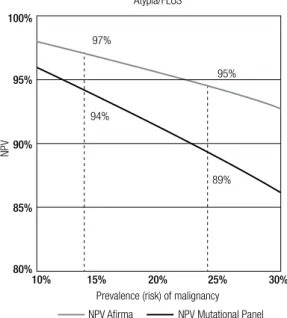
ade-quately excluded to avoid surgery. For Bethesda cate-gory III test sensitivity was 63%, speciicity 99%, and NPV 94% with a 14% prevalence of malignancy. When applied to the 24% prevalence of malignancy seen in the large, multicenter (academic and community), prospec-tive, and blinded study of Alexander and cols. (20) the resultant NPV declines to 89% (Figure 3). High PPV tests with inadequate NPV performance can “rule in” cancer and perhaps alter the extent of surgery, but they cannot “rule out” cancer, so surgery cannot be avoi-ded. Currently, the strongest argument for their use is where the decision has been made for surgery, but the extent of surgery may be determined by the test result (38,40). Based on the potential ability to prevent com-pletion thyroidectomies by performing an initial total thyroidectomy in patients with indeterminate cytology when a DNA mutation is present, Yip and cols. repor-ted the possibility of cost savings by mutation panel tes-ting in a decision-tree model (40). Still, no prospective study using patients has assessed the clinical utility of DNA mutation panels on thyroid surgery. The avai-lable clinical validity data are limited to retrospective analyses of methodologies from academic laboratories (38,39), and no data are available to assess the analyti-cal or clinianalyti-cal validity of commercially available DNA mutation panels. BRAF mutations are uncommon in Bethesda categories III and IV (37,38), and RET/PTC
and PAX8/PPARG translocations are rare in all cyto-logically indeterminate categories (29,34,38). BRAF
Figure 3. Negative predictive value (NPV) of Airma (20) and a Mutational Panel (38) according to the prevalence of malignancy.
100%
95%
90%
85%
80%
10% 15%
89% 94%
95% 97%
20% 25% 30%
Prevalence (risk) of malignancy Atypia/FLUS
NPV Airma NPV Mutational Panel
Cop
yright
© ABE&M t
odos os dir
eit
os r
eser
vados
.
mutations are more prevalent in Bethesda category V (38) where the ~62% ROM (4,20) is high enough that most of these patients are already treated with a total thyroidectomy. Kleiman and cols. reported that preo-perative screening for BRAF mutations was unlikely to alter patient management at their center (37).
Veracyte Airma gene expression Classiier (geC)
The Airma GEC, based on the measurement of mRNA expression, was developed and clinically validated to identify pre-operatively histologically benign nodules amongst those with indeterminate cytology. By pre--operatively identifying these patients, clinical and so-nographic follow-up may be recommended in lieu of diagnostic surgery, thus ending the diagnostic odyssey (Figure 4) (41,42). The Airma GEC analysis is indica-ted only for nodules with indeterminate cytology, and is not performed on cytologically benign, malignant or nondiagnostic (insuficient) FNA samples.
The Airma GEC is provided by Veracyte’s CLIA--certiied clinical laboratory. The molecular classiier proceeds in a step‐wise fashion, irst applying 6 casset-tes before applying the inal benign versus malignant classiier. These cassettes differentiate speciic rare neo-plasm subtypes and act as ilters that halt further sample processing if any cassette returns a “suspicious” result. This prevents some of the rare, nonfollicular cell‐deri-ved neoplasms from being scored by the main classiier. These cassettes classify samples representing (1) ma-lignant melanoma, (2) renal cell carcinoma, (3) breast carcinoma, (4) parathyroid tissue, and (5) medullary thyroid carcinoma. A inal cassette (6) was also trained
using Hürthle cell adenomas and carcinomas. Failing to trigger one of these rare neoplasm cassettes, the GEC evaluates the expression of 142 genes that are used in a proprietary algorithm to classify indeterminate thyroid nodule FNAs as either “Benign” or “Suspicious”.
Analytical validity
The GEC performance was evaluated in a series of forty-three individual reagent and analytical veriication studies. Extensive reagent and analytical performance studies were conducted to evaluate the reliability and reproducibility of the GEC under a variety of experi-mental and clinical conditions, with robust and highly reproducible results (43). Interfering substances inclu-ding human blood and genomic DNA were not found to interfere with extraction or ampliication steps of the assay. Analytical sensitivity studies demonstrated tole-rance to variations in RNA input across the range of 5 ng to 25 ng, as well as dilution of malignant FNA mate-rial down to 20% with FNA matemate-rial from lymphocytic thyroiditis and nodule hyperplasia. Analytical sensitivity and speciicity studies with blood (up to 83%) and ge-nomic DNA (30%) demonstrated negligible assay in-terference, although false positive results could result from very bloody FNAs. FNA preservative solution maintained high quality and quantity of RNA material under various stressed time, temperature, and shipping conditions with no signiicant effect on GEC scores, or “Benign” versus “Suspicious” calls (100% concordan-ce). Based on these data, room-temperature storage at the clinical site and chilled-box shipping was veriied for routine practice.
Clinical validity
A whole genome approach identiied candidate genes, and support vector machine learning methods were used to develop the classiier algorithm. The initial pu-blication included test validation on an independent sample set of cytologically indeterminate thyroid nodu-le FNAs with a prospective multicenter, doubnodu-le blind study design (44). The Airma GEC achieved high sen-sitivity and NPV. After further optimization, the 142 gene classiier was validated in a second prospective multicenter validation study. The study included the largest ever prospectively collected set of thyroid FNA biopsies from 3,789 unique patients. Based on the ex-pected 24% prevalence of malignancy in cytologically indeterminate samples in clinical practice, a 95% NPV
Figure 4. Implementing the Airma gene expression classiier (GEC) into clinical practice. Indeterminate is Bethesda categories III and IV (26). Superscript references: a (60), b (1), c (4), d (18), e (17), f (5), g (42), h (20).
450,000 FNAs on U.S. population
Routine cytopathology +
Airma Gene Expression Classiier (GEC) collection
8% Non-diagnostica
Repeat FNAa,b With 6%-8%
risk of malignancyc,d,e
follow with ultrasound
GEC Benign (~5% risk of malignancyh)
GEC suspicious (~40% risk of
malignancyh)
Airma GEC Without Airma,
66%-80% prove benign with surgical resectionc,f
Thyroid surgeryb,g
66% Benigna
15%-30% Indeterminateb
Cop
yright
© ABE&M t
odos os dir
eit
os r
eser
vados
.
for the Airma GEC was achieved on an independent sample set of 265 cytologically indeterminate nodules when the molecular results were compared to blinded gold standard histopathology diagnosis (20). Thus, the ROM for a thyroid nodule with Bethesda categories III and IV indeterminate cytology but an Airma GEC Be-nign classiier result is about 5%. This risk is comparable to the 6%-8% cancer risk for a thyroid nodule with a be-nign cytology diagnosis (Figures 1-4) (4,16-21).This suggests that GEC Benign cytologically indeterminate (Bethesda categories III and IV) nodules can be mana-ged as would a cytologically benign nodule, as sugges-ted by the NCCN Thyroid Carcinoma Guideline (42). Speciicity is the percentage of truly benign nodules identiied by the test. The GEC raised speciicity from 0% for cytologically indeterminate categories to 52% when the GEC was Benign. This indicates that just over half of the benign nodules from Bethesda categories III and IV can be identiied and removed from the surgical pool. For Bethesda category V nodules (suspicious for malignancy), the sensitivity and speciicity performance of the GEC was comparable, but the high prevalence of malignancy in this category lowers the NPV to 85% (Figure 2). Thus, while the ROM is reduced from an initial 62% based on the cytological category to 15% when the GEC is benign, surgery may not be avoidable based on the residual ROM and therefore these nodu-les are not routinely relexed to GEC testing. However, some physicians request the GEC to be performed in this cytological category to screen the sample with the rare neoplasm cassettes, and because they may alter the extent of surgery from a total thyroidectomy to a diag-nostic hemithyroidectomy when the GEC is benign.
Optimally, physicians routinely collect the Airma GEC with every FNA they perform, or have on-site rapid cytological assessment so that the GEC can be collected on every patient with indeterminate cytology during one patient visit (Figure 4). This avoids the inconvenience, delayed diagnosis, and costs associated with repeating the FNA when the irst FNA comes back indeterminate. Additionally, it is well known that cytologically indeter-minate nodules may not be categorized as indetermina-te if they undergo a repeat FNA (28). At irst blush, a repeat FNA would seem like a good idea to potentially re-stratify cytologically indeterminate patients as either cytologically benign or malignant. Unfortunately, this creates a conundrum for patients whose repeat FNA is cytologically benign as their ROM may not be fully re-duced to the same risk as if their irst FNA had been
cyto-logically benign (28,31). The conundrum is accentuated by the fact that the GEC is not indicated for cytologically benign material as this is not cost-effective due to the low PPV that results from the low prevalence of malignancy in this setting, and because the GEC speciicity of 70% for cytologically benign nodules will predictably result in a false-positive GEC Suspicious call in many cytology benign nodules (20). For these reasons, the GEC speci-men must be collected at the same time as paired cyto-logy, optimally collected during the irst thyroid FNA. When GEC testing is desired in a patient for whom only cytology was previously collected, the cytology must be repeated along with the GEC collection.
Rare neoplasms
Additio-Cop
yright
© ABE&M t
odos os dir
eit
os r
eser
vados
.
nally, surgical management is altered to include a mi-nimum of total thyroidectomy and central neck dissec-tion. Finally, pre-operative RET proto-oncogene status alters management of unintentionally devascularized parathyroid glands (57).
Experience with the other rare neoplasm cassettes is more limited, however, the renal cell carcinoma, breast carcinoma, and parathyroid tissue cassettes have all been triggered and surgical conirmation of these diag-noses have been conirmed in nearly all cases (unpubli-shed data).
Clinical utility and cost-effectiveness
The impact of the Airma GEC on the physician deci-sion to refer to surgery when the FNA cytology is in-determinate but the Airma GEC result is benign has been studied. Duick and cols. reported on the initial 2,040 consecutive indeterminate thyroid FNA biopsies collected in clinical practice through March 2012 (58). Fifty-two percent of these cytologically indetermina-te samples were GEC benign. Fifty-one physicians (46 community-based, 5 academic) at 21 practice locations participated. Decisions were analyzed for 368 patients (395 nodules). Physicians adopted watchful waiting in lieu of diagnostic thyroid surgery 92.4% of the time when the Airma GEC result reclassiied the cytologi-cally indeterminate nodule as benign. In contrast to the 74% historical rate of diagnostic surgery on cytologi-cally indeterminate thyroid nodules (4), 7.6% of those that were Airma GEC benign proceeded to surgery, a dramatic 90% reduction in the decision to operate (p < 0.001) (58). The decision to operate was not statistically different from the 9% operative rate on patients with be-nign cytology diagnoses reported in a recent meta-re-view (4). Physicians who choose to proceed to surgery with an Airma GEC benign result reported similar rea-sons to operate as those found for operated cytologically benign nodules (e.g. large nodule, compression, other suspicious nodule, rapid nodule growth) (58).
An independent study found no difference in missed cancers between the current care paradigm and the Air-ma GEC in a Markov model employing 10,000 Monte Carlo simulations of the expected range of probabilities for different potential outcomes (59). They also mode-led the cost effectiveness and impact on quality adjus-ted life years (QALYs) over a ive year period for usual care versus utilization of the Airma GEC in the United States. Compared to the traditional care paradigm, the Airma GEC modestly improved quality of life while
re-ducing direct healthcare costs by $4,653 per ive year episode of care (allowing $1,453 in direct savings assu-ming the test cost $3,200) (59). This cost savings esti-mate was conservative because it projected a 14% rate of operation on GEC benign thyroid nodules (based on expert opinion), a rate almost twice the 7.6% rate subse-quently reported in actual clinical practice (58). Using the actual rate of thyroidectomy when the GEC is be-nign, each test would have saved $2,600 (58).
CoNClUSioN
The introduction of thyroid FNA dramatically reduced unnecessary diagnostic thyroid surgery by rendering an actionable ROM when the cytological diagnosis was be-nign, suspicious for malignancy, or malignant. Among patients with cytologically benign lesions the ROM is low, and this combined with the generally indolent na-ture of thyroid cancer has led clinicians to follow these patients conservatively and avoid diagnostic surgery. In the minority of the patients where FNA cytology is indeterminate, most patients have been referred for diagnostic surgery given the higher ROM (3). The ap-plication of molecular diagnostics to these cytologically indeterminate samples offers the opportunity impro-ve patient care by clarifying this diagnostic ambiguity. Immunohistochemistry, microRNA, DNA mutation/ rearrangement testing, and RNA gene expression clas-siication have attempted to end the diagnostic odyssey of these patients. Immunohistochemical and mutational approaches have inadequate sensitivity and NPV for ma-lignancy, such that diagnostic surgery cannot be avoided (3). When a mutation/rearrangement is present, howe-ver, the high positive predictive value for malignancy can “rule-in” malignancy and alter patient management by commanding an initial total thyroidectomy as oppo-sed to a diagnostic hemithyroidectomy (34). Currently, the most validated and effective approach to “rule-out” cancer and avoid diagnostic surgery in patients with in-determinate cytology is the Airma GEC based on its high NPV (34). Opportunities clear exist in the literatu-re and Guidelines to clarify these concepts of “rule-in”
indetermi-Cop
yright
© ABE&M t
odos os dir
eit
os r
eser
vados
.
nate nodules. This increases the diagnostic speciicity of indeterminate cytology from 0% to 52%. Thus, about half of those with truly benign lesions can be removed from the surgical pool and conservatively followed as if they were cytologically benign. The remaining GEC “suspicious” patients are enriched for malignancy with a positive predictive value of about 40%. Given this in-termediate ROM other factors are needed to decide if these patients should undergo a diagnostic hemithyroi-dectomy or total thyroihemithyroi-dectomy. The low frequency of
BRAF, RET/PTC, PPARG mutations in AUS/FLUS and FN/SFN patients suggests that testing for these mutations/rearrangements to make this decision may be of low yield (37,38), although testing for RAS mu-tations may be the most productive (34,38), especially if one found it acceptable to perform a total thyroidec-tomy in the setting of a benign follicular adenoma with a RAS mutation (3,38). Analytic validity and clinical utility studies, as well as prospective multicenter clini-cal validation of this gene mutation testing paradigm against blinded histopathology experts remain to be performed.
Disclosure: RTK is a stockholder and employee of Veracyte, Inc. LSW has no conlict of interest.
ReFeReNCeS
1. Cooper DS, Doherty GM, Haugen BR, Kloos RT, Lee SL, Mandel SJ, et al. Revised American Thyroid Association management guidelines for patients with thyroid nodules and differentiated thyroid cancer. Thyroid. 2009;19(11):1167-214.
2. Hegedus L. Clinical practice. The thyroid nodule. N Engl J Med. 2004;351(17):1764-71.
3. Melillo RM, Santoro M. Molecular biomarkers in thyroid FNA samples. J Clin Endocrinol Metab. 2012;97(12):4370-3.
4. Wang CC, Friedman L, Kennedy GC, Wang H, Kebebew E, Steward DL, et al. A large multicenter correlation study of thyroid nodule cytopathology and histopathology. Thyroid. 2011;21(3):243-51. 5. Bryson PC, Shores CG, Hart C, Thorne L, Patel MR, Richey L, et
al. Immunohistochemical distinction of follicular thyroid adeno-mas and follicular carcinoadeno-mas. Arch Otolaryngol Head Neck Surg. 2008;134(6):581-6.
6. Saravanan P, Chau WF, Roberts N, Vedhara K, Greenwood R, Day-an CM. Psychological well-being in patients on “adequate” doses of l-thyroxine: results of a large, controlled community-based questionnaire study. Clin Endocrinol (Oxf). 2002;57(5):577-85. 7. Cooper DS, Biondi B. Subclinical thyroid disease. Lancet.
2012;379(9821):1142-54.
8. Shrime MG, Goldstein DP, Seaberg RM, Sawka AM, Rotstein L, Freeman JL, et al. Cost-effective management of low-risk pap-illary thyroid carcinoma. Arch Otolaryngol Head Neck Surg. 2007;133(12):1245-53.
9. Esnaola NF, Cantor SB, Sherman SI, Lee JE, Evans DB. Optimal treatment strategy in patients with papillary thyroid cancer: a de-cision analysis. Surgery. 2001;130(6):921-30.
10. Hundahl SA, Cady B, Cunningham MP, Mazzaferri E, McKee RF, Rosai J, et al. Initial results from a prospective cohort study of 5,583 cases of thyroid carcinoma treated in the United States during 1996. U.S. and German Thyroid Cancer Study Group. An American College of Surgeons Commission on Cancer Patient Care Evaluation study. Cancer. 2000;89(1):202-17.
11. Bergenfelz A, Jansson S, Kristoffersson A, Martensson H, Reihner E, Wallin G, et al. Complications to thyroid surgery: results as re-ported in a database from a multicenter audit comprising 3,660 patients. Langenbecks Arch Surg. 2008;393(5):667-73.
12. Ernandes-Neto M, Tagliarini JV, Lopez BE, Padovani CR, Marques Mde A, Castilho EC, et al. Factors inluencing thyroidectomy com-plications. Braz J Otorhinolaryngol. 2012;78(3):63-9.
13. Sosa JA, Bowman HM, Tielsch JM, Powe NR, Gordon TA, Udelsman R. The importance of surgeon experience for clini-cal and economic outcomes from thyroidectomy. Ann Surg. 1998;228(3):320-30.
14. Saunders BD, Wainess RM, Dimick JB, Doherty GM, Upchurch GR, Gauger PG. Who performs endocrine operations in the Unit-ed States? Surgery. 2003;134(6):924-931; discussion 931. 15. Gonzalez-Sanchez C, Franch-Arcas G, Gomez-Alonso A.
Morbid-ity following thyroid surgery: does surgeon volume matter? Lan-genbecks Arch Surg. 2012 Nov 6.
16. Borget I, Vielh P, Leboulleux S, Allyn M, Iacobelli S, Schlumberger M, et al. Assessment of the cost of ine-needle aspiration cytol-ogy as a diagnostic tool in patients with thyroid nodules. Am J Clin Pathol. 2008;129(5):763-71.
17. Renshaw A. An estimate of risk of malignancy for a benign di-agnosis in thyroid ine-needle aspirates. Cancer Cytopathol. 2010;118(4):190-5.
18. Lewis CM, Chang KP, Pitman M, Faquin WC, Randolph GW. roid ine-needle aspiration biopsy: variability in reporting. Thy-roid. 2009;19(7):717-23.
19. Shrestha M, Crothers BA, Burch HB. The impact of thyroid nod-ule size on the risk of malignancy and accuracy of ine-needle aspiration: a 10-year study from a single institution. Thyroid. 2012;22(12):1251-6.
20. Alexander EK, Kennedy GC, Baloch ZW, Cibas ES, Chudova D, Diggans J, et al. Preoperative diagnosis of benign thyroid nod-ules with indeterminate cytology. N Engl J Med. 2012;367:705-15. 21. Marchevsky AM, Walts AE, Bose S, Gupta R, Fan X, Frishberg D,
et al. Evidence-based evaluation of the risks of malignancy pre-dicted by thyroid ine-needle aspiration biopsies. Diagn Cytopa-thol. 2010;38(4):252-9.
22. Davies L, Welch HG. Thyroid cancer survival in the United States: observational data from 1973 to 2005. Arch Otolaryngol Head Neck Surg. 2010;136(5):440-4.
23. Ito Y, Miyauchi A, Inoue H, Fukushima M, Kihara M, Higashiyama T, et al. An observational trial for papillary thyroid microcarcino-ma in Japanese patients. World J Surg. 2010;34(1):28-35. 24. Renshaw AA. Subclassiication of atypical cells of undetermined
signiicance in direct smears of ine-needle aspirations of the thy-roid: distinct patterns and associated risk of malignancy. Cancer Cytopathol. 2011;119(5):322-7.
25. Piana S, Frasoldati A, Ferrari M, Valcavi R, Froio E, Barbieri V, et al. Is a ive-category reporting scheme for thyroid ine needle as-piration cytology accurate? Experience of over 18,000 FNAs re-ported at the same institution during 1998-2007. Cytopathology. 2011;22(3):164-73.
26. Cibas ES, Ali SZ. The Bethesda System for Reporting Thyroid Cy-topathology. Thyroid. 2009;19(11):1159-65.
in-Cop
yright
© ABE&M t
odos os dir
eit
os r
eser
vados
.
ter-observer diagnostic agreement and provides non-overlapping estimates of malignancy risks. Diagn Cytopathol. 2012;40 Suppl 1:E62-8.
28. VanderLaan PA, Marqusee E, Krane JF. Clinical outcome for atypia of undetermined signiicance in thyroid ine-needle aspirations: should repeated fna be the preferred initial approach? Am J Clin Pathol. 2011;135(5):770-5.
29. Bongiovanni M, Krane JF, Cibas ES, Faquin WC. The atypical thy-roid ine-needle aspiration: past, present, and future. Cancer Cy-topathol. 2012;120(2):73-86.
30. Faquin WC, Baloch ZW. Fine-needle aspiration of follicular pat-terned lesions of the thyroid: diagnosis, management, and fol-low-up according to National Cancer Institute (NCI) recommen-dations. Diagn Cytopathol. 2010;38(10):731-9.
31. Renshaw AA. Does a repeated benign aspirate change the risk of malignancy after an initial atypical thyroid ine-needle aspiration? Am J Clin Pathol. 2010;134(5):788-92.
32. Na DG, Kim JH, Sung JY, Baek JH, Jung KC, Lee H, et al. Core-needle biopsy is more useful than repeat ine-Core-needle aspiration in thyroid nodules read as nondiagnostic or atypia of undetermined signiicance by the Bethesda system for reporting thyroid cytopa-thology. Thyroid. 2012;22(5):468-75.
33. de Matos PS, Ferreira AP, de Oliveira Facuri F, Assumpcao LV, Metze K, Ward LS. Usefulness of HBME-1, cytokeratin 19 and ga-lectin-3 immunostaining in the diagnosis of thyroid malignancy. Histopathology. 2005;47(4):391-401.
34. Kim MI, Alexander EK. Diagnostic use of molecular markers in the evaluation of thyroid nodules. Endocr Pract. 2012;18(5):796-802. 35. Lodewijk L, Prins AM, Kist JW, Valk GD, Kranenburg O, Rinkes IH,
et al. The value of miRNA in diagnosing thyroid cancer: a system-atic review. Cancer Biomark. 2012;11(6):229-38.
36. Dettmer M, Vogetseder A, Durso MB, Moch H, Komminoth P, Per-ren A, et al. MicroRNA expression array identiies novel diagnos-tic markers for conventional and oncocydiagnos-tic follicular thyroid car-cinomas. J Clin Endocrinol Metab. 2013;98(1):E1-7.
37. Kleiman DA, Sporn MJ, Beninato T, Crowley MJ, Nguyen A, Uc-celli A, et al. Preoperative BRAF(V600E) mutation screening is unlikely to alter initial surgical treatment of patients with indeter-minate thyroid nodules: a prospective case series of 960 patients. Cancer. 2012 Dec 21.
38. Nikiforov YE, Ohori NP, Hodak SP, Carty SE, LeBeau SO, Ferris RL, et al. Impact of mutational testing on the diagnosis and manage-ment of patients with cytologically indeterminate thyroid nod-ules: a prospective analysis of 1056 FNA samples. J Clin Endocri-nol Metab. 2011;96(11):3390-7.
39. Ferraz C, Eszlinger M, Paschke R. Current state and future per-spective of molecular diagnosis of ine-needle aspiration biopsy of thyroid nodules. J Clin Endocrinol Metab. 2011;96(7):2016-26. 40. Yip L, Farris C, Kabaker AS, Hodak SP, Nikiforova MN, McCoy KL,
et al. Cost impact of molecular testing for indeterminate thyroid nodule ine-needle aspiration biopsies. J Clin Endocrinol Metab. 2012;97(6):1905-12.
41. Jameson JL. Minimizing unnecessary surgery for thyroid nod-ules. N Engl J Med. 2012;367(8):765-7.
42. NCCN Clinical Practice Guidelines in Oncology. Thyroid Carcino-ma. Available: http://www.nccn.org/professionals/physician_gls/ pdf/thyroid.pdf. Accessed on: Jan 9, 2013.
43. Walsh PS, Wilde JI, Tom EY, Reynolds JD, Chen DC, Chudova DI, et al. Analytical performance veriication of a molecular diagnos-tic for cytology-indeterminate thyroid nodules. J Clin Endocrinol Metab. 2012;97(12):E2297-306.
44. Chudova D, Wilde JI, Wang ET, Wang H, Rabbee N, Egidio CM, et al. Molecular classiication of thyroid nodules using high-dimensionality genomic data. J Clin Endocrinol Metab. 2010;95(12):5296-304.
45. Lew JI, Snyder RA, Sanchez YM, Solorzano CC. Fine needle as-piration of the thyroid: correlation with inal histopathology in a surgical series of 797 patients. J Am Coll Surg. 2011;213(1):188-194; discussion 194-85.
46. Papaparaskeva K, Nagel H, Droese M. Cytologic diagnosis of medullary carcinoma of the thyroid gland. Diagn Cytopathol. 2000;22(6):351-8.
47. Shah SS, Faquin WC, Izquierdo R, Khurana KK. FNA of misclassi-ied primary malignant neoplasms of the thyroid: impact on clini-cal management. Cytojournal. 2009;6:1.
48. Jo VY, Stelow EB, Dustin SM, Hanley KZ. Malignancy risk for ine-needle aspiration of thyroid lesions according to the Bethesda System for Reporting Thyroid Cytopathology. Am J Clin Pathol. 2010;134(3):450-6.
49. Choi N, Moon WJ, Lee JH, Baek JH, Kim DW, Park SW. Ultraso-nographic indings of medullary thyroid cancer: differences ac-cording to tumor size and correlation with ine needle aspiration results. Acta Radiol. 2011;52(3):312-6.
50. Bugalho MJ, Santos JR, Sobrinho L. Preoperative diagnosis of medullary thyroid carcinoma: ine needle aspiration cytology as compared with serum calcitonin measurement. J Surg Oncol. 2005;91(1):56-60.
51. Kudo T, Miyauchi A, Ito Y, Takamura Y, Amino N, Hirokawa M. Diagnosis of medullary thyroid carcinoma by calcitonin mea-surement in ine-needle aspiration biopsy specimens. Thyroid. 2007;17(7):635-8.
52. Dustin SM, Jo VY, Hanley KZ, Stelow EB. High sensitivity and positive predictive value of ine-needle aspiration for uncommon thyroid malignancies. Diagn Cytopathol. 2012;40(5):416-21. 53. Elisei R, Bottici V, Luchetti F, Di Coscio G, Romei C, Grasso L, et
al. Impact of routine measurement of serum calcitonin on the di-agnosis and outcome of medullary thyroid cancer: experience in 10,864 patients with nodular thyroid disorders. J Clin Endocrinol Metab. 2004;89(1):163-8.
54. Fischer AH, Clayton AC, Bentz JS, Wasserman PG, Henry MR, Souers RJ, et al. Performance differences between conven-tional smears and liquid-based preparations of thyroid ine-needle aspiration samples: analysis of 47,076 responses in the College of American Pathologists Interlaboratory Comparison Program in Non-Gynecologic Cytology. Arch Pathol Lab Med. 2013;137(1):26-31.
55. Kloos RT, O’Reilly K, Traweek ST, Alexander EK, Harrell RM, Hau-gen BR, et al. Novel Hau-gene expression classiier raises pre-opera-tive suspicion of medullary thyroid cancer (abstract). AACE 21st Annual Scientiic and Clinical Congress. Philadelphia, PA; 2012. 56. Costante G, Durante C, Francis Z, Schlumberger M, Filetti S.
Determination of calcitonin levels in C-cell disease: clinical in-terest and potential pitfalls. Nat Clin Pract Endocrinol Metab. 2009;5(1):35-44.
57. Kloos RT, Eng C, Evans DB, Francis GL, Gagel RF, Gharib H, et al. Medullary thyroid cancer: management guidelines of the Ameri-can Thyroid Association. Thyroid. 2009;19(6):565-612.
58. Duick DS, Klopper JP, Diggans JC, Friedman L, Kennedy GC, Lan-man RB, et al. The Impact of Benign Gene Expression Classiier Test Results on the Endocrinologist-Patient Decision to Operate on Patients with Thyroid Nodules with Indeterminate Fine-Needle Aspiration Cytopathology. Thyroid. 2012 Jul 11.
59. Li H, Robinson KA, Anton B, Saldanha IJ, Ladenson PW. Cost-ef-fectiveness of a novel molecular test for cytologically indetermi-nate thyroid nodules. J Clin Endocrinol Metab. 2011;96(11):E1719-26.
Cop
yright
© ABE&M t
odos os dir
eit
os r
eser
vados
.
Correção do Artigo
Molecular markers in the diagnosis of thyroid nodules
Laura S. Ward, Richard T. KloosArq Bras Endocrinol Metab. 2013;57(2):89-97
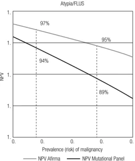
Figure 3. Negative predictive value (NPV) of Airma (20) and a Mutational
Panel (38) according to the prevalence of malignancy.
Figure 3. Negative predictive value (NPV) of Airma (20) and a Mutational
Panel (38) according to the prevalence of malignancy.
Leia-se:
100%
95%
90%
85%
80%
10% 15%
89% 94%
95% 97%
20% 25% 30%
Prevalence (risk) of malignancy Atypia/FLUS
NPV Airma NPV Mutational Panel
NPV
1.
1.
1.
1.
1.
0. 0.
89% 94%
95% 97%
0. 0. 0.
Prevalence (risk) of malignancy Atypia/FLUS
NPV Airma NPV Mutational Panel
NPV