Cop
yright
© ABE&M t
odos os dir
eit
os r
eser
vados
.
144
original article
Arq Bras Endocrinol Metab. 2013;57/2
Radioiodine therapy in elderly
patients with subclinical
hyperthyroidism due to
non-voluminous nodular goiter and
its effect on bone metabolism
Terapia com radioiodo em pacientes idosos com hipertireoidismo subclínico por bócio nodular não volumoso e efeito sobre o metabolismo ósseo
Pedro Weslley Rosario1,2
ABSTRACT
Objective: To evaluate 131I therapy in elderly patients with subclinical hyperthyroidism (SCH) due to nodular disease and who did not receive antithyroid drugs (ATDs), and the effect of the treat-ment on bone metabolism.Subjects and methods: Thirty-six patients with TSH ≤ 0.1 mIU/L and non-voluminous goiter (< 60 cm3) were studied. Bone mineral density (BMD) was assessed in 17 women with osteopenia.Results: Mean 24-h 131I uptake was 17.5%. Symptoms of thyrotoxicosis were reported by two (5.5%) patients in the irst week after therapy. One year after radioiodine treatment, SCH was resolved in 30 (83.3%) patients, and hypothyroidism was detected in one (2.7%). In the patients in whom TSH returned to normal, femoral and lumbar spine BMD incre-ased by 1.9% and 1.6%, respectively, in average.Conclusions: In elderly patients with SCH and non-voluminous goiter, radioiodine not preceded by ATDs is a safe and effectivetherapeutic alternative. Resolution of SCH has beneicial effects on BMD in postmenopausal women with osteopenia. Arq Bras Endocrinol Metab. 2013;57(2):144-7
Keywords
Subclinical hyperthyroidism; radioiodine; nodular disease
RESUMO
Objetivo: Avaliar a terapia com 131I em idosos com hipertireoidismo subclínico (HSC) por doença nodular que não receberam drogas antitireoidianas (DATs) e o efeito no metabolismo ósseo.Sujeitos e métodos: Trinta e seis pacientes com TSH ≤ 0,1 mUI/L e bócio não volumoso (< 60 cm3) foram estudados. Dezessete mulheres com osteopenia foram submetidas à avaliação da densidade mineral óssea (DMO).Resultados: Captação média de 131I em 24 h foi 17,5%. Sin-tomas de tireotoxicose foram reportados por dois pacientes (5,5%) na primeira semana após a terapia. Um ano após o radioiodo, HSC foi resolvido em 30 pacientes (83,3%) e hipotireoidismo ocorreu em 1 (2,7%). Nas pacientes que normalizaram o TSH, DMO em fêmur e coluna lombar incrementou em média 1,9% e 1,6%, respectivamente.Conclusões: Em idosos com HSC e bócio não volumoso, radioiodo, não precedido de DATs, é uma alternativa terapêutica segura e eicaz. Resolução do HSC tem benefício na DMO em mulheres menopausadas com osteopenia. Arq Bras Endocrinol Metab. 2013;57(2):144-7
Descritores
Hipertireoidismo subclínico; radioiodo; doença nodular
1 Postgraduation Program, Santa Casa de Belo Horizonte, Belo Horizonte, MG, Brazil 2 Endocrinology Service, Santa Casa de Belo Horizonte, Belo Horizonte, MG, Brazil
Correspondence to:
Pedro Weslley Rosario Instituto de Ensino e Pesquisa, Santa Casa de Belo Horizonte Rua Domingos Vieira, 590 30150-240 – Belo Horizonte, MG, Brazil
Cop
yright
© ABE&M t
odos os dir
eit
os r
eser
vados
.
145
Arq Bras Endocrinol Metab. 2013;57/2
INTRODUCTION
A
lthough controversy exists regarding the indica-tions for treatment of subclinical hyperthyroidism (SCH), there is consensus that therapy is necessary in elderly patients with TSH ≤ 0.1 mIU/L (1). In this age group, SCH is almost always caused by nodular di-sease (2). Radioiodine therapy is an excellent option for patients with a contraindication to or who refuse surgery; it is also an alternative to thyroidectomy in pa-tients with non-voluminous goiter without compressi-ve symptoms or suspicion of malignancy(1). If 131I is administered in the presence of undetectable levels of TSH, normal thyroid tissue does not take up, or takes up very little radioiodine, reducing the risk of hypo-thyroidism (3,4). In addition, the chance of success (re-solution of hyperthyroidism) may be greater in patients who did not receive antithyroid drugs (ATDs) before the administration of 131I (5). On the other hand, treat-ment with ATDs reduces the risk of acute exacerbation of thyrotoxicosis (5), which is a matter of concern, par-ticularly in older patients.One of the repercussions of SCH is a decline of bone mineral density (BMD), notably in postmeno-pausal women and in patients with TSH ≤ 0.1 mIU/L (1,6). However, few studies have evaluated the effect of treatment of SCH on bone metabolism and bone mass (7-9).
We report here our experience with 131I therapy in elderly patients with SCH (TSH ≤ 0.1 mIU/L) due to non-voluminous nodular goiter who did not receive ATDs prior to radioiodine administration. In addition, we evaluated the effect of this treatment on bone turn-over and BMD in women with osteopenia.
SUBJECTS AND METHODS
Thirty-six patients (24 with multinodular disease and 12 with uninodular disease; 29 women, age: 66 to 80 years) were studied. SCH was diagnosed when TSH was ≤ 0.1 mIU/L in two measurements obtained at a 3-month interval, associated with normal free T4 and total T3 levels. Autonomous nodular disease was diagnosed based on the demonstration of one or more accumulating areas upon 131I scintigraphy and sup-pression of the remaining parenchyma. None of the patients presented compressive symptoms due to volu-minous goiter.
For the treatment with 131I, it was ensured that pa-tients had not used amiodarone or had been recently
ex-posed to iodinated contrast. In addition, patients were asked to consume a low-iodine diet 7 days prior to ther-apy. Patients who had been treated with ATDs, those prepared with recombinant human TSH (rhTSH), or those previously undergoing 131I treatment were ex-cluded from the study. All patients received 50 mg/ day atenolol from the irst week before 131I administra-tion until normalizaadministra-tion of TSH. A ixed activity of 15 mCi 131I was administered. Higher 131I activities were not used since the group mainly consisted of women (80%) older than 40 years with non-voluminous goiter and mild hyperthyroidism, all predictors of a better re-sponse to radioiodine treatment (10).
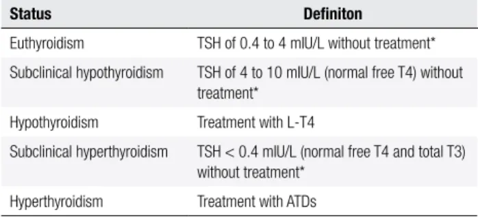
Patients were asked to report symptoms of thyro-toxicosis (11) when they occurred, and were also asked about the presence of symptoms 2 and 7 days after 131I therapy. TSH, free T4 and total T3 were measured one week, and 1, 3, 6, 9, and 12 months after radioiodine administration. L-T4 therapy was introduced if TSH > 10 mIU/L in the presence of low free T4, or if TSH continued to be > 10 mIU/L in two measurements, even when free T4 was normal. ATDs were indicated only if TSH was undetectable and free T4 or total T3 was elevated one month after treatment, or if TSH continued to be undetectable and free T4 and total T3 were normal one year after therapy. Patients were classi-ied one year after radioiodine administration as shown in table 1.
Table 1. Classiication of the patients one year after radioiodine therapy
Status Deiniton
Euthyroidism TSH of 0.4 to 4 mIU/L without treatment* Subclinical hypothyroidism TSH of 4 to 10 mIU/L (normal free T4) without
treatment* Hypothyroidism Treatment with L-T4
Subclinical hyperthyroidism TSH < 0.4 mIU/L (normal free T4 and total T3) without treatment*
Hyperthyroidism Treatment with ATDs
ATD: antithyroid drug. * Use of L-T4 or ATD.
For the evaluation of the beneit of SCH treatment, a subgroup of 17 women was studied. These women had osteopenia [no history of fractures and lumbar and/or femoral BMD - 1 to - 2.5 standard deviations of the young adult mean (T-score)], normal serum PTH and no vitamin D deiciency [25 (OH) vitamin D > 20 ng/mL (12,13) in all patients and > 30 ng/dL in 14 women], and did not use estrogens, corticosteroids, antiresorptive agents or bone-building drugs. These
Cop
yright
© ABE&M t
odos os dir
eit
os r
eser
vados
.
146 Arq Bras Endocrinol Metab. 2013;57/2
patients were submitted to the assessment of BMD, serum carboxyterminal telopeptide (CTx), and osteo-calcin before, and one year after radioiodine treatment. The study was approved by the Ethics Committee of the institution.
TSH, free T4, total T3 and PTH were measured with a chemiluminescent assay with reference values of 0.4-4 mIU/L, 0.8-2.0 ng/dL, 80-180 ng/dL, and 12-65 pg/mL, respectively. CTx and osteocalcin were measured by an electrochemiluminescence assay, with reference values of 0.104 to 1.008 ng/mL (post-menopause), and 12 to 46 ng/mL, respectively. High--performance liquid chromatography was used for the measurement of 25(OH) vitamin D.
BMD was measured by dual energy x-ray absorpti-ometry (DEXA) in the femoral neck and lumbar spine (L1-L4). Scintigraphy was carried out 24 hours after the administration of 100-300 µCi 131I. Sonography was performed with a linear multifrequency 10-12 MHz transducer for morphological analysis (B-mode).
The two-tailed unpaired Student t-test and Mann-Whitney U test were used for statistical analysis. P-value less than 0.05 was considered to be signiicant.
RESULTS
In the 12 patients with uninodular disease, the volume of the nodule estimated by ultrasound ranged from 3.6 to 20 cm3, and total gland volume ranged from 12.5 to 32.1 cm3. Uptake of 131I in the 24th hour ranged from 11.5% to 32.1% (mean: 18.7%). In the 24 patients with multinodular disease, thyroid volume ranged from 28 to 58 cm3. Uptake of 131I in the 24th hour ranged from 9.5% to 30.2% (mean: 17.1%).
Symptoms of thyrotoxicosis (palpitation and tre-mors) were reported by two (5.5%) patients in the irst week after radioiodine treatment, but they were tran-sient and did not require speciic treatment or hospital admission. There was no case of arrhythmia after treat-ment. Laboratory tests revealed elevated free T4 in four (11%) patients in the irst week after therapy, but total T3 was elevated in only two of these patients. Free T4 and total T3 returned to normal in all four patients one month after radioiodine administration.
One year after radioiodine therapy, SCH was re-solved in 30 (83.3%) patients, and hypothyroidism was detected in one (2.7%). None of the patients progressed to overt thyrotoxicosis. Six (16.6%) patients contin-ued to present SCH, but hyperfunction improved in
three of these patients, with TSH increasing from ≤ 0.1 mIU/L to 0.2, 0.22, and 0.3 mIU/L, respectively.
Twelve of the 17 women evaluated before and one year after radioiodine therapy were euthyroid. In the patients in whom thyroid function returned to normal, femoral BMD increased by 1.9%, and lumbar spine BMD by 1.6%, in average. In addition, there was a sig-niicant decrease of serum CTx levels [mean of 45%, p < 0.01], but no signiicant change in serum osteocal-cin concentration was observed. Four women contin-ued to present SCH. In these patients, BMD decreased by 2% in the femur and by 1.8% in the lumbar spine, in average. No changes in serum osteocalcin or CTx were observed.
DISCUSSION
A high rate of resolution (83.3%) of hyperthyroidism was achieved in the present study. This success rate, even with an activity of 15 mCi, can be explained by the population studied, i.e., a predominance of women > 40 years with non-voluminous goiter and mild hyper-thyroidism, since all of these factors are known predic-tors of a better response to radioiodine (10).Although a long-term increase is possible, the rate of hypothyroi-dism seen in the irst year (3%) was low. The adminis-tration of 131I during TSH suppression impairs or re-duces uptake by normal (non-autonomous) thyroid tissue, decreasing the risk of hypothyroidism. Some observations support this hypothesis. First, in Graves’ disease, in which the whole gland takes up iodine, the frequency of hypothyroidism in the irst year is high, ranging from 55% to 80% (14,15). Second, patients with toxic nodular disease who receive 131I in the pre-sence of detectable levels of TSH show higher rates of hypothyroidism than those with undetectable TSH at the time of treatment (3,4). Finally, the administration of rhTSH before 131I therapy to patients with nodular disease, which increases the tissue area that takes up io-dine, notably increases the risk of hypothyroidism (16). Thus, normalization of TSH with ATDs before radio-iodine, restoring the uptake by normal thyroid tissue, increases the risk of hypothyroidism. However, many authors do not consider hypothyroidism an undesirable effect of radioiodine treatment since the main objective of therapy, i.e., reversal of thyrotoxicosis, is achieved.
During 131I therapy of elderly patients with clinical hyperthyroidism, prior thyroid function compensation (normalization of T4 and T3) with ATDs should be
Cop
yright
© ABE&M t
odos os dir
eit
os r
eser
vados
.
147
Arq Bras Endocrinol Metab. 2013;57/2
considered to reduce the risk of exacerbation of thy-rotoxicosis, which can have important clinical conse-quences in these patients (1). This risk appears to be very low in SCH, and compensation (normalization of TSH) before 131I does not seem to be necessary (1). On the other hand, administration of beta-blockers is re-commended since, as observed for patients with clinical thyrotoxicosis, these drugs minimize the symptoms in patients with SCH who develop exacerbation of thyro-toxicosis after radioiodine (1). The use of beta-blockers may explain the absence of important symptoms in the present patients, even in those who showed transient increase of thyroid hormone levels after radioiodine treatment. Therefore, preparation with beta-blockers before 131I therapy seems to be suficient in SCH, even in the case of elderly patients and TSH ≤ 0.1 mIU/L.
With respect to the effect of TSH normalization on BMD, the present study showed an increase of both femoral and lumbar spine BMD in as early as the irst year, in agreement with the indings of Faber and cols. (8) after treatment of SCH with 131I. Muddle and cols. (7) also observed an increase of distal forearm BMD after treatment of SCH with methimazole. In addition to the increase in BMD seen in treated patients, BMD loss was observed in patients who did not receive treat-ment (8), a inding that supports the beneicial effect of this therapy. This effect seems to be the conse-quence of reduced bone resorption, since plasma CTx levels were also signiicantly decreased. No signiicant change in serum osteocalcin after TSH normalization was observed in the present investigation or in a previ-ous study (7), supporting the point of view that the increase in BMD was indeed due to reduced bone re-sorption. Finally, it should be emphasized that all of these studies involved postmenopausal women with endogenous SCH due to nodular disease and TSH ≤ 0.1 mIU/L (7, present study), or < 0.2 mIU/L (8), and the results, therefore, cannot be readily extrapo-lated to men, premenopausal women, patients with SCH whose TSH levels are only slightly reduced, or elderly patients with low TSH and no structural thy-roid disease. For example, Yonem and cols. (9) found no increase in BMD in premenopausal women after 6 months of treatment with ATDs.
The present results suggest that 131I therapy not preceded by ATDs in elderly patients with SCH and non-voluminous goiter is, in fact, safe, effective, and associated with low rates of short-term hypothyroid-ism. In addition, resolution of SCH seems to have
be-neicial effects on bone mass in postmenopausal wom-en with osteopwom-enia.
Disclosure: no potential conlict of interest relevant to this article was reported.
REFERENCES
1. Bahn Chair RS, Burch HB, Cooper DS, Garber JR, Greenlee MC, Klein I, et al. Hyperthyroidism and other causes of thyrotoxico-sis: management guidelines of the American Thyroid Association and American Association of Clinical Endocrinologists. Thyroid. 2011;21:593-646.
2. Rosario PW. Natural history of subclinical hyperthyroidism in el-derly patients with TSH between 0.1 and 0.4 mIU/l: a prospective study. Clin Endocrinol (Oxf). 2010;72:685-8.
3. Pedersen-Bjergaard U, Kirkegaard C. Relationship between serum TSH and the responsiveness of toxic solitary autono-mous thyroid nodules to radioiodine therapy. Eur J Endocrinol. 1998;139:587-90.
4. Pedersen-Bjergaard U, Kirkegaard C. Serum TSH and the respon-se to radioiodine treatment of toxic multinodular goitre. Eur J Endocrinol. 1997;137:365-9.
5. Walter MA, Briel M, Christ-Crain M, Bonnema SJ, Connell J, Co-oper DS, et al. Effects of antithyroid drugs on radioiodine treat-ment: systematic review and meta-analysis of randomised con-trolled trials. BMJ. 2007;334:514.
6. Rosario PW. Bone and heart abnormalities of subclinical hyper-thyroidism in women below the age of 65 years. Arq Bras Endo-crinol Metabol. 2008;52:1448-51.
7. Muddle AH, Houben AJ, Nieuwenhuijzen Kruseman AC. Bone metabolism during anti-thyroid drug treatment of endogenous subclinical hyperthyroidism. Clin Endocrinol (Oxf). 1994;41:421-4. 8. Faber J, Jensen IW, Petersen L, Nygaard B, Hegedus L, Siersbaek--Nielsen K. Normalization of serum thyrotropin by mean of radio-iodine treatment in subclinical hyperthyroidism: effect of bone loss in postmenopausal women. Clin Endocrinol (Oxf). 1998;48:285-90. 9. Yonem O, Dokmetas HS, Aslan SM, Erselcan T. Is antithyroid tre-atment really relevant for young patients with subclinical hyper-thyroidism? Endocr J. 2002;49:307-14.
10. Allahabadia A, Daykin J, Sheppard MC, Gough SC, Franklyn JA. Radioiodine treatment of hyperthyroidism-prognostic factors for outcome. J Clin Endocrinol Metab. 2001;86:3611-7.
11. Klein I, Trzepacz PT, Roberts M, Levey GS. Symptom rating scale for assessing hyperthyroidism. Arch Intern Med. 1988;148:387-90. 12. Rosen CJ, Abrams SA, Aloia JF, Brannon PM, Clinton SK, Durazo-Ar-vizu RA, et al. IOM committee members respond to Endocrine So-ciety vitamin D guideline. J Clin Endocrinol Metab. 2012;97:1146-52. 13. Aloia JF. Clinical Review: The 2011 report on dietary reference in-take for vitamin D: where do we go from here? J Clin Endocrinol Metab. 2011;96:2987-96.
14. Alexander EK, Larsen PR. High dose of (131)I therapy for the tre-atment of hyperthyroidism caused by Graves’ disease. J Clin En-docrinol Metab. 2002;87:1073-7.
15. Andrade VA, Gross JL, Maia AL. The effect of methimazole pre-treatment on the eficacy of radioactive iodine therapy in Graves’ hyperthyroidism: one-year follow-up of a prospective, randomi-zed study. J Clin Endocrinol Metab. 2001;86:3488-93.
16. Fast S, Nielsen VE, Grupe P, Boel-Jørgensen H, Bastholt L, Ander-sen PB, et al. Prestimulation with Recombinant Human Thyrotro-pin (rhTSH) improves the long-term outcome of radioiodine the-rapy for multinodular nontoxic goiter. J Clin Endocrinol Metab. 2012;97:2653-60.