Cop
yright
© ABE&M t
odos os dir
eit
os r
eser
vados
.
Assessment of the elasticity properties
of the ascending aorta in patients
with subclinical hypothyroidism
by tissue Doppler imaging
Avaliação das propriedades de elasticidade da aorta ascendente por Doppler tecidual em pacientes com hipotireoidismo subclínico
Mustafa YurtdasÇ1, Ramazan Gen2, Turkay Özcan3, Mehmet Kasım Aydın4
ABSTRACT
Objective: We aimed to investigate whether aortic elastic properties were affected in subclinical hypothyroidism (SCH) by using tissue Doppler imaging (TDI). Subjects and methods: Forty-three patients with newly diagnosed SCH and forty-eight healthy controls were included to the study. Systolic and diastolic diameters of the ascending aorta were measured by M-mode transthoracic echocardiography, and the upper wall velocities of ascending aorta and mitral annulus velocities were measured by TDI. Aortic stiffness index (ASI) and aortic distensibility were computed using the formulas accepted in literature. Results: The clinical and demographic features of both groups were comparable. Aortic distensibility was signiicantly lower, and ASI was signiicantly higher in SCH patients than in controls. Systolic aortic upper wall velocity (Sao) was also signiicantly lower in SCH patients. Early (Eao) and late diastolic aortic upper wall (Aao) velocities did not differ be-tween the two groups. Mitral annulus (Sm, Em, and Am) velocities were also similar bebe-tween the groups. Sao was negatively correlated with ASI, and positively correlated with aortic distensibility. TSH level was positively correlated with ASI, total cholesterol and low-density lipoprotein-choles-terol, and negatively correlated with aortic distensibility and Sao. Conclusions: In this study, our re-sults showed that SCH is associated with impaired elasticity of the ascending aorta. Elastic proper-ties of the ascending aorta can be directly evaluated by the reproducibly measurement of the upper wall movements of the ascending aorta by TDI in SCH patients. Arq Bras Endocrinol Metab. 2013;57(2):132-8
Keywords
Tissue Doppler imaging; subclinical hypothyroidism; stiffness
RESUMO
Objetivo:Nosso objetivo foi investigar se as propriedades elásticas da aorta são afetadas no hipotireoidismo subclínico (HSC), utilizando o Doppler tecidual (UDT). Sujeitos e métodos: Qua-renta e três pacientes com diagnóstico recente de HSC e 48 indivíduos saudáveis foram incluí-dos no estudo. Os diâmetros sistólico e diastólico da aorta foram mediincluí-dos por ecocardiograia transtorácica modo M e as velocidades de luxo da parede superior da aorta ascendente e de luxo transvalvar mitral foram medidas por UDT. O índice de rigidez da aorta (IRA) e a distensi-bilidade aórtica foram calculados usando fórmulas aceitas na literatura. Resultados: As caracte-rísticas clínicas e demográicas dos dois grupos foram comparáveis. A distensibilidade aórtica foi signiicativamente menor e IRA signiicativamente maior nos pacientes com HSC do que nos controles. A velocidade de luxo sistólico na parede aórtica superior (Sao) foi signiicantemente menor em pacientes com HSC. A velocidade de luxo diastólico inicial (Eao) e tardio (Aao) na parede aórtica superior e as velocidades de luxo transvalvar (Sm, Em e Am) não diferiram entre os dois grupos. Sao foi negativamente correlacionada com IRA e positivamente correlacionada com a distensibilidade aórtica. O nível de TSH foi positivamente correlacionado com IRA, coles-terol total e lipoproteína de baixa densidade-colescoles-terol e negativamente correlacionado com a distensibilidade aórtica e Sao. Conclusões: Os resultados do presente estudo demonstraram que o HSC é associado com elasticidade deiciente da aorta ascendente. Propriedades elásticas da aorta ascendente podem ser diretamente avaliadas por medições reprodutíveis dos movimentos da parede superior da aorta por UDT em pacientes com HSC. Arq Bras Endocrinol Metab. 2013;57(2):132-8
Descritores
Doppler tissular; hipotireoidismo subclínico; rigidez 1 Lokman Hekim Hospital,
Department of Cardiology, Van, Turkey
2 Mersin University, Department of
Endocrinology, Mersin, Turkey
3 Mersin University, Department of
Cardiology, Mersin, Turkey
4 Lokman Hekim Hospital,
Department of Internal Medicine, Van, Turkey
Correspondence to:
Mustafa YurtdasÇ
Lokman Hekim Van Hastanesi, Kardiyoloji Bölümü
65200, Van, Turkey [email protected]
Cop
yright
© ABE&M t
odos os dir
eit
os r
eser
vados
.
INTRODUCTION
S
ubclinical hypothyroidism (SCH) is deined as an elevated serum level of thyroid stimulating hormo-ne (TSH) with normal free thyroid values (1,2). The prevalence of SCH has been reported to be between 4% and 10% in adult population samples (2,3). Coronary artery disease (CAD) and cardiovascular mortality are increased in patients with SCH, particularly in those with higher TSH levels (4,5).Functional properties of large arteries are impor-tant both for the normal function of the artery itself and for left ventricular function (6). Previous studies have shown that increased aortic stiffness and decrea-sed distensibility, which are impairments in the elastic properties of the aorta, are presented together with premature atherosclerosis and CAD (7-10). Several procedures have been used for the determination of aortic stiffness and/or distensibility, such as magne-tic resonance imaging, angiography,applanation to-nometry, velocity vector imaging (VVI) (9-12). But the vast majority of these techniques are invasive and time-consuming and may require complex equipment and training. Conventional and tissue Doppler ima-ging (TDI) echocardiography have been shown to be useful methods in the evaluation of global ventricular functions and elastic properties of the aorta (13-17). Although increased arterial stiffness index has been formerly shown in various patient groups, including those with CAD, diabetes mellitus (DM), overt hypo-thyroidism, and SCH on different vascular beds and in different sites such as the radial artery, carotid artery, and aorta (14-20), as far as we know, no report has evaluated elastic properties of the ascending aorta by TDI in patients with SCH.
To clarify this issue, we aimed to investigate, using TDI, whether elastic properties of the ascending aorta are affected in patients with SCH.
SUBJECTS AND METHODS
Study population
Newly detected asymptomatic or mild symptomatic (such as those presenting dry hair or skin, cold into-lerance, forgetfulness, constipation, depression, and weakness) SCH patients with increased serum TSH (4.2<TSH<20.0 mIU/mL), normal free T3 (FT3) and free T4 (FT4) levels were recruited for the study.
Forty--three patients with SCH in at least two thyroid hormo-nal proiles measured 3 months before study were exa-mined prospectively. The control group (11 males, 37 females; mean age, 42 ± 11 years old) consisted of heal-thy volunteers with normal FT3 (normal range: 2.0-4.4 pg/mL), FT4 (normal range: 0.93-1.7 ng/dL), and TSH (normal range: 0.27-4.2 mIU/mL) levels ma-tched for age, sex, and body mass index (BMI). The ex-clusion criteria were previous history of thyroid disease, valvular heart disease, cardiac rhythm disturbance, heart failure, arterial hypertension, pulmonary hypertension, pregnancy, hepatic or renal disease, respiratory disease, DM, neurological or psychological disease, and malig-nancy. Also, CAD was excluded based on normal results of the exercise stress echo test applied to all study mem-bers. None of the subjects received medications that could inluence heart rate and serum thyroid hormone levels. Physical examination, medical history, inlamma-tory markers, and electrocardiogram were normal for all participants. The study protocol was approved by the Research Review Board of Mersin University, and infor-med consent was obtained from all participants.
Echocardiographic study
Cop
yright
© ABE&M t
odos os dir
eit
os r
eser
vados
.
examination of the ascending aorta. BP was measured three times on each occasion at 3-minute intervals and averaged. Pulse pressure (PP) was obtained by subtrac-ting diastolic blood pressure (DBP) from systolic blood pressure (SBP). Systolic and diastolic inner diameters of the ascending aorta were recorded by M-mode echocar-diography 3 cm above the aortic valve, in a parasternal long axis image. Aortic systolic diameter (AoS) was me-asured at the maximum anterior motion of the aorta, and aortic diastolic diameter (AoD) was measured at the peak of the QRS complex on the recorded electrocar-diogram, simultaneously (Figure 1). Distensibility and stiffness index of the ascending aorta were calculated by using the following formulaes (14-17).
Aortic distensibility = 2 X (AoS - AoD) / (SBP -
DBP) X AoD (10-6.cm2.dyn-1),
Aortic stiffness index = ln (SBP / DBP) / [(AoS
- AoD) / AoD] (pure number), ln = natural logarithm. The indexes of aortic systolic and diastolic diameters, and pulsatile change (cm/m2) for each subject were
es-timated by dividing AoS, AoD and the pulsatile change in aortic diameter (PAod) by BSA, respectively. Then, the echocardiographic device was switched to theTDI mode, and aortic superior wall velocities in systole (Sao), early diastole (Eao) and late diastole (Aao) were estimated at the same point used in the M-mode measurements (Figure 2). In addition, from the apical window, mitral annulus velo-cities in systole (Sm), early diastole (Em) and late diastole (Am) were calculated. All the parameters were computed and the average of ive consecutive cycles was calculated.
Figure 1. Systolic and diastolic diameter measurements of the ascending
aorta with M-mode echocardiography. AoS: aortic systolic diameter; AoD: aortic diastolic diameter.
Figure 2. Aortic upper wall velocity measurements with tissue Doppler
imaging. Sao: systolic aortic upper wall velocity; Eao: early diastolic aortic upper wall velocity; Aao: late diastolic aortic upper wall velocity.
Biochemical and hormonal analysis
Blood samples were withdrawn after 12 hours of overnight fasting for FT3, FT4, TSH, total choleste-rol (TC), triglyceride (TG), high-density lipoprotein--cholesterol (HDL-C). Assays for TC, TG, HDL-C were performed using a Cobas Integra 800 automated analyzer (Roche Diagnostics, Manheim, Germany). Se-rum low-density lipoprotein-cholesterol (LDL-C) was calculated according to Friedewald’s Formula (24). Se-rum FT3, FT4, and TSH levels were measured using the Modular E170 automated analyzer (Roche Diag-nostics, Manheim, Germany). The sensitivity of the as-says for FT3, FT4, and TSH was 0.260 pg/mL, 0.023 ng/dL, and 0.005 mIU/mL, respectively.
Reproducibility
Interobserver variability of aortic elastic measurements was measured as the difference between two measure-ments of the same patient by two different cardiologists divided by the mean value. Intraobserver variability was calculated as the difference between two measurements of the same patient by a single cardiologist divided by the mean value. Intraobserver and interobserver varia-bility were less than 5% for all aortic elasticity measure-ments in the total study population.
Statistical analysis
Cop
yright
© ABE&M t
odos os dir
eit
os r
eser
vados
.
USA). Kolmogorov-Smirnov test was used to inves-tigate whether the distribution of measurements was normal or not. Categorical variables were expressed as percentages, and compared using the Chi-square test. Continuous variables were expressed as means ± SD. To compare the variables that showed normal distribu-tion in the two groups, independent t-test was used. In contrast, for the variables that did not show normal dis-tribution, Mann-Whitney U test was used. Linear cor-relations between measurements in both groups were determined by Pearson’s correlation. Two-tailed tests were used to test all hypotheses, and p-value of < 0.05 was considered signiicant.
RESULTS
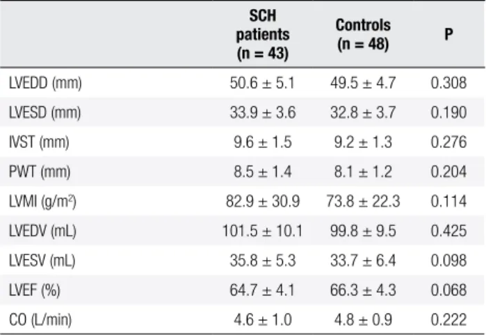
The baseline clinical and demographic features, and FT3, FT4, TSH levels of the patients and controls are shown in Table 1. Demographic characteristics of the two groups were similar. There were no signiicant di-fferences in SBP, DBP, PP, and heart rate between the two groups. The participants with SCH demonstrated an increase in serum TSH above the normal upper li-mit (8.9 ± 2.8 mUI/mL, p < 0.001), and had higher TC and LDL-C than those of controls (p = 0.009, p = 0.003; respectively). Echocardiographic variables of the groups are shown in Table 2. Mean left ventricular di-mensions and volumes, IVS, and PW thickness were not signiicantly different between the groups. In addition, LVMI, LVEF, and cardiac output of both groups were also similar.
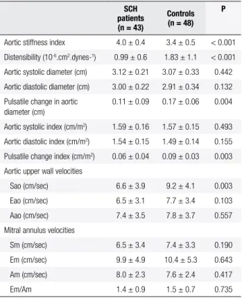
Aortic stiffness index, aortic distensibility, aortic diameters, and TDI measurements of the two groups are summarized in Table 3. Aortic stiffness index was signiicantly higher (4.0 ± 0.4 vs. 3.4 ± 0.5; p < 0.001) and aortic distensibility was signiicantly lower (0.99 ± 0.6 vs. 1.83 ± 1.1 x 10-6.cm2.dynes-1; p < 0.001) in
patients with SCH than in those who were euthyroid. The values of AoS and AoD were comparable between SCH patients and controls. PAod, however, was sig-niicantly lower in patients with SCH compared with the controls (0.11 ± 0.09 vs. 0.17 ± 0.06; p = 0.004). Sao was also signiicantly lower in patients with SCH than in controls (6.6 ± 3.9 vs. 9.2 ± 4.1 cm/sec; p = 0.003). Early diastolic aortic upper wall velocity (Eao) and late diastolic aortic upper wall velocity (Aao), and mitral annulus systolic (Sm), early diastolic (Em), and late diastolic wave (Am) velocities were similar in the two groups.
Table 1. Clinical and demographic characteristics and thyroid hormone
levels of both groups
SCH patients (n = 43)
Controls (n = 48) P
Sex (male/female) 7/36 11/37 0.599
Age (years) 43 ± 9 42 ± 11 0.539
Body mass index (kg/m2) 28 ± 3 27 ± 4 0.449
Smoking (%) 23 35 0.253
SBP (mmHg) 128 ± 9 126 ± 9 0.514
DBP (mmHg) 75 ± 7 73 ± 8 0.106
Pulse pressure (mmHg) 52 ± 9 53 ± 13 0.734
Mean arterial pressure (mmHg) 93 ± 6 90 ± 7 0.107
Heart rate (per/min) 70 ± 13 73 ± 11 0.179
Total cholesterol (mg/dL) 217 ± 18 207 ± 17 0.009
Triglycerides (mg/dL) 153 ± 30 143 ± 31 0.112
LDL-C (mg/dL) 141 ± 20 130 ± 17 0.003
HDL-C (mg/dL) 46 ± 6 48 ± 7 0.147
Free T3 (pg/mL) 2.79 ± 0.63 2.91 ± 0.72 0.408
Free T4 (ng/dL) 1.13 ± 0.22 1.21 ± 0.29 0.145
TSH (mUI/mL) 8.9 ± 2.8 1.4 ± 0.3 < 0.001
SBP: systolic blood pressure; DBP: diastolic blood pressure; LDL-C: low-density lipoprotein cholesterol; HDL-C: high-density lipoprotein cholesterol; TSH: thyroid stimulating hormone; SCH: subclinical hypothyroidism.
Table 2. Echocardiographic variables of SCH patients and controls
SCH patients (n = 43)
Controls (n = 48) P
LVEDD (mm) 50.6 ± 5.1 49.5 ± 4.7 0.308
LVESD (mm) 33.9 ± 3.6 32.8 ± 3.7 0.190
IVST (mm) 9.6 ± 1.5 9.2 ± 1.3 0.276
PWT (mm) 8.5 ± 1.4 8.1 ± 1.2 0.204
LVMI (g/m2) 82.9 ± 30.9 73.8 ± 22.3 0.114
LVEDV (mL) 101.5 ± 10.1 99.8 ± 9.5 0.425
LVESV (mL) 35.8 ± 5.3 33.7 ± 6.4 0.098
LVEF (%) 64.7 ± 4.1 66.3 ± 4.3 0.068
CO (L/min) 4.6 ± 1.0 4.8 ± 0.9 0.222
LVEDD: left ventricular end-diastolic diameter; LVESD: left ventricular end-systolic diameter; IVST: interventricular septum thickness; PWT: posterior wall thickness; LVMI: left ventricular mass index; LVEDV: left ventricular end-diastolic volume; LVESV: left ventricular end-systolic volume; LVEF: left ventricular ejection fraction; CO: cardiac output; SCH: subclinical hypothyroidism.
Cop
yright
© ABE&M t
odos os dir
eit
os r
eser
vados
.
DISCUSSION
The indings of our study demonstrated that patients with SCH have higher stiffness index, lower distensibility of the ascending aorta, and lower Sao than the controls. The pulse wave is observable and measurable in an arterial system throughout the circulation. During the cardiac systole, a certain volume of blood is ejected and propagates via arteries due to the transformation of kine-tic to potential energy in each segment of ejected blood. In each site, the pulse wave progresses, pressure and blood low velocity change, and ultimately, aortic wall activities are observed (6,25). Thus, aortic wall move-ments, which bring about differentiation in the diameter of the proximal aortic lumen, enable us capture images of the changes in aortic elastic properties (14-17,25). Arterial stiffness index determines the elastic properties of the arterial wall, in a manner relatively independent of blood pressure, and aortic distensibility evaluates the ability of the arteries to dilate during the cardiac cycle, and measures the function of the artery (6,7,25).
Aortic stiffness and aortic distensibility have been examined with some methods, such as VVI and pulse wave velocity (PWV) (12,26). However, VVI is a new and invasive method, requiring transesophageal echocar-diography, which limits its routine clinical practice. Also, PWV is not the ideal procedure to evaluate aortic elasti-city properties since it is affected by many factors inclu-ding hematological and physiological characteristics, as well as heart rate and blood pressure variations (27-29).
The change in the diameter of the aorta detected by echocardiography can be related to the distending pres-sure and provides a meapres-sure of stiffness. However, this echocardiographic method of determining aortic stiff-ness using mathematical equations may have some limi-tations (30,31). First, blood pressure and pulse pressure estimated at the level of brachial artery may not exactly relect aortic pulse pressure. Secondly, blood pressure measurement and aortic echocardiographic assessment cannot be carried out simultaneously.
Direct measurement of aortic elasticity by TDI, which is a practical method for the measurement of diameter changes related to wall movements, may pro-vide further help than other methods described above, because it is not affected by hematological and cardio-vascular physiology. A number of studies have shown that elastic properties of the ascending aorta could be directly and reproducibly evaluated by measuring aor-ta movements in patients with CAD, DM, and hyper-tension (HT) by TDI (14-17).To our knowledge, the
Table 3. Aortic stiffness index, distensibility, M-mode, and TDI
measure-ments in SCH patients and controls
SCH patients (n = 43)
Controls (n = 48)
P
Aortic stiffness index 4.0 ± 0.4 3.4 ± 0.5 < 0.001
Distensibility (10-6.cm2.dynes-1) 0.99 ± 0.6 1.83 ± 1.1 < 0.001
Aortic systolic diameter (cm) 3.12 ± 0.21 3.07 ± 0.33 0.442
Aortic diastolic diameter (cm) 3.00 ± 0.22 2.91 ± 0.34 0.132
Pulsatile change in aortic diameter (cm)
0.11 ± 0.09 0.17 ± 0.06 0.004
Aortic systolic index (cm/m2) 1.59 ± 0.16 1.57 ± 0.15 0.493
Aortic diastolic index (cm/m2) 1.54 ± 0.15 1.49 ± 0.14 0.155
Pulsatile change index (cm/m2) 0.06 ± 0.04 0.09 ± 0.03 0.003
Aortic upper wall velocities
Sao (cm/sec) 6.6 ± 3.9 9.2 ± 4.1 0.003
Eao (cm/sec) 6.5 ± 3.1 7.7 ± 3.4 0.103
Aao (cm/sec) 7.4 ± 3.5 7.8 ± 3.7 0.557
Mitral annulus velocities
Sm (cm/sec) 6.5 ± 3.4 7.4 ± 3.3 0.190
Em (cm/sec) 9.9 ± 4.9 10.4 ± 5.3 0.643
Am (cm/sec) 8.0 ± 2.3 7.6 ± 2.4 0.417
Em/Am 1.4 ± 0.9 1.5 ± 0.7 0.735
Sao: systolic aortic upper wall velocity; Eao: early diastolic aortic upper wall velocity; Aao: late diastolic aortic upper wall velocity; Sm: systolic mitral annulus velocity; Em: early diastolic mitral annulus velocity; Am: late diastolic mitral annulus velocity; TDI: tissue Doppler imaging; SCH: subclinical hypothyroidism.
Table 4. Relationships between aortic stiffness index, aortic distensibility,
Sao, TC, LDL-C and TSH value in SCH patients
TSH value
r p
Aortic stiffness index 0.524 < 0.001
Aortic distensibility -0.436 0.003
Sao -0.582 < 0.001
TC 0.631 < 0.001
LDL-C 0.653 < 0.001
Sao: systolic aortic upper wall velocity; TC: total cholesterol; LDL-C: low-density lipoprotein-cholesterol; TSH: thyroid stimulating hormone; SCH: subclinical hypothyroidism.
Cop
yright
© ABE&M t
odos os dir
eit
os r
eser
vados
.
present study is the irst one assessing elasticity indices of the ascending aorta using TDI in patients with SCH. Elastic properties and wall movements of the ascen-ding aorta can be affected by several risk factors, such as aging and smoking status (6,8,9). However, these risk factors cannot account for the differences in aortic stiff-ness obtained in the present study, because the distribu-tions of these parameters were the same for both groups. Also, it is known that DM and HT have an unfavorable effect on arterial stiffness (15,16) and, accordingly, dia-betic and hypertensive patients were not recruited in the presented study. Thus, in the current trial, none of these risk factors appeared to inluence arterial stiffness.
Dyslipidemia may cause some alterations in the elas-ticity of arterial wall, such as increase in central pulse pressure and endothelial dysfunction (6,32). In addi-tion, several reports in the literature indicate that SCH is associated with atherogenic lipid proile (33,34). Similar to these results, we found that serum TC and LDL-C were signiicantly elevated in patients with SCH compa-red with euthyroid subjects. Also, we found signiicant positively correlation between serum TSH levels, and TC and LDL-C values in patients with SCH, which is in line with the indings of study by Gen and cols. (34). Al-though no correlation was found between the stiffness index or distensibility and lipid values, a signiicant cor-relation was observed between stiffness index (positive) or distensibility (negative) and TSH values. The most plausible explanation for this association is that TSH may have a direct effect on the arterial wall.
Endothelial-dependent vasodilation is inversely cor-related with the administration of recombinant human TSH and serum TSH levels (35,36).In concordance with the results of these trials, our indings made us think that high TSH concentration may make SCH pa-tients more prone to dyslipidemia and endothelial dys-function, and thus, lead to impaired wall movements in the ascending aorta.
One of the important indings of our study was the reduced Sao, Eao, and Aao in patients with SCH com-pared with controls. From these measurements, only the decrease in Sao reached statistically signiicance. Moreo-ver, we also found a negative correlation between Sao and aortic stiffness index and TSH value, and a positive correlation between Sao and distensibility. Reduced Sao is associated with arterial stiffness in patients with CAD and type-1 DM(14,15), a inding consistent with the results of the current trial. During the cardiac cycle, the wave proile observed in the aortic wall movements has the same shape as the one found for the ventricle
pa-rallel to the pressure changes in the aortic lumen, that is, Sao is observed while ventricle contracts, and Eao and Aao occured during ventricular diastole (6,14,15). Another important point is whether Sao derives from the movement of the aorta or myocardial activation. Since we found no correlation between Sao and mitral annulus (Sm, Em, Am) velocities, we consider that Sao originates from aortic wall activities. Therefore, all these indings suggest that SCH may make the aortic wall di-late less during ventricular systole, as a consequence of stiffness observed in the ascending aorta.
A variety of studies have shown that arterial stiffness is the most signiicant reason for cardiovascular compli-cations, and that decreased elasticity of the aorta could be attributed to the presence of atherosclerotic lesions in the aortic wall, even in the absence of evident cardio-vascular disease (6,7,9). Thus, impairment of arterial wall elasticity may pave the way for atherosclerosis.
The knowledge about the relationship between car-diovascular disease and SCH shows conlicting informa-tion. The most convincing data supporting greater car-diovascular risk in patients with SCH come from several recent meta-analyses, in which SCH has been reported to be associated with an elevated risk of cardiovascular di-sease and mortality (4,5). Moreover, some studies have demonstrated that patients with SCH had increased bra-chial-ankle PWV, a useful index of arterial stiffness, and increased carotid arterial stiffness, a useful predictor of cardiovascular risk (18-20). Elevated arterial stiffness and reduced Sao are important predictors of CAD (14). Based on these data and our indings, it can be concluded that SCH could have a deleterious effect on vascular elasticity, and hence, could be an important risk factor for atheros-clerosis by means of the mechanisms discussed above.
The present study had several limitations. The most important one was the small number of patients, which eliminate the ability to apply subgroup analysis according to the levels of TSH and/or the severity of SCH. The second limitation was the absence of data after norma-lization of SCH with levothyroxine treatment. Another limitation was that cardiac catheterization had not been carried out in our study population to exclude CAD and to evaluate intraventricular pressure, which may inluence aortic wall TDI. This limitation can be omitted because it would be dificult, due to ethical concerns, to perform an invasive procedure in asymptomatic individuals with normal exercise stress echo results, as well as similar stroke volume obtained by echocardiography in both groups.
Cop
yright
© ABE&M t
odos os dir
eit
os r
eser
vados
.
euthyroid control subjects. Reduced Sao was associa-ted with the increased aortic stiffness, lipid proiles, and TSH levels. We also concluded that elastic properties of the ascending aorta evaluated by TDI are simple and practical to determine the level of stiffness of the aorta in patients with SCH.
Disclosure: no potential conlict of interest relevant to this article was reported.
REFERENCES
1. Surks MI, Ortiz E, Daniels GH, Sawin CT, Col NF, Cabin RH, et al. Subclinical thyroid disease. Scientiic review and guidelines for diagnosis and management. JAMA. 2004;291:228-38.
2. Canaris GJ, Manowitz NR, Mayor G, Ridgway EC. The Colorado thyroid disease prevalence study. Arch Intern Med. 2000;160:526-34. 3. Hollowell JG, Staehling NW, Flanders WD, Hannon WH, Gunter
EW, Spencer CA, et al. Serum TSH, T4, and thyroid antibodies in the United States population (1988 to 1994): National Health and Nutrition Examination Survey (NHANES III). J Clin Endocrinol Metab. 2002;87:489-99.
4. Ochs N, Auer R, Bauer DC, Nanchen D, Gussekloo J, Cornuz J, et al. Meta-analysis: subclinical thyroid dysfunction and the risk for coronary heart disease and mortality. Ann Intern Med. 2008;148:832-45.
5. Imaizumi M, Akahoshi M, Ichimaru S, Nakashima E, Hida A, Soda M, et al. Risk for ischemic heart disease and all-cause mor-tality in subclinical hypothyroidism. J Clin Endocrinol Metab. 2004;89:3365-70.
6. Wagenseil JE, Mecham RP. Vascular extracellular matrix and arte-rial mechanics. Physiol Rev. 2009;89:957-89.
7. McEniery CM, Wilkinson IB, Avolio AP. Does arterial stiffness pre-dict atherosclerotic coronary events? Clin Exp Pharmacol Physiol. 2007;34:665-71.
8. O’Rourke MF, Hashimoto J. Mechanical factors in arterial aging: a clinical perspective. J Am Coll Cardiol. 2007;50:1-13.
9. Malayeri AA, Natori S, Bahrami H, Bertoni AG, Kronmal R, Lima JA, et al. Relation of aortic wall thickness and distensibility to car-diovascular risk factors (from the multi-ethnic study of atheros-clerosis [MESA]). Am J Cardiol. 2008;102:491-6.
10. Stefanadis C, Stratos C, Vlachopoulos C, Marakas S, Boudoulas H, Kallikazaros I, et al. Pressure-diameter relation of the human aorta. A new method of determination by the application of a spe-cial ultrasonic dimension catheter. Circulation. 1995;92:2210-9. 11. Chen CH, Ting CT, Nussbacher A, Nevo E, Kass DA, Pak P, et al.
Validation of carotid artery tonometry as a means of estimating augmentation index of ascending aortic pressure. Hypertension. 1996;27:168-75.
12. Kim KH, Park JC, Yoon HJ, Yoon NS, Hong YJ, Park HW, et al. Use-fulness of aortic strain analysis by velocity vector imaging as a new echocardiographic measure of arterial stiffness. J Am Soc Echocardiogr. 2009;22:1382-8.
13. Martins RM, Fonseca RHA, Duarte MMT, Reuters VS, Ferreira MM, Almeida C, et al. Impact of subclinical hypothyroidism treatment in systolic and diastolic cardiac function. Arq Bras Endocrinol Me-tab. 2011;55:460-7.
14. Eryol NK, Topsakal R, Cicek Y, Abacı A, Oguzhan A, Basar E, et al. Colour Doppler tissue imaging in assessing the elastic properties of the aorta and in predicting coronary artery disease. Jpn Heart J. 2002;43:219-30.
15. Karamitsos TD, Karvounis HI, Didangellos TP, Papadopoulos CE, Dalamanga EG, Karamitsos DT, et al. Usefulness of colour tissue Doppler imaging in assessing aortic elastic properties in type 1 diabetic patients. Diabet Med. 2006;23:1201-6.
16. Vitarelli A, Giordano M, Germano G, Pergolini M, Cicconetti P, Tomei F, et al. Assessment of ascending aorta wall stiffness in hypertensive patients by tissue Doppler imaging and strain Dop-pler echocardiography. Heart. 2010;96:1469-74.
17. Ozhan H, Yazıcı M, Albayrak S, Erbilen E, Bulur S, Akdemir R, et al. Elastic properties of the ascending aorta and left ventricular function in patients with hypothyroidism. Echocardiography. 2005;22:649-56.
18. Nagasahi T, Inaba M, Kumeda M, Hiura Y, Shirakawa K, Yamada S, et al. Increased pulse wave velocity in subclinical hypothyroi-dism. J Clin Endocrinol Metab. 2006;91:154-8.
19. Valentina VN, Marijan B, Chedo D, Branka K. Subclinical hypo-thyroidism and risk to carotid atherosclerosis. Arq Bras Endocri-nol Metab. 2011;55:475-80.
20. Tian L, Gao C, Liu J, Zhang X. Increased carotid arterial stiffness in subclinical hypothyroidism. Eur J Intern Med. 2010;21:560-3. 21. Devereux RB, Reichek N. Echocardiographic determination of left
ventricular mass in man: Anatomic validation of the method. Cir-culation. 1977;55:613-7.
22. DuBois D, DuBois EF. Clinical calorimetry: a formula to estima-te the approximaestima-te surface area if height and weight be known. Arch Intern Med. 1916;17:863-71.
23. Devereux RB. Detection of left ventricular hypertrophy by M-mo-de echocardography. Anatomic validation, standardization, and comparison to other methods. Hypertension. 1987;9:II19-26. 24. Friedewald WT, Levy RI, Fredrickson DS. Estimation of the
con-centration of low density lipoprotein in plasma, without use of the preparative ultracentrifuge. Clin Chem. 1972;18:499-502. 25. Korpas D, Halec J. Pulse wave variability within two short-term
measurements. Biomed Pap Med Fac Univ Palacky Olomouc Czech Repub. 2006;150:339-44.
26. Asmar R, Benetos A, Topouchian J, Laurent P, Pannier B, Brisac AM, et al. Assessment of arterial distensibility by automatic pulse wave velocity measurement. Validation and clinical application studies. Hypertension. 1995;26:485-90.
27. Bia D, Aguirre I, Zocalo Y, Devera L, Cabrera Fischer E, Armentano R. [Regional differences in viscosity, elasticity and wall buffering function in systemic arteries: pulse wave analysis of the arterial pressure–diameter relationship]. Rev Esp Cardiol. 2005;58:167-74. 28. Lehmann ED. Noninvasive measurements of aortic stiffness:
me-thodological considerations. Pathol Biol. 1999;47:716-30. 29. Lantelme P, Mestre C, Lievre M, Gressard A, Milon H. Heart rate.
An important confounder of pulse wave velocity assessment. Hypertension. 2002;39:1083-7.
30. Benetos A, Laurent S, Hoeks AP, Boutouyrie PH, Safar ME. Arte-rial alterations with ageing and high blood pressure: a non-inva-sive study of carotid and femoral arteries. Arterioscler Thromb. 1993;13:90-7.
31. O’Rourke M, Frohlich ED. Pulse pressure: is it a clinically useful risk factor? Hypertension. 1999;34:372-4.
32. Plana N, Ferre R, Merino J, Aragones G, Girona J, Hears M, et al. Heterozygous familial hypercholesterolaemic patients have in-creased arterial stiffness, as determined using the augmentation Index. J Atheroscler Thromb. 2011;18:1110-6.
33. Duntas LH, Mantzou E, Koutras DA. Circulating levels of oxidi-zed low-density lipoprotein in overt and mild hypothyroidism. Thyroid. 2002;12:1003-7.
34. Gen R, Akbay E, Sezer K. Insulin resistance and cardiovascular risk factors in patients with mild and severe subclinical hypo-thyroidism. Endocrinologist. 2010;20:128-30.
35. Dardano A, Ghiadoni L, Plantinga Y, Caraccio N, Bemi A, Duranti E, et al. Recombinant human thyrotropin reduces endothelium--dependent vasodilation in patients monitored for differentiated thyroid carcinoma. J Clin Endocrinol Metab. 2006;91:4175-8. 36. Taddei S, Carraccio N, Virdis A, Dardano A, Versari D, Ghiadoni L,