Cop
yright
© ABE&M t
odos os dir
eit
os r
eser
vados
.
398
case report
Arq Bras Endocrinol Metab. 2014;58/4
An uncommon case of
Marine-Lenhart syndrome
Um caso incomum de síndrome de Marine-Lenhart
Giuseppe Giuffrida1, Salvatore Giovinazzo1, Rosaria Certo1, Teresa Manuela
Vicchio1, Sergio Baldari2, Alfredo Campennì2, Rosaria Maddalena Ruggeri1
SUMMARY
The term Marine-Lenhart syndrome describes the association between Graves’ disease and autonomously functioning thyroid nodules (AFTN), such as toxic adenoma or toxic multinodu-lar goiter. The two diseases may coexist or may be present at different moments in the same patient. In the literature, there are many reports on the development of Graves’ disease after radioiodine treatment for AFTN, but very little information may be found on the occurrence of AFTN after radioiodine therapy for Graves’ disease. We describe here the case of a female pa-tient with Graves’ disease who was successfully treated with radioiodine for Graves’ disease, returning to normal thyroid function. Three years later, biochemical analysis and ultrasound examination identiied a thyroid nodule that progressively increased in size. The 99m Tc-pertech-netate scintigraphy showed avid uptake in the right lobule, which corresponded to a nodular lesion consistent with AFTN. Arq Bras Endocrinol Metab. 2014;58(4):398-401
SUMÁRIO
O termo “síndrome de Marine-Lenhart” descreve a associação da doença de Graves e nódulos tireoidianos de funcionamento autônomo (AFTN), como no adenoma tóxico ou bócio multino-dular tóxico. As duas doenças podem coexistir ou podem estar presentes em diferentes mo-mentos no mesmo paciente. Na literatura, existem muitos relatos sobre o desenvolvimento da doença de Graves após radioiodoterapia para AFTN, mas muito poucos dados podem ser en-contrados em relação ao aparecimento de AFTN após radioiodoterapia para doença de Graves. Descrevemos o caso de uma paciente do sexo feminino com doença de Graves que realizou com sucesso o tratamento com iodo radioativo, com a normalização da função da tireoide. Três anos depois, uma análise bioquímica e um exame de ultrassonograia identiicaram o apareci-mento de um nódulo na tireoide que progressivamente auapareci-mentou de tamanho. A cintilograia com 99mTc-pertecnetato revelou uma captação ávida no lóbulo direito, correspondente à lesão nodular, consistente com uma AFTN. Arq Bras Endocrinol Metab. 2014;58(4):398-401
1 Department of Clinical and
Experimental Medicine, Unit of Endocrinology, University of Messina, Italy
2 Department of Biomedical
Sciences and of Morphological and Functional Images, Unit of Nuclear Medicine, University of Messina, Italy
Correspondence to:
Rosaria Maddalena Ruggeri Dipartimento di Medicina Clinica e Sperimentale, UOC di Endocrinologia Padiglione H, 4 piano, Policlinico Universitario “G. Martino” 98125 – Messina, Italy [email protected]
Received on Dec/10/2013 Accepted on Mar/3/2014
DOI: 10.1590/0004-2730000003173
INTRODUCTION
G
raves’ disease and autonomously-functioning thyroid nodules (AFTN) both cause thyrotoxico-sis by different pathophysiological mechanisms (1,2). The coexistence of both diseases has been termed “Marine-Lenhart syndrome”. Since the irst descrip-tion in 1911 by Marine and Lenhart (3), the presence of focal autonomy in patients with Graves’ disease has been reported by numerous authors, and with diffe-rent presentations (4-14), with an overall prevalence of such association ranging from 2.7% to 4.1% (4).Mo-reover, during the last years, several papers have been published on the development of Graves’ disease shor-tly after radioiodine therapy for AFTNs (15-18). The incidence of this event is signiicantly higher, especially in patients with elevated serum thyroid peroxidase an-tibodies (TPO-Ab) levels at baseline (15,16), as well as in patients TPOAb-negative at baseline who became TPOAb-positive after treatment (17,18).
Cop
yright
© ABE&M t
odos os dir
eit
os r
eser
vados
.
399 Arq Bras Endocrinol Metab. 2014;58/4
CASE REPORT
A 42-year-old woman came to our outpatient clinic in December 2008 because of fatigue, palpitations, tre-mors, nervousness and irritability, insomnia, oligo-ame-norrhea, sweating, and weight loss for three months. Graves’ disease was diagnosed based on clinical symp-toms/signs, a TSH level of < 0.001 mIU/L (normal values, 0.27-4.2) with elevated free triiodothyronine (FT3, 17.39 pg/mL, n.v. 2-4.4) and free thyroxine (FT4, 38.3 pmol/L; n.v. 12-22), and positivity for TSH receptor antibodies (TRAb, 19 IU/L, n.v. < 1.5), as well as thyroid peroxidase antibodies (TPOAb, 158 U/L; n.v. < 35). Thyroid ultrasound (US) examination showed a diffuse enlargement of the gland, associated with hypoechogenicity and increased vascularity. The
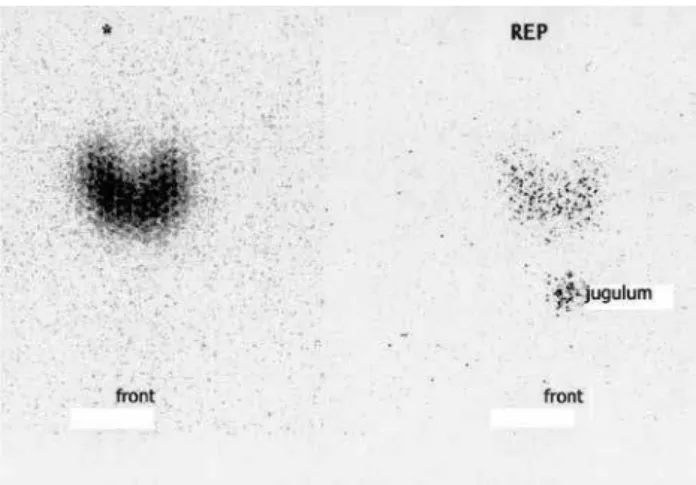
131I thyroid scan revealed an enlarged gland with
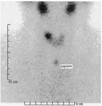
diffu-se increadiffu-sed uptake of radioiodine at 6 and 24 hours (Figure 1). Therapy with methimazole (MMI, 30 mg/ day) was started, and the patient was referred for radio-active iodine treatment (RIT) in March 2009. Within six weeks from RIT, her thyroid function tests norma-lized with TSH of 1.64 mIU/L and FT4 of 16 pm/L. TPO-Ab were 364 UI/L. Six months later, thyroid US examination showed a 7-mm hypoechoic nodule with regular margins and an increased intranodular blood low in color-Doppler in the upper portion of the right lobe. Over the next 18 months, the nodule increased in size up to a maximum diameter of 12 mm (Figure 2). Changes in serum TSH levels during the follow-up after radioiodine treatment are shown in igure 3. In the last evaluation, serum TSH was low-normal (0.67 mIU/L) with normal levels of FT3 and FT4. TRAb were nega-tive. 99mTc-Pertechnetate scintigraphy revealed an avid
tracer uptake in the right lobe, corresponding to the nodular lesion demonstrated by US, consistently with an AFTN (Figure 4). Thus, our patient developed an AFTN three years after the onset of hyperthyroidism due to Graves’ disease that was successfully treated with radioiodine. Looking at the pertinent literature (15-18), we present our case as a variant of classic Marine--Lenhart syndrome. When the “hot” nodule was dis-covered, there was no diffuse increase in radiotracer uptake by the gland and TRAb were negative, because hyperthyroidism due to Graves’ disease – diagnosed three years before – was successfully cured by radioio-dine. On the other hand, the two diseases developed in the same individual and there was a close temporal relationship between radioiodine treatment of Graves’ disease and the occurrence of the “hot” thyroid nodule.
Marine-Lenhart syndrome
Figure 1. The 131I thyroid scan revealed an enlarged gland with diffuse,
increased uptake of radioiodine at 6 (left panel) and 24 (right panel) hours.
Figure 2. Ultrasonographic appearance of a hypoechoic nodule with
regular margins in the upper portion of the right lobe: over 18 months, it increased in size from 7 mm originally to a maximum diameter of about 12 mm.
3
2.5
2
1.5
TSH (mIU/L)
1
0.5
0
0 6
0.001
0.67
TSH (mIU/L)
0.7 1.28 2.7
2
1.64
12 24 1
Weeks Years
2 3
Figure 3. Serum TSH levels during the three-year long follow-up after
Cop
yright
© ABE&M t
odos os dir
eit
os r
eser
vados
.
400 Arq Bras Endocrinol Metab. 2014;58/4
DISCUSSION
Coexistence of Graves’ disease and AFTN was irst des-cribed by Marine and Lenhart, in a study about thyroid histopathology and iodine content in exophthalmic goiter (3). Additional reports followed, and most of these articles describe only one or few patients (4-14). The overall prevalence of such association was repor-ted between 2.7% to 4.1% (4). Different mechanisms are implicated in the pathogenesis of Graves’ disease and in the nodular formation of thyroid tissue with functional autonomy. Graves’ disease is caused by an autoimmune process that involves the whole thyroid gland and is characterized by the presence of stimula-ting TSH receptor antibodies (1). AFTNs are clonal in origin and virtually independent from TSH for growth and function (2). When a thyroid nodule is recorded in the context of Graves’ disease, it is assumed to be scintigraphically “cold”. As it emerges from some data in the literature, a nodular variant of Graves’ disease can be deined as Marine-Lenhart syndrome when the following criteria are met: (i) the thyroid scan shows an enlarged gland and one or more poorly functio-ning nodules; (ii) the nodule is TSH-dependent and the peri-nodular tissue is TSH-independent; (iii) after endogenous or exogenous TSH stimulation, the
re-turn of function can be demonstrated in the nodule; and (iv) the nodule is histologically benign (4). In this case, if one or more autonomous nodules are present in the context of Graves’ disease, they are suppressed by the over-activity of the remaining gland and, the-refore, there is no radioiodine uptake. Once the most part of the gland has been treated with oral medica-tion or radioiodine and, as a consequence, has beco-me progressively less active, the nodules increase their activity in a TSH-dependent way (11). In a unifying pathogenetic hypothesis, it has also been proposed that autoimmunity, such as presence, intrinsic function and concentration of TRAb may inluence the preferential development of diffuse or nodular follicular hyperplasia (19), and further enhance nodules activity (20). But several authors believe that Marine-Lenhart syndrome may be due to different pathological mechanisms that occur independently of each other in the same patients, without any relationship between TRAb positivity, and nodular growth and/or function (21). Therefore, the diagnosis of Marine-Lenhart syndrome remains difi-cult to be determined, and the simultaneous occurren-ce of the two diseases is still matter of debate (21,22). As it occurred in some case reports (7,8,22), there has been also controversy regarding how to diagnose Ma-rine-Lenhart syndrome, depending on which imaging techniques have been used to identify the thyroid no-dule. In fact, although palpation is highly suggestive, it needs conirmation by ultrasonography, in order to exclude an asymmetrical enlargement of one lobe that may give the false sensation of a nodular lesion (23). Moreover, although there is ultrasound detection, it is necessary to determine nodule uptake in a thyroid scan. In most cases, a 99mTc-pertechnetate thyroid scan
ena-bles the identiication of focal abnormal uptake of the tracer, corresponding to the AFTN, even in the context of a diffuse, intense uptake by the gland (7-14).
Besides the coexistence of thyroid autonomy (Plummer’s disease) and Graves’ disease, which is sometimes questionable (21,22), it is also possible that the two diseases may occur in the same patient years apart. There is literature on the risk that Graves-like hy-perthyroidism may develop after radioiodine treatment in patients with elevated serum TPO-Ab levels at base-line (15,16), as well as in TPOAb-negative patients at baseline who became TPOAb-positive after treatment (17,18). Therefore, it could be hypothesized that, in a subject genetically susceptible to thyroid autoim-munity, follicular cell damage caused by radioiodine
Marine-Lenhart syndrome
Figure 4. 99mTc-Pertechnetatethyroid scintigraphy revealed an avid tracer
Cop
yright
© ABE&M t
odos os dir
eit
os r
eser
vados
.
401 Arq Bras Endocrinol Metab. 2014;58/4
could trigger an autoimmune response against TSH receptors, thus explaining the occurrence of Graves’ disease after radioiodine therapy (15-18). Unlike most cases reported in the literature, our particular case of Marine-Lenhart syndrome shows the appearance of an AFTN as a consequence of Graves’ disease treatment with radioiodine. First, Waldherr and cols. described a 46-year-old woman who developed AFTNs within 13 years of radioiodine treatment for Graves’ disease, with strongly positive thyroid antibodies. The authors suggested that the autonomous nodules were a con-sequence of Graves’ disease treatment with radioio-dine (6). Similarly, our patient developed an AFTN three years after the onset of a hyperthyroidism due to Graves’ disease successfully treated with radioiodine. A
99mTc thyroid scan showed an area of increased focal
uptake in the right lobe, corresponding to the palpable nodule, and the existence of the nodule was conirmed by thyroid ultrasonography. The nodule developed in the context of the thyroid gland few months after the radioiodine treatment had been performed, as TSH level started to rise.
In conclusion, the possibility of an association be-tween autoimmune thyroid diseases, namely Graves’ disease, and AFTN emerges from several data in the literature (4,24). The most intriguing and interesting aspect of this association is represented not so much by the co-existence of the two diseases, often not easy to ascertain, as by the possibility that they can develop in the same patient over a lifetime. Clinicians should be aware of such a possibility, especially in those patients who are candidate to radioiodine treatment.
Statement of authorship:each author gave a substantial
contribu-tion to the paper, and approved the inal version to be published.
Funding: this study was not supported by any grant.
Disclosure: no potential conlict of interest relevant to this article was reported.
REFERENCES
1. Kahaly GJ, Bartalena L, Hegedüs L. The American Thyroid Asso-ciation/American Association of Clinical Endocrinologists guide-lines for hyperthyroidism and other causes of thyrotoxicosis: an European perspective. Thyroid. 2011;21:585-91.
2. Krohn K, Paschke R. Progress in understanding the etiology of thyroid autonomy. J Clin Endocrinol Metab. 2001;86:3336-45. 3. Marine D, Lenhart CH. Pathological anatomy of exophthalmic
goi-ter. Arch Intern Med. 1911;8:265-316.
4. Charkes ND. Graves’ disease with functioning nodules (Marine--Lenhart syndrome). J Nucl Med. 1972;13:885-92.
5. Nishikawa M, Yoshimura M, Yoshikawa N, Toyoda N, Yonemoto T, Ogawa Y, et al. Coexistence of an autonomously functioning thyroid nodule in a patient with Graves’ disease: an unusual pre-sentation of Marine-Lenhart syndrome. Endocr J. 1997;44(4):571-4. 6. Waldherr C, Otte A, Haldemann A, Müller-Brand J. Marine-Le-nhart syndrome: a case observation upon 18 years. Nuklearme-dizin. 1999;38(8):345-8.
7. Braga-Basaria M, Basaria S. Marine-Lenhart syndrome. Thyroid. 2003;13:991.
8. El-Kaissi S, Kotowicz MA, Goodear M, Wall JR. An unusual case of Marine-Lenhart syndrome. Thyroid. 2003;13:993-4.
9. Paunkovic N, Paunkovic J. Associated Graves’ disease and Plum-mer disease. Hellenic J Nucl Med. 2003;6:44-7.
10. Cakir M. Marine-Lenhart syndrome. J Natl Med Assoc. 2005;97:1036-8.
11. Chatzopoulos D, Iakovou I, Moralidis E. Images in thyroidolo-gy: Marine-Lenhart syndrome and radioiodine-131 treatment. Thyroid. 2007;17:373-4.
12. Brahma A, Beadsmoore C, Dhatariya K. The oldest case of Mari-ne-Lenhart syndrome? JRSM Short Rep. 2012;3:21.
13. Damle N, Mishra R. Identifying Marine-Lenhart syndrome on a 99mTc-pertechnetate thyroid scan. Indian J Endocrinol Metab. 2013;17(2):366.
14. Scherer T, Wohlschlaeger-Krenn E, Bayerle-Eder M, Passler C, Rei-ner-Concin A, Krebs M, et al. A case of simultaneous occurrence of Marine-Lenhart syndrome and a papillary thyroid microcarci-noma. BMC Endocrine Disorders. 2013;13:16.
15. Chiovato L, Santini F, Vitti P, Bendinelli G, Pinchera A. Appearan-ce of thyroid stimulating antibody and Graves’ disease after ra-dioiodine therapy for toxic nodular goiter. Clin Endocrinol (Oxf). 1994;40:803-6.
16. Schmidt M, Gorbauch E, Dietlein M, Faust M, Stützer H, Eschner W, et al. Incidence of postradioiodine immunogenic hyperthyroi-dism/Graves’ disease in relation to a temporary increase in thyro-tropin receptor antibodies after radioiodine therapy for autono-mous thyroid disease. Thyroid. 2006;16:281-8.
17. Boddenberg B, Voth E, Schicha H. Immunogenic hyperthyroidism following radioiodine ablation of a focal autonomy. Nuklearme-dizin. 1993;32:18-22.
18. Custro N, Ganci A, Scaidi V. Relapses of hyperthyroidism in pa-tients treated with radioiodine for nodular toxic goiter: relation to thyroid autoimmunity. J Endocrinol Invest. 2003;26:106-10. 19. Studer H, Huber G, Derwahl M, Frey P. Die Umwandlung von
Basedowstrumen in Knotenkroepfe. Schweiz Med Wochenschr. 1989;119:203-8.
20. Poertl S, Kirner J, Saller B, Mann K, Hoermann R. T3-release from autonomously functioning thyroid nodules in vitro. Exp Clin En-docrinol Diabetes. 1998;106:489-93.
21. Biersack HJ, Biermann K. The Marine-Lenhart syndrome revisi-ted. Wien Klin Wochenschr. 2011;123:459-62.
22. Cakir M. Diagnosis of Marine-Lenhart syndrome. Letter to the Edi-tor. Thyroid. 2004;14:555.
23. Wiest PW, Hartshorne MF, Inskip PD, Crooks LA, Vela BS, Tele-pak RJ, et al. Thyroid palpation versus high-resolution thyroid ultrasonography in the detection of nodules. J Ultrasound Med. 1998;17:487-96.
24. Ruggeri RM, Campennì A, Sindoni A, Baldari S, Trimarchi F, Ben-venga S. Association of autonomously functioning thyroid nodu-les with Hashimoto’s thyroiditis: study on a large series of pa-tients. Exp Clin Endocrinol Diabetes. 2011;119:621-7.