Cop
yright
© ABE&M t
odos os dir
eit
os r
eser
vados
.
Effects of supplementation with
omega-3 on insulin sensitivity
and non-esteriied free fatty acid
(NEFA) in type 2 diabetic patients
Efeito da suplementação com ômega-3 sobre a sensibilidade à insulina e aos ácidos graxos livres não esteriicados
(AGNE) em pacientes com diabetes tipo 2
Payam Farahbakhsh Farsi1,2, Abolghassem Djazayery2,3,
Mohammad Reza Eshraghian4, Fariba Koohdani1,2, Ali Akbar
Saboor-Yaraghi1,2, Hoda Derakhshanian1,2, Mahnaz Zarei1,2,
Mohammad Hassan Javanbakht1,2, Mahmoud Djalali1,2
ABSTRACT
Objective: The aim of this study was to determine the role of omega-3 supplementation on NEFA concentration, insulin sensitivity and resistance, and glucose and lipid metabolism in type 2 diabetic patients. Subjects and methods: Forty-four type 2 diabetic patients were ran-domly recruited into two groups. Group A received 4 g/day omega-3 soft gels, and group B received a placebo for 10 wks. Blood samples were collected after 12-h fast. Physical activity records, three-day food records, and anthropometric measurements were obtained from all participants at the beginning and end of the study. Results: Omega-3 supplementation caused a signiicant reduction in NEFA in the intervention group compared with the placebo group (P = 0.009). Additionally, the administration of omega-3 resulted in signiicantly greater chan-ges (Diff) for the intervention group in various parameters, such as insulin and Quicki indices compared with the placebo group (P < 0.05). Conclusions: Omega-3 fatty acid supplementation in type 2 diabetic patients improved insulin sensitivity, probably due to the decrease in NEFA concentrations. Arq Bras Endocrinol Metab. 2014;58(4):335-40
Keywords
Non-esteriied fatty acids; omega-3 fatty acids; insulin; diabetes mellitus
RESUMO
Objetivo: O objetivo deste estudo foi analisar o papel da suplementação com ácidos graxos ômega-3 sobre a concentração de ácidos graxos não esteriicados (AGNE), resistência e sensi-bilidade à insulina e metabolismo de lipídios em pacientes com diabetes melito tipo 2. Sujeitos e métodos: Quarenta e quatro pacientes com diabetes tipo 2 foram recrutados aleatoriamente e alocados em um de dois grupos. O Grupo A recebeu 4 g/dia de ômega-3 na forma de cápsu-las gelatinosas e o grupo B recebeu placebo durante 10 semanas. Amostras de sangue foram coletadas após 12 horas de jejum. Registros da atividade física, da dieta de três dias e medidas antropométricas foram obtidos de todos os participantes no início e no inal do estudo. Resul-tados: A suplementação com ômega-3 causou uma redução signiicativa na AGNE em compa-ração com grupo placebo (P = 0,008). Além disso, a administração de ômega-3 resultou em alte-rações signiicativamente maiores (Dif) em vários parâmetros, tais como a insulina, HOMA-IR e QUICKI, comparando com placebo (P < 0,05). Conclusões: A suplementação com ácidos graxos ômega-3 em pacientes diabéticos tipo 2 melhorou a sensibilidade à insulina, provavelmente devido à diminuição da concentração de AGNE. Arq Bras Endocrinol Metab. 2014;58(4):335-40
Descritores
Ácidos graxos não esteriicados; ácidos graxos ômega-3; insulina; diabetes melito
1 Department of Cellular and
Molecular Nutrition, School of Nutritional Sciences and Dietetics, Tehran University of Medical Sciences, Tehran, Iran
2 Department of Nutrition and
Biochemistry, School of Public Health, Tehran University of Medical Sciences, Tehran, Iran
3 Department of Community
Nutrition, School of Nutritional Sciences and Dietetics, Tehran University of Medical Sciences, Tehran, Iran
4 Department of Biostatistics, School
of Public Health, Tehran University of Medical Sciences, Tehran, Iran
Correspondence to:
Mahmoud Djalali
Department of Nutrition and Biochemistry, School of Public Health, Tehran University of Medical Sciences, Poorsina Street, Enghelab Avenue 14155-6446 – Tehran, Iran [email protected]
Received on Jul/2/2013 Accepted on Oct/24/2013
Cop
yright
© ABE&M t
odos os dir
eit
os r
eser
vados
.
INTRODUCTION
H
yperglycemia and type 2 diabetes are causedby insulin resistance and beta-cell dysfunction. Beta-cell dysfunction can be caused by genetic and en-vironmental factors, including inlammation and stress agents, such as glucose and NEFA. Saturated NEFAs are detrimental to beta-cell function. Their effect is exacerbated in the presence of high levels of glucose and may cause a condition called glucolipotoxicity (1). As fatty acids can stimulate insulin secretion, they have an important role in the mechanism of beta-cell com-pensation to insulin resistance (2). In diabetics with a positive family history, an increase in lipid turnover rate can prevent diabetes (2,3). Increased NEFA stimulates insulin secretion, although omega-3 cannot stimulate insulin secretion (4,5). NEFA concentrations as co-mo-dulators of plasma glucose levels and insulin secretion have been used in mathematical models (6,7). Studies on rodents have shown that ish oil may improve insu-lin sensitivity or reduce glucose levels (8). The effects of ish oil on insulin sensitivity and resistance in type 2 diabetic patients are not fully understood. NEFA inter-feres with pancreatic beta-cells contributing to hyperin-sulinemia, hyperglycemia, and diabetes. In addition, it leads to insulin resistance in muscles and the liver. The-refore the aim of this study was to determine the role of omega-3 supplementation on NEFA concentration, insulin sensitivity and resistance, and glucose and lipid metabolism in type 2 diabetic patients.
SUBJECTS AND METHODS
Study population
This study was a randomized controlled double-blind clinical trial. Patients were enrolled from January 2012 through May 2012. All patients came from the Iranian Diabetic Association, Tehran, Iran. The criteria for in-clusion were: willingness to participate, 30-65 years of age, T2DM diagnosis, and a body mass index of 18.5 to 40 kg/m2. Patients were required to cease
consump-tion of dietary supplements at least 2 wks. before the beginning of the test period and throughout the inter-vention. Subjects who had consumed omega-3 supple-ments in the three months before the beginning of the study were excluded. None of the patients patient had chronic renal, hepatic, gastrointestinal, or hematologi-cal disease or a thyroid disorder. None of the patients
had used orlistat, sibutramine, or any other weight-loss drugs. None was pregnant or lactating. None was re-ceiving thiazolidinediones or insulin therapy. All par-ticipants were requested to maintain their usual exer-cise and dietary habits. A total of 45 patients with type 2 diabetes mellitus who met the inclusion criteria were randomly allocated in one of two groups. Permuted-block randomization was used for grouping, and the two groups were matched according to BMI. The inter-vention group received 4 soft gels of omega-3 (Maxepa Forte Capsules, Seven Seas, UK) per day, and the con-trol group received 4 g/d placebo soft gels containing corn oil, which had the same appearance (Zahravi, Iran). The protocol was approved by the Ethics Review Board of Tehran University of Medical Sciences (TUMS), and each patient signed an informed consent form.
From the 23 patients allocated to the intervention group, one patient was excluded due to non-willing-ness, and all of the 22 patients in the control group completed the study. Therefore, 44 patients inished the study.
Study design
To assess baseline values, blood samples were collected for analysis after 12-h overnight fast. Thereafter, for 10 wks., the intervention group received omega-3 soft gels, while the control group received the placebo. An-ti-diabetic medicine and other medications were kept stable in all patients. Patient compliance was monitored every two weeks.
Cop
yright
© ABE&M t
odos os dir
eit
os r
eser
vados
.
Statistical analysis
Data were expressed as the means ± standard error of mean (SEM). Student’s t test was used to compare the mean of the responses. If normal assumption was not applicable, non-parametric tests were applied. Analysis of covariance (ANCOVA) was also used to control the effect of baseline values of different variables in the two groups. P values < 0.05 were considered statistically signiicant. Statistical analysis was performed using The Statistical Package for Social Sciences (version 18.0; SPSS Inc., Chicago, USA).
RESULTS
Effect of omega-3 supplementation on general parameters
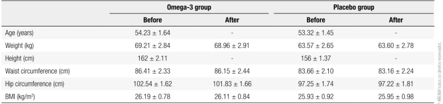
A total of 44 diabetic patients (17 males, 27 females) completed the study. Anthropometric measurements were done as shown in table 1. No signiicant differen-ces between the groups were observed at the beginning of the intervention. Comparisons of weight between the groups showed no difference before (P = 0.15) or after (P = 0.19) 10 wks. Waist and hip circumferences between the groups were not statistically different be-fore (P = 0.38 and 0.32) or after the intervention (P = 0.37 and 0.68). BMI did not change signiicantly be-fore (P = 0.83) or after (P = 0.91) the study.
Effects of omega-3 supplementation on NEFA
NEFA values had normal distribution. To control the confounding effect of baseline NEFA values, ANCOVA was used (P = 0.57). At baseline, there were no differ-ences in NEFA levels between the groups (P = 0.955). After 10 wks., a signiicant decrease in NEFA was observed for the omega-3 group (P = 0.009).
Howe-ver, no change occurred in the placebo group (P = 0.99). Comparisons of the groups at the end of the study showed a signiicant change in NEFA levels in the intervention group compared with that of the con-trol (P = 0.008). The mean difference between the two groups was not signiicant (P = 0.11).
Effects of omega-3 supplementation on plasma lipid levels
Concentrations of lipid markers (total cholesterol, TG, LDL-C, and HDL-C) had normal distribution. After 10 wks., total cholesterol, HDL-C, LDL-C, and TG decreased slightly, but not signiicantly, for the inter-vention group. The mean difference between the two groups for HDL-C was signiicant (P = 0.03), and was marginally signiicant (P = 0.05) for TG.
Effects of omega-3 supplementation on insulin
After 10 wks. of intervention, insulin levels decreased in the omega-3 group compared with baseline values (P = 0.02). Although the difference between groups at the end of the study was not statistically signiicant, the mean difference between the two groups for insulin was signiicant (P = 0.03).
Effects of omega-3 supplementation on HOMA-IR and QUICKI
In contrast to QUICKI, HOMA-IR did not have nor-mal distribution. Accordingly, we log-transformed the data to obtain a normal distribution. After 10 wks. of supplementation, within-group analysis showed a sig-niicant decrease in HOMA-IR for the intervention group (P = 0.011), and the QUICKI signiicantly in-creased (P = 0.002).
Table 1. General characteristic of participants at the baseline (week 0) and at the end of the study (week 10)
Omega-3 group Placebo group
Before After Before After
Age (years) 54.23 ± 1.64 - 53.32 ± 1.45
-Weight (kg) 69.21 ± 2.84 68.96 ± 2.91 63.57 ± 2.65 63.60 ± 2.78
Height (cm) 162 ± 2.11 - 156 ± 1.37
-Waist circumference (cm) 86.41 ± 2.33 86.15 ± 2.44 83.66 ± 2.10 83.16 ± 2.24 Hip circumference (cm) 102.54 ± 1.62 101.83 ± 1.66 97.25 ± 1.74 97.22 ± 1.81 BMI (kg/m2) 26.19 ± 0.78 26.11 ± 0.84 25.93 ± 0.92 25.95 ± 0.98
Cop
yright
© ABE&M t
odos os dir
eit
os r
eser
vados
.
Table 2. Comparison of various parameters within and between groups
Omega-3 group Placebo group P-value
between groups Before
P-value between groups After
Before After P-value Before After P-value
Insulin (µIU/mL) 10.68 ± 0.86 8.51 ± 0.59 0.028 7.6 ± 0.59 7.51 ± 0.56 0.695 0.005 0.228 NEFA (ng/mL) 1936.73 ± 90.91 1737.72 ± 51.01 0.09 1929.64 ± 87.43 1997 ± 77.17 0.58 0.955 0.008
TG (mg/100 mL) 156.55 ± 15.25 131.59 ± 12.92 0.076 136.77 ± 21.34 145.86 ± 17.24 0.39 0.455 0.511 Cholesterol (mg/dL) 228.64 ± 12.75 204.36 ± 8.27 0.055 222.50 ± 9.97 215.27 ± 10.72 0.365 0.707 0.425 LDL-C (mg/dL) 107.74 ± 6.81 104.50 ± 5.00 0.634 92.05 ± 5.99 98.00 ± 4.89 0.229 0.098 0.358
HDL-C (mg/dL) 45.45 ± 2.34 43.14 ± 2.31 0.068 45.77 ± 2.04 47.18 ± 2.35 0.260 0.919 0.227 HOMA-IR 4.15 ± 0.47 3.07 ± 0.35 0.011 3.15 ± 0.43 3.02 ± 0.33 0.553 0.125 0.922 QUICKI 0.31 ± 0.007 0.32 ± 0.005 0.002 0.32 ± 0.004 0.32 ± 0.005 0.982 0.083 0.867
Data are presented as mean ± SEM. Baseline and inal values were compared with paired t-test. The differences between groups were assessed by Independent t-test. P < 0.05 was considered as signiicant. NEFA: non-esteriied free fatty acid; TG; triglyceride; HOMA-IR: homeostatic model assessment for insulin resistance; QUICKI: quantitative insulin sensitivity check Index.
After 10 wks., there were no signiicant changes
in HOMA-IR and QUICKI between the groups (P
= 0.92, and P = 0.86, respectively). The mean dif-ference between the two groups for HOMA-IR (P = 0.054) was marginally signiicant, and was signiicant for QUICKI (P = 0.009).
DISCUSSION
To our knowledge, this study represents the irst analy-sis of the effect of omega-3 supplementation on NEFA in diabetic patients. The present study showed a detri-mental effect of omega-3 on NEFA reduction. Previous studies explained that long time exposure to NEFA led to impaired insulin secretion and contributed to beta-cell dysfunction and death (10). Plasma NEFA found in obese individuals was increased compared with
normal-Table 3. Comparison of changes in various parameters (Diff) between
groups
Change in omega-3 group
Change in
placebo group P-value
Insulin (µIU/mL) -2.17 ± 0.92 -0.09 ± 0.24 0.035
NEFA (ng/mL) 199.0 ± 113.94 -67.36 ± 122.14 0.11
TG (mg/100 mL) -24.95 ± 13.39 9.09 ± 10.35 0.051 Total cholesterol (mg/dL) -24.27 ± 11.92 -7.22 ± 7.80 0.238 LDL-C (mg/dL) -2.90 ± 6.01 5.95 ± 4.80 0.256
HDL-C (mg/dL) -2.31 ± 1.20 1.40 ± 1.21 0.035
HOMA-IR -1.08 ± 0.42 -0.1306 ± 0.21 0.054 QUICKI 0.0143 ± 0.004 -0.000 ± 0.003 0.009
Data are presented as mean ± SEM. The differences between groups were assessed by Independent t-test. P < 0.05 was considered as signiicant. NEFA: non-esteriied free fatty acid; TG; triglyceride; HOMA-IR: homeostatic model assessment for insulin resistance; QUICKI: quantitative insulin sensitivity check Index.
weight individuals (11). Lipolysis led to an increment in plasma NEFA concentration, which caused insulin resistance in other tissues. Furthermore, even in healthy people, increased levels of plasma NEFA contributed to insulin resistance in the liver and skeletal muscles (12-15). Previous studies have shown that acute exposure to elevated plasma NEFA enhances glucose and non-glucose stimulated insulin secretion (16).
Acute elevations in plasma levels of long-chain fatty acids enhance plasma insulin levels by stimulating in-sulin secretion or by decreasing inin-sulin clearance. In normal individuals, long-term increases of fatty acids also stimulate insulin secretion. Inversely, pre-diabetic subjects cannot properly compensate for the free fatty acids that induce insulin resistance (4). Our results are in line with those of Boden and cols., who showed that omega-3 supplementation in diabetic patients cannot induce insulin secretion. It seems that a longer period of supplementation may contribute to decreasing insu-lin clearance.
Cop
yright
© ABE&M t
odos os dir
eit
os r
eser
vados
.
and glucose sensitivity, their changes were in the same direction.
We observed that insulin sensitivity increased sig-niicantly. It was formerly shown that, in early-onset of diabetes in rats, fasting plasma triglyceride decreases, fasting plasma NEFA increases, and ish oil or EPA ad-ministration diminish plasma NEFA concentration in type 2 diabetic rats (8). Exposure to high NEFA con-centrations causes dysfunction in glucose-stimulated insulin secretion (23). In vivo studies have shown that insulin gene expression and insulin content decreased following an infusion of NEFA and glucose in rats (24). Hemodialysis patients with very low levels of NEFA at baseline showed no decrease in NEFA after receiving omega-3 (25). It seems that baseline levels of NEFA are the cause of differences between hemodialysis pa-tients and diabetic subjects.
In previous studies, in subjects with normal trigly-ceride levels, NEFA decreased about 25% with ish oil supplementation (26). Subjects of the present study had normal TG and showed an 11% reduction in NEFA.
Moreover, in a previous study, 30 g/day ish oil re-duced NEFA levels and TG while subjects had normal triglycerides at baseline (27). It is important to note that the subjects were healthy, and the omega-3 dosage was higher than that of our study.
Fish oil administration to high triglyceride, insulin-resistant, or obese patients showed different and incon-clusive changes in TG and NEFA, depending on the study population (28,29). So far, studies have not ap-proved the effect of omega-3 on NEFA, but the reduc-tion in TG levels has been explained (30). Koh and cols. showed that consumption of 2g/d omega-3 fatty acids did not signiicantly change insulin and insulin sensiti-vity (determined by QUICKI) in hypertriglyceridemia patients (31). It is necessary to recall that QUICKI is inluenced by glucose and insulin, which may not rise in hypertriglyceridemia patients.
In conclusion, our data suggested that omega-3 fatty acid supplementation decreases HOMA-IR and increases QUICKI, while NEFA decreases only insigni-icantly when compared with the placebo group.
Acknowledgements: this study was supported by Tehran Uni-versity of Medical Sciences (grant nº 15177). We would like to thank the Iranian Diabetes Society for their collaboration in refer-ring diabetic patients to our group.
Disclosure: no potential conlict of interest relevant to this article was reported.
REFERENCES
1. Maris M, Robert S, Waelkens E, Derua R, Hernangomez MH, D’Hertog W, et al. Role of the saturated nonesteriied fatty acid pal-mitate in beta cell dysfunction. J Proteome Res. 2013;12(1):347-62.
2. Nolan CJ, Madiraju MS, Delghingaro-Augusto V, Peyot ML,
Prent-ki M. Fatty acid signaling in the beta-cell and insulin secretion. Diabetes. 2006;55 Suppl 2:S16-23.
3. Bunt JC, Krakoff J, Ortega E, Knowler WC, Bogardus C. Acute
insu-lin response is an independent predictor of type 2 diabetes melli-tus in individuals with both normal fasting and 2-h plasma glucose concentrations. Diabetes Metab Res Rev. 2007;23(4):304-10. 4. Boden G. Free fatty acids and insulin secretion in humans. Curr
Diab Rep. 2005;5(3):167-70.
5. Mostad IL, Bjerve KS, Basu S, Sutton P, Frayn KN, Grill V. Addi-tion of n-3 fatty acids to a 4-hour lipid infusion does not affect insulin sensitivity, insulin secretion, or markers of oxidative stress in subjects with type 2 diabetes mellitus. Metabolism. 2009;58(12):1753-61.
6. Mari A, Camastra S, Toschi E, Giancaterini A, Gastaldelli A, Min-grone G, et al. A model for glucose control of insulin secretion during 24 h of free living. Diabetes. 2001;50 Suppl 1:S164-8.
7. Mari A, Schmitz O, Gastaldelli A, Oestergaard T, Nyholm B,
Fer-rannini E. Meal and oral glucose tests for assessment of beta -cell function: modeling analysis in normal subjects. Am J Physiol En-docrinol Metab. 2002;283(6):E1159-66.
8. Cummings BP, Stanhope KL, Graham JL, Griffen SC, Havel PJ. Supplementation with EPA or ish oil for 11 months lowers circu-lating lipids, but does not delay the onset of diabetes in UC Davis-type 2 diabetes mellitus rats. Br J Nutr. 2010;104(11):1628-34. 9. Navas-Carretero S, Perez-Granados AM, Schoppen S, Vaquero
MP. An oily ish diet increases insulin sensitivity compared to a red meat diet in young iron-deicient women. Br J Nutr. 2009;102(4):546-53.
10. Kang ZF, Deng Y, Zhou Y, Fan RR, Chan JC, Laybutt DR, et al. Phar-macological reduction of NEFA restores the eficacy of incretin-based therapies through GLP-1 receptor signalling in the beta cell in mouse models of diabetes. Diabetologia. 2013;56(2):423-33. 11. Cusi K, Kashyap S, Gastaldelli A, Bajaj M, Cersosimo E. Effects on
insulin secretion and insulin action of a 48-h reduction of plasma free fatty acids with acipimox in nondiabetic subjects genetically predisposed to type 2 diabetes. Am J Physiol Endocrinol Metab. 2007;292(6):E1775-81.
12. Boden G. Role of fatty acids in the pathogenesis of insulin resis-tance and NIDDM. Diabetes. 1997;46(1):3-10.
13. Kelley DE, Mandarino LJ. Fuel selection in human skeletal muscle in insulin resistance: a reexamination. Diabetes. 2000;49(5):677-83. 14. Magnan C, Cruciani C, Clement L, Adnot P, Vincent M, Kergoat M, et al. Glucose-induced insulin hypersecretion in lipid-infused healthy subjects is associated with a decrease in plasma norepi-nephrine concentration and urinary excretion. J Clin Endocrinol Metab. 2001;86(10):4901-7.
15. Boden G. Free fatty acids-the link between obesity and insulin resistance. Endocr Pract. 2001;7(1):44-51.
16. Kashyap S, Belfort R, Gastaldelli A, Pratipanawatr T, Berria R, Pratipanawatr W, et al. A sustained increase in plasma free fatty acids impairs insulin secretion in nondiabetic subjects genetically predisposed to develop type 2 diabetes. Diabetes. 2003;52(10):2461-74.
17. Mingrone G, Manco M, Granato L, Calvani M, Scarfone A, Mora EV, et al. Leptin pulsatility in formerly obese women. FASEB J. 2005;19(10):1380-2.
adi-Cop
yright
© ABE&M t
odos os dir
eit
os r
eser
vados
.
pose tissue in post-obese patients submitted to biliopancreatic diversion. Eur J Endocrinol. 2003;148(5):543-50.
19. Greco AV, Mingrone G, Vettor R, Manco M, Rosa G, Capristo E, et al. Lowering of circulating free-fatty acids levels and reduced expression of leptin in white adipose tissue in postobesity status. J Investig Med. 2002;50(3):207-13.
20. Camastra S, Manco M, Mari A, Greco AV, Frascerra S, Mingrone G, et al. Beta-cell function in severely obese type 2 diabetic patients: long-term effects of bariatric surgery. Diabetes Care. 2007;30(4):1002-4. 21. Guidone C, Manco M, Valera-Mora E, Iaconelli A, Gniuli D, Mari A,
et al. Mechanisms of recovery from type 2 diabetes after malab-sorptive bariatric surgery. Diabetes. 2006;55(7):2025-31.
22. Nolan CJ, Leahy JL, Delghingaro-Augusto V, Moibi J, Soni K, Peyot ML, et al. Beta cell compensation for insulin resistance in Zucker fatty rats: increased lipolysis and fatty acid signalling. Dia-betologia. 2006;49(9):2120-30.
23. Poitout V, Robertson RP. Glucolipotoxicity: fuel excess and beta-cell dysfunction. Endocr Rev. 2008;29(3):351-66.
24. Hagman DK, Latour MG, Chakrabarti SK, Fontes G, Amyot J, Tremblay C, et al. Cyclical and alternating infusions of glucose and intralipid in rats inhibit insulin gene expression and Pdx-1 binding in islets. Diabetes. 2008;57(2):424-31.
25. Delarue J, Guillodo MP, Guillerm S, Elbaz A, Marty Y, Cledes J. Fish oil attenuates adrenergic overactivity without altering
glu-cose metabolism during an oral gluglu-cose load in haemodialysis patients. Br J Nutr. 2008;99(5):1041-7.
26. Bordin P, Bodamer OA, Venkatesan S, Gray RM, Bannister PA, Halliday D. Effects of ish oil supplementation on apolipoprotein B100 production and lipoprotein metabolism in normolipidaemic males. Eur J Clin Nutr. 1998;52(2):104-9.
27. Dagnelie PC, Rietveld T, Swart GR, Stijnen T, van den Berg JW. Effect of dietary ish oil on blood levels of free fatty acids, ketone bodies and triacylglycerol in humans. Lipids. 1994;29(1):41-5. 28. Abate N, Chandalia M, Snell PG, Grundy SM. Adipose tissue
me-tabolites and insulin resistance in nondiabetic Asian Indian men. J Clin Endocrinol Metab. 2004;89(6):2750-5.
29. Vistisen B, Hellgren LI, Vadset T, Scheede-Bergdahl C, Helge JW, Dela F, et al. Effect of gender on lipid-induced insulin resistance in obese subjects. Eur J Endocrinol. 2008;158(1):61-8.
30. Pownall HJ, Brauchi D, Kilinc C, Osmundsen K, Pao Q, Payton-Ross C, et al. Correlation of serum triglyceride and its reduction by omega-3 fatty acids with lipid transfer activity and the neutral lipid compositions of high-density and low-density lipoproteins. Atherosclerosis. 1999;143(2):285-97.