Cop
yright
© ABE&M t
odos os dir
eit
os r
eser
vados
.
Cop
yright
© ABE&M t
odos os dir
eit
os r
eser
vados
.
The role of imaging in congenital
adrenal hyperplasia
O papel dos métodos de imagem em hiperplasia congênita de suprarrenal
Sara Reis Teixeira1, Paula Condé Lamparelli Elias2, Marco Túlio Soares Andrade1, Andrea Farias Melo1, Jorge Elias Junior1
ABSTRACT
Congenital adrenal hyperplasia (CAH) is an autossomic recessive disorder caused by impaired steroidogenesis. Patients with CAH may present adrenal insuficiency with or without salt-wast-ing, as well as various degrees of virilization and fertility impairment, carrying a high incidence of testicular adrenal rest tumors and increased incidence of adrenal tumors. The diagnosis of CAH is made based on the adrenocortical proile hormonal evaluation and genotyping, in se-lected cases. Follow-up is mainly based on hormonal and clinical evaluation. Utility of imaging in this clinical setting may be helpful for the diagnosis, management, and follow-up of the pa-tients, although recommendations according to most guidelines are weak when present. Thus, the authors aimed to conduct a narrative synthesis of how imaging can help in the manage-ment of patients with CAH, especially focused on genitography, ultrasonography, computed tomography, and magnetic resonance imaging. Arq Bras Endocrinol Metab. 2014;58(7):701-8
Keywords
Adrenal hyperplasia, congenital; magnetic resonance imaging; computed tomography; ultrasonography; diagnostic imaging
RESUMO
Hiperplasia congênita de suprarrenal (CAH) é uma doença autossômica recessiva causada por deiciências enzimáticas na esteroidogênese. Clinicamente, os pacientes com CAH podem apresentar insuiciência adrenal com ou sem perda de sal, vários graus de virilização e di-minuição na fertilidade, alta incidência de restos adrenais testiculares e de tumores adrenais. O diagnóstico de CAH é feito baseado nos resultados da avaliação hormonal e genotípica, em casos selecionados. O seguimento dos pacientes é principalmente feito com avaliação clínica e hormonal. Métodos de diagnóstico por imagem podem ser muito úteis não só no diagnóstico como no manejo e seguimento dos pacientes com CAH. Porém, as recomendações, de acordo com a maioria dos consensos, quando existem, são escassas. Nesse contexto, com base em uma revisão sistemática, o objetivo deste artigo foi sintetizar a literatura em relação a como os métodos de diagnóstico por imagem podem ser úteis no manejo dos pacientes com CAH, com foco em genitograia, ultrassonograia, tomograia computadorizada e ressonância magnética.
Arq Bras Endocrinol Metab. 2014;58(7):701-8
Descritores
Hiperplasia adrenal congênita; ressonância magnética; tomograia computadorizada; ultrassonograia; diagnóstico por imagem
1 Department of Internal
Medicine, Division of Radiology, Clinical Hospital, Ribeirao Preto Medical School, University of Sao Paulo (FMRP-USP), Ribeirao Preto, SP, Brazil
2 Department of Internal Medicine,
Division of Endocrinology, Clinical Hospital, FMRP-USP, Ribeirao Preto, SP, Brazil
Correspondence to: Jorge Elias Junior Radiology Division, Ribeirao Preto Medical School University of Sao Paulo Av. Bandeirantes, 3900
14049-090 – Ribeirao Preto, SP, Brazil [email protected]
Received on Mar/21/2014 Accepted on Jun/17/2014
DOI: 10.1590/0004-2730000003371
INTRODUCTION
C
ongenital adrenal hyperplasia (CAH) is an autos-somic recessive disorder caused by impaired ste-roidogenesis, in approximately 95% of the cases, secon-dary to 21-hydroxylase deiciency. Patients may present adrenal insuficiency with or without salt-wasting, as well as various degrees of virilization and fertilityCop
yright
© ABE&M t
odos os dir
eit
os r
eser
vados
.
nal imaging is suggested only for patients with an atypical course (2). Conversely, CAH must be excluded in cases of adrenal incidentaloma supposedly asymptomatic or oligo-symptomatic. To evaluate gonads, ultrasonography (US) is recommended for screening males from adolescence, but there is no recommendation to screen females (2).
Thus, the authors aim to conduct a narrative syn-thesis of how imaging can help in the management of patients with CAH, especially focused on genitography (GX), US, computed tomography (CT), and magnetic resonance imaging (MRI).
SEARCH STRATEGY AND SELECTION OF ARTICLES
A systematic search was conducted in MedLine® (from
1950 to July 2013) and in Web of Science® (from 1965 to
July 2013) databases for articles published in English, Spa-nish, Portuguese, and French. On MedLine®, the MeSH
term “adrenal hyperplasia, congenital” was searched with the other imaging related MeSH terms with AND at a time using “ultrasonography”, or “magnetic resonance imaging”, or “diffusion magnetic resonance imaging”, or “tomography, X-ray computed”, or “multidetector com-puted tomography”, or “positron-emission tomography and computed tomography”, or “tomography scanners, X-ray computed radiography”, or “diagnostic imaging”. Web of Science® was searched for articles with the
sear-ch terms “congenit* and adren* and hyperpl*” “AND imag*”. Books and other selected references cited in the most relevant retrieved articles were also reviewed.
Studies that were conducted in animals and in which only scintigraphy or nuclear imaging modalities were used as imaging modalities were not within the scope of this review. Also, studies that only used X-ray to evalua-te bone age or any imaging modalities to assess bone mine ral density were not extensively reviewed, as it is well established by the guidelines that bone age should be assessed annually after 2 years of age and regular eval-uation of bone mineral density is not recommended (2).
Imaging
Genitography
The urogenital sinus is the embryologic precursor of the bladder, urethra, and distal third of the vagina in
tures (one of them is the anus) in the perineal region as-sociated with ambiguous genitalia (3). Typically, female patients with classic CAH have ambiguous genitalia at birth. Thus an anatomic detailed image plays an impo-rtant role in planning strategy for feminizing surgery. Genitography shows the urethra, the level of external sphincter, the presence or absence of the vagina, the urethrovaginal conluence, and the cervical impression of the uterus (3) (Figure 1).
Genitography associated with voiding cistography has also the ability to show upper genitourinary abnor-malities, present in 21%-80% (4) of the patients with CAH, mostly seen in girls.
Although some authors concluded that genitogra-phy did not add information to endoscopic (5) or sur-gical indings, many advocate its use as part of routine investigation in female patients with CAH (4), particu-larly in those infants with ambiguous genitalia.
Ultrasonography
US is the modality of choice to image abdominal and pelvic organs in children and fetuses. It is widely availa-ble, versatile, and portaavaila-ble, with lack of ionizing radia-tion, there is no need for sedaradia-tion, and provides high-resolution images in any required plane.
Cop
yright
© ABE&M t
odos os dir
eit
os r
eser
vados
.
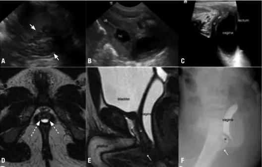
Figure 1. Newborn with “non palpable testicles”. Ultrasonography (A-C) showed enlarged and cerebriform pattern of the adrenal glands (arrows in A).
Sagittal view of the pelvis through the abdominal wall (B): the uterus (“ut”) and the presence of hydrocolpus (“hc”) are well depicted. The ovaries (not
shown) were also present. Sagittal ultrasonography view of the pelvis through the perineum (C) was not suficient to show with high conidence the
conluence of the urethra and the vagina, which was better viewed on magnetic resonance imaging (MRI) (E, sagittal T2-weighetd MRI of the pelvis) and
genitography (F). The arrows in (E) and (F) are pointing the conluence of the vagina and the urethra (“u”). Axial T2-weighted MRI of the pelvis showing
prostate tissue (dashed arrows in D) in this patient. The pattern of the adrenal glands and presence of mullerian derivatives allowed institution of therapy
while laboratory tests were done to conirm congenital adrenal hyperplasia in this XX neonate. MRI and genitography were requested for planning feminizing surgery.
A
D
B
E
C
F
Although the clinical features of CAH are usually present, testicular adrenal rest tumor (TART) may be the only clinical inding at presentation (11). Based on microscopic studies, TARTs are reported to be pre sent in all males with CAH (12). On US, it has been docu-mented with a prevalence of up to 94% (13). The most common sonographic features are bilateral spikelike appearance intratesticular hypoechoic masses with no sound attenuation, surrounding the mediastinal testis (11). On color Doppler they are hypo or avascular (14) and there is no deviation or changes in caliber of the vessels that course the lesions (11,14). However, TART may also appear as heterogeneous or hyperechoic nod-ules (11,14,15), and even as an epididymal nodule (11). TART echogenicity is related to the size of the lesions, being hypoechoic in lesions smaller than 2 cm and het-erogeneous or hyperechoic in lesions larger than 2 cm (14). These hyperechoic areas may represent ibrosis or calciications (14). In addition, larger lesions may not be conined to the mediastinal testis and smaller lesions are more often seen unilaterally (14). On follow-up, TART can vary in size (15), but there is no correlation between hormonal control or hormonal markers and TART (15-17). TARTs are thought to be responsible
for testicular parenchymal damage that contributes to reduced fertility (17,18). It can be found even in young children with a prevalence of 21% (19) and it is suggested that gonadal dysfunction is already present before puberty (19). Therefore, early detection of tes-ticular lesions is advised (20) to improve treatment and prevent longstanding gonadal impairment function. Thus, some authors advocate that not only adolescents should undergo US (21) (Figure 2).
High prevalence of impaired fertility is not restric-ted to men as it was reporrestric-ted also in women with CAH (13,17). The prevalence of polycystic ovaries is increased in women with classical and nonclassical CAH (22). Bi-lateral enlarged ovaries (23), biBi-lateral ovarian cysts, and ovarian adrenal rest tumors (OART) (24) may occur and can also be depicted by US. OART may present as hypoechoic nodules on US (24,25), similar to TART.
Cop
yright
© ABE&M t
odos os dir
eit
os r
eser
vados
.
curately the size and morphology of the Mullerian struc-tures, the uterus, the vagina, and the gonads (3). In ad-dition to the adrenal glands indings described above, the presence of a uterus in a patient with ambi guous genitalia indicates that the diagnosis is mostly likely CAH (9). Ultrasound evaluation of the pelvic structures is not only performed for diagnosis but also as part of the preoperative approach for surgery, often in conjunction with other exams, such as genitography and MRI (3). US provides adequate information about the vagina and urogenital sinus for preoperative decision-making (28).
Many other abnormalities in patients with CAH can also be demonstrated by US: cardiac dysfunction that reverses with therapy (29), vascular dysfunction and increased carotid intima media thickness (30), ske-letal and midface malformations associated with P450 oxidoreductase deiciency in prenatal diagnosed fetuses (31), hydrops of placental stem villi in a 46,XX fetus (32), association with increased nuchal translucency detected in the prenatal period (33), bilateral ovarian
Adrenocortical tumors in patients with CAH are not rare. A prevalence of up to 83% of adrenocortical masses in homozygote patients is reported (36). Despite this high frequency, adrenocortical tumors in this setting are most likely to be benign, as malignant lesions are rare (36). In many reports CT scans showed nodules (36-38) that may regress with adequate therapy (37), adenomas (23), mye-lolipomas (39), and the typical pattern of diffuse enlarge-ment (38) with a heterogeneous enhanceenlarge-ment (Figure 3). Positron emission tomography with CT scan (PET-CT) was used in 3 case reports. In one, PET-CT was used to evaluate an adrenal mass in an untreated pa-tient and showed a mass proved to be an adrenocortical tumor of uncertain prognosis (40). In the other two, PET-CT depicted OARTs (25,41), interestingly, in one of these reports both MRI and CT could not shown this inding. On the other hand, adrenal rest tumors have already been described on conventional CT as a soft tissue masse in the ovary, OART (42), and in the perirenal region (43).
Magnetic resonance imaging
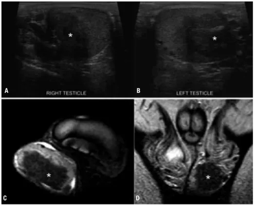
Figure 2. Testicular adrenal rest tumor (TART) in a 14 year-old male, with simple virilizing congenital adrenal hyperplasia. On ultrasonography (A-B), there is a round heterogeneous, predominantly hypoechoic nodule (*) within both testicles, in the region of the mediastinal testis. On magnetic resonance
imaging (C, axial; D, coronal) the TARTs are hypointense on T2-weighted images.
A B
Cop
yright
© ABE&M t
odos os dir
eit
os r
eser
vados
.
Magnetic resonance imaging
Studies based on MRI are in accordance with others performed with CT (36) demonstrating a high pre-valence of adrenal nodules (73%) in adults with CAH (16). Moreover, adrenal MRI imaging in CAH patients showed indings such as normal or diffuse enlarged adrenal glands (44), adrenal nodules (16,37,45), and myelolipomas (46,47), which is consistent with US and CT indings (Figure 3). The size of the adrenal glands and presence of nodules may relate to hormonal control status; a signiicant correlation between adrenal and nodule sizes and hormonal parameters has been described (16). Moreover, these morphological adre-nal features are more prevalent in patients with a poor hormonal control status (48), even as the prevalence of adrenal masses increases with adrenal volume (16), which may regress with adequate treatment (49).
Excellent soft-tissue contrast, spatial resolution, and capability of multiplanar imaging make MRI more sensitive than other imaging modalities to evaluate the pelvis. MRI is indicated when US fails to adequately demonstrate the morphology, size, and relationship between Mullerian duct derivatives in virilized female infants (3,27) (Figure 1). It is the primary imaging modality when evaluation of pelvic organs and mor-phology in older children, adolescents, and adults are
needed. In addition, although rare, prostatic tissue has been shown in females with CAH with a prevalence of up to 15% (50).
The prevalence of ectopic adrenal rest tumors in the testicles showed by MRI is high, ranging from around 29% (32) to 94% (14). Despite this high incidence, presence of TART did not show any correlation with short hormonal parameters in adults (16). On MRI, TARTs are typically isointense relatively to parenchyma on T1-weighted images, hypointense on T2-weighted images (14,51), and present well-deined margins (14). After intravenous injection of gadolinium they have a signiicant enhancement (51). Although MRI has the same sensitivity as US in detecting TART (51), when testis sparing surgery is considered MRI is recommen-ded due to its better depiction of the tumor margins (14). Adrenal rest tissue was also documented in the retroperitoneum encasing the aorta with regression of the size after glucocorticoid treatment (52).
Concerning brain changes in patients with CAH, MRI showed white matter abnormalities (53), smaller amygdalas (54), and temporal lobe atrophy in young population (55). White matter abnormalities may also be secondary to electrolytic complications of the dis-ease (56). Pituitary abnormalities (57) and hypotha-lamic hamartoma (58) were described in CAH patients Figure 3. Adrenal glands in three different patients with congenital adrenal hyperplasia (CAH). Enlarged adrenal glands (arrows) can be seen on computed
tomography (A) and magnetic resonance imaging (B), in different patients. On “A”, the left adrenal gland had nodular margins (dashed arrow). On “B” the
left adrenal gland (dashed arrow) is larger than the right adrenal gland, which was within normal limits. Another adult patient with abdominal pain in which
ultrasonography (not shown) depicted an adrenal mass. Computed tomography (C) and magnetic resonance imaging (D-E) showed a right adrenal
myelolipoma (circles). CAH was conirmed posteriorly. Axial contrast-enhanced computed tomography (A); coronal T1-weighted magnetic resonance
imaging (B); axial computed tomography (C) and T1-in-phase (D) and out-of-phase (E) magnetic resonance imaging.
A B
Cop
yright
© ABE&M t
odos os dir
eit
os r
eser
vados
.
CONCLUSIONS
Although the diagnosis of CAH is based on hormonal dosages and genetic analysis, imaging still has an impor-tant role in the management of these patients regarding a proper clinical setting. In addition to radiographs evaluating bone age included as a tool in the clinical follow-up, genitography, US, MRI, CT, and other ima-ging modalities add important information for diagno-sis, follow-up, and surgical planning (Table 1).
The detection of TART is essential to be done as early as possible, as the patients can be monitored and treated more intensively, in order to prevent fertility impairment and testicles damage (20). US igures as the modality of choice for this purpose (20,51). Therefore, it should
of CAH. Furthermore, US is the irst modality to evalu-ate neonevalu-ates and young infants with ambiguous genitalia (27), a feature frequently present in virilized females. As a tool to help early detection of risk for cardiovascular diseases, US may be used to evaluate carotid vessels (30).
MRI is a problem solving for detailed depiction of pelvic structures when US in not suficient, mainly in cases of ambiguous genitalia (3,27). It is recommended to evaluate TART before testes sparing surgery (14). Also, MRI can evaluate in detail adrenal nodules. Due to its lack of radiation and better soft tissue contrast, MRI may be considered as the method of choice to follow-up patients, as adrenal changes on MRI corre-late to hormonal parameters (16). CT is mainly used to evaluate the adrenal glands but with the drawback of
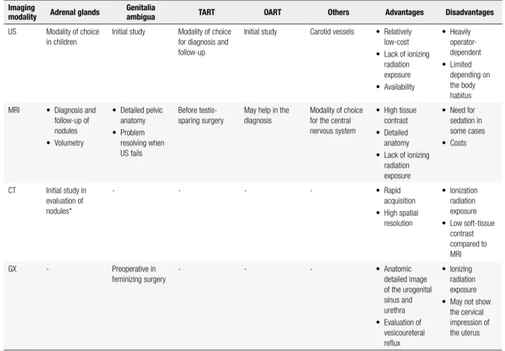
Table 1. Summary of the imaging approach in evaluation of variable conditions related to congenital adrenal hyperplasia
Imaging
modality Adrenal glands
Genitalia
ambigua TART OART Others Advantages Disadvantages
US Modality of choice in children
Initial study Modality of choice for diagnosis and follow-up
Initial study Carotid vessels • Relatively low-cost
• Lack of ionizing radiation exposure
• Availability
• Heavily operator-dependent
• Limited depending on the body habitus
MRI • Diagnosis and follow-up of nodules
• Volumetry
• Detailed pelvic anatomy
• Problem resolving when US fails
Before testis-sparing surgery
May help in the diagnosis
Modality of choice for the central nervous system
• High tissue contrast
• Detailed anatomy
• Lack of ionizing radiation exposure
• Need for sedation in some cases
• Costs
CT Initial study in evaluation of nodules*
- - - - • Rapid
acquisition
• High spatial resolution
• Ionization radiation exposure
• Low soft-tissue contrast compared to MRI
GX - Preoperative in feminizing surgery
- - - • Anatomic
detailed image of the urogenital sinus and urethra
• Evaluation of vesicoureteral relux
• Ionizing radiation exposure
• May not show the cervical impression of the uterus
Cop
yright
© ABE&M t
odos os dir
eit
os r
eser
vados
.
ionizing radiation. To assess pelvic anatomy, genitogra-phy is used for planning feminizing surgery (3).
For radiologists and sonographers, the detection of enlarged adrenal glands, adrenal gland nodules, and coiled adrenal glands in any imaging study should prompt raise the possibility of CAH. Also, they must keep in mind the possibility of CAH in cases of am-biguous genitalia, testis lesions, ovarian solid nodules, abnormal retroperitoneum solid tissues, and even white matter abnormalities seen in brain MRI.
Disclosure: no potential conlict of interest relevant to this article was reported.
REFERENCES
1. Merke DP, Bornstein SR. Congenital adrenal hyperplasia. Lancet. 2005;365(9477):2125-36.
2. Speiser PW, Azziz R, Baskin LS, Ghizzoni L, Hensle TW, Merke DP, et al. Congenital adrenal hyperplasia due to steroid 21-hydroxy-lase deiciency: an Endocrine Society clinical practice guideline. J Clin Endocrinol Metab. 2010;95(9):4133-60.
3. Chavhan GB, Parra DA, Oudjhane K, Miller SF, Babyn PS, Pippi Salle FL. Imaging of ambiguous genitalia: classiication and diag-nostic approach. Radiographics. 2008;28(7):1891-904.
4. Nabhan ZM, Eugster EA. Upper-tract genitourinary malforma-tions in girls with congenital adrenal hyperplasia. Pediatrics. 2007;120(2):e304-7.
5. Vanderbrink BA, Rink RC, Cain MP, Kaefer M, Meldrum KK, Mis-seri R, et al. Does preoperative genitography in congenital ad-renal hyperplasia cases affect surgical approach to feminizing genitoplasty? J Urol. 2010;184(4 Suppl):1793-8.
6. Sivit CJ, Hung W, Taylor GA, Catena LM, Brown-Jones C, Kushner DC. Sonography in neonatal congenital adrenal hyperplasia. AJR Am J Roentgenol. 1991;156(1):141-3.
7. Hauffa BP, Menzel D, Stolecke H. Age-related changes in adrenal size during the irst year of life in normal newborns, infants and patients with congenital adrenal hyperplasia due to 21-hydroxy-lase deiciency: comparison of ultrasound and hormonal param-eters. Eur J Pediatr. 1988;148(1):43-9.
8. Avni EF, Rypens F, Smet MH, Galetty E. Sonographic demonstra-tion of congenital adrenal hyperplasia in the neonate: the cerebri-form pattern. Pediatr Radiol. 1993;23(2):88-90.
9. Hernanz-Schulman M, Brock JW, 3rd, Russell W. Sonographic indings in infants with congenital adrenal hyperplasia. Pediatr Radiol. 2002;32(2):130-7.
10. Saada J, Grebille AG, Aubry MC, Raii A, Dumez Y, Benachi A. So-nography in prenatal diagnosis of congenital adrenal hyperpla-sia. Prenat Diagn. 2004;24(8):627-30.
11. Avila NA, Premkumar A, Shawker TH, Jones JV, Laue L, Cutler GB, Jr. Testicular adrenal rest tissue in congenital adrenal hyper-plasia: indings at Gray-scale and color Doppler US. Radiology. 1996;198(1):99-104.
12. Shanklin DR, Richardson AP Jr, Rothstein G. Testicular hilar nod-ules in adrenogenital syndrome. The nature of the nodnod-ules. Am J Dis Child. 1963;106:243-50.
13. Stikkelbroeck NM, Otten BJ, Pasic A, Jager GJ, Sweep CG, Noor-dam K, et al. High prevalence of testicular adrenal rest tumors, impaired spermatogenesis, and Leydig cell failure in adolescent
and adult males with congenital adrenal hyperplasia. J Clin Endo-crinol Metab. 2001;86(12):5721-8.
14. Stikkelbroeck NM, Suliman HM, Otten BJ, Hermus AR, Blickman JG, Jager GJ. Testicular adrenal rest tumours in postpubertal males with congenital adrenal hyperplasia: sonographic and MR features. Eur Radiol. 2003;13(7):1597-603.
15. Avila NA, Shawker TS, Jones JV, Cutler GB Jr, Merke DP. Tes-ticular adrenal rest tissue in congenital adrenal hyperplasia: serial sonographic and clinical indings. AJR Am J Roentgenol. 1999;172(5):1235-8.
16. Reisch N, Scherr M, Flade L, Bidlingmaier M, Schwarz HP, Mül-ler-Lisse U, et al. Total adrenal volume but not testicular adrenal rest tumor volume is associated with hormonal control in pa-tients with 21-hydroxylase deiciency. J Clin Endocrinol Metab. 2010;95(5):2065-72.
17. Reisch N, Flade L, Scherr M, Rottenkolber M, Pedrosa Gil F, Bidlingmaier M, et al. High prevalence of reduced fecundity in men with congenital adrenal hyperplasia. J Clin Endocrinol Metab. 2009;94(5):1665-70.
18. Claahsen-van der Grinten HL, Otten BJ, Hermus AR, Sweep FC, Hulsbergen-van de Kaa CA. Testicular adrenal rest tumors in pa-tients with congenital adrenal hyperplasia can cause severe tes-ticular damage. Fertil Steril. 2008;89(3):597-601.
19. Martinez-Aguayo A, Rocha A, Rojas N, et al. Testicular adrenal rest tumors and Leydig and Sertoli cell function in boys with clas-sical congenital adrenal hyperplasia. J Clin Endocrinol Metab. 2007;92(12):4583-9.
20. Claahsen-van der Grinten HL, Otten BJ, Stikkelbroeck MM, Sweep FC, Hermus AR. Testicular adrenal rest tumours in congen-ital adrenal hyperplasia. Best Pract Res Clin Endocrinol Metab. 2009;23(2):209-20.
21. Dieckmann K, Lecomte P, Despert F, Maurage C, Sirinelli D, Rol-land JC. [Congenital adrenal hyperplasia and testicular hypertro-phy]. Arch Pediatr. 1995;2(12):1167-72.
22. New MI. Nonclassical congenital adrenal hyperplasia and the polycystic ovarian syndrome. Ann N Y Acad Sci. 1993;687:193-205. 23. Forsbach G, Guitron-Cantu A, Vazquez-Lara J, Mota-Morales
M, Diaz-Mendoza ML. Virilizing adrenal adenoma and primary amenorrhea in a girl with adrenal hyperplasia. Arch Gynecol Ob-stet. 2000;263(3):134-6.
24. Russo G, Paesano P, Taccagni G, Del Maschio A, Chiumello G. Ovarian adrenal-like tissue in congenital adrenal hyperplasia. N Engl J Med. 1998;339(12):853-4.
25. Tiosano D, Vlodavsky E, Filmar S, Weiner Z, Goldsher D, Bar-Sha-lom R. Ovarian adrenal rest tumor in a congenital adrenal hyper-plasia patient with adrenocorticotropin hypersecretion following adrenalectomy. Horm Res Paediatr. 2010;74(3):223-8.
26. Sivan E, Koch S, Reece EA. Sonographic prenatal diagnosis of ambiguous genitalia. Fetal Diagn Ther. 1995;10(5):311-4.
27. Garel L. Abnormal sex differentiation: who, how and when to im-age. Pediatr Radiol. 2008;38 Suppl 3:S508-11.
28. Chertin B, Hadas-Halpern I, Fridmans A, Kniznik M, Abu-Arafeh W, Zilberman M, et al. Transabdominal pelvic sonography in the preoperative evaluation of patients with congenital adrenal hy-perplasia. J Clin Ultrasound. 2000;28(3):122-4.
29. Minette MS, Hoyer AW, Pham PP, DeBoer MD, Reller MD, Boston BA. Cardiac function in congenital adrenal hyperplasia: a pattern of reversible cardiomyopathy. J Pediatr. 2013;162(6):1193-8, 8 e1. 30. Wasniewska M, Balsamo A, Valenzise M, Manganaro A, Faggioli
G, Bombaci S, et al. Increased large artery intima media thickness in adolescents with either classical or non-classical congenital adrenal hyperplasia. J Endocrinol Invest. 2013;36(1):12-5. 31. Reisch N, Idkowiak J, Hughes BA, Ivison HE, Abdul-Rahman OA,
hyper-Cop
yright
© ABE&M t
odos os dir
eit
os r
eser
vados
.
irst-trimester nuchal translucency as a prenatal manifestation of salt-wasting congenital adrenal hyperplasia. Ultrasound Obstet Gynecol. 2002;20(4):392-4.
34. Baş F, Saka N, Darendeliler F, Tuzlali S, Ilhan R, Bundak R, et al. Bilateral ovarian steroid cell tumor in congenital adrenal hyper-plasia due to classic 11beta-hydroxylase deiciency. J Pediatr En-docrinol Metab. 2000;13(6):663-7.
35. Souverijns G, Peene P, Keuleers H, Vanbockrijck M. Ectopic locali-sation of adrenal cortex. Eur Radiol. 2000;10(7):1165-8.
36. Jaresch S, Kornely E, Kley HK, Schlaghecke R. Adrenal inciden-taloma and patients with homozygous or heterozygous congeni-tal adrenal hyperplasia. J Clin Endocrinol Metab. 1992;74(3):685-9. 37. Giacaglia LR, Mendonca BB, Madureira G, Melo KF, Suslik CA,
Arnhold IJ, et al. Adrenal nodules in patients with congenital ad-renal hyperplasia due to 21-hydroxylase deiciency: regression after adequate hormonal control. J Pediatr Endocrinol Metab. 2001;14(4):415-9.
38. Harinarayana CV, Renu G, Ammini AC, Khurana ML, Ved P, Kar-markar MG, et al. Computed tomography in untreated congenital adrenal hyperplasia. Pediatr Radiol. 1991;21(2):103-5.
39. Mermejo LM, Elias Junior J, Saggioro FP, Tucci Junior S, Castro Md, Moreira AC, et al. Giant adrenal myelolipoma associated with 21-hydroxylase deiciency: unusual association mimicking an androgen-secreting adrenocortical carcinoma. Arq Bras Endo-crinol Metabol. 2010;54(4):419-24.
40. Chevalier N, Carrier P, Piche M, Chevallier A, Wagner K, Tardy V, et al. Adrenocortical incidentaloma with uncertain prognosis as-sociated with an inadequately treated congenital adrenal hyper-plasia. Ann Endocrinol (Paris). 2010;71(1):56-9.
41. Crocker MK, Barak S, Millo CM, Beall SA, Niyyati M, Chang R, et al. Use of PET/CT with cosyntropin stimulation to identify and localize adrenal rest tissue following adrenalectomy in a woman with congenital adrenal hyperplasia. J Clin Endocrinol Metab. 2012;97(11):E2084-9.
42. Al-Ahmadie HA, Stanek J, Liu J, Mangu PN, Niemann T, Young RH. Ovarian ‘tumor’ of the adrenogenital syndrome: the irst reported case. Am J Surg Pathol. 2001;25(11):1443-50.
43. Claahsen-van der Grinten HL, Duthoi K, Otten BJ, d’Ancona FC, Hulsbergen-vd Kaa CA, Hermus AR. An adrenal rest tumour in the perirenal region in a patient with congenital adrenal hyperplasia due to congenital 3beta-hydroxysteroid dehydrogenase deicien-cy. Eur J Endocrinol. 2008;159(4):489-91.
44. Azziz R, Kenney PJ. Magnetic resonance imaging of the adrenal gland in women with late-onset adrenal hyperplasia. Fertil Steril. 1991;56(1):142-4.
45. Chervin RA, Danilowicz K, Pitoia F, Gomez RM, Bruno OD. [A study of 34 cases of adrenal incidentaloma]. Medicina (B Aires). 2007;67(4):341-50.
48. Bachega TAM J, Madureira G, Suslik CA, Gomes GC, Secaf E, Bloise W, et al. Adrenal image studies in patients with congenital adrenal hyperplasia due to 21-hydroxylase deiciency. 7th Annual Meeting of the Sociedad Latinoamericana de Endocrinologia Pe-diatrica. Itapanema, Brasil. 1993. p. 678.
49. Mokshagundam S, Surks MI. Congenital adrenal hyperplasia di-agnosed in a man during workup for bilateral adrenal masses. Arch Intern Med. 1993;153(11):1389-91.
50. Paulino Mda C, Steinmetz L, Menezes Filho HC, Kuperman H, Della Manna T, Vieira JG, et al. [Search of prostatic tissue in 46,XX congenital adrenal hyperplasia]. Arq Bras Endocrinol Metabol. 2009;53(6):716-20.
51. Avila NA, Premkumar A, Merke DP. Testicular adrenal rest tissue in congenital adrenal hyperplasia: comparison of MR imaging and sonographic indings. AJR Am J Roentgenol. 1999;172(4):1003-6. 52. Storr HL, Barwick TD, Snodgrass GA, Booy R, Morel Y, Reznek RH,
et al. Hyperplasia of adrenal rest tissue causing a retroperitoneal mass in a child with 11 beta-hydroxylase deiciency. Horm Res. 2003;60(2):99-102.
53. Sinforiani E, Livieri C, Mauri M, Bisio P, Sibilla L, Chiesa L, et al. Cognitive and neuroradiological indings in congenital adrenal hyperplasia. Psychoneuroendocrinology. 1994;19(1):55-64. 54. Rose AB, Merke DP, Clasen LS, Rosenthal MA, Wallace GL,
Vaituz-is AC, et al. Effects of hormones and sex chromosomes on stress-inluenced regions of the developing pediatric brain. Ann N Y Acad Sci. 2004;1032:231-3.
55. Nass R, Heier L, Moshang T, Oberield S, George A, New MI, et al. Magnetic resonance imaging in the congenital adrenal hyperpla-sia population: increased frequency of white-matter abnormali-ties and temporal lobe atrophy. J Child Neurol. 1997;12(3):181-6. 56. Lee S, Sanefuji M, Watanabe K, Uematsu A, Torisu H, Baba H, et
al. Clinical and MRI characteristics of acute encephalopathy in congenital adrenal hyperplasia. J Neurol Sci. 2011;306(1-2):91-3. 57. Speiser PW, Heier L, Serrat J, New MI, Nass R. Failure of steroid
replacement to consistently normalize pituitary function in con-genital adrenal hyperplasia: hormonal and MRI data. Horm Res. 1995;44(6):241-6.
58. Pasquino AM, Pucarelli I, Cambiaso P, Cappa M. Precocious pu-berty with hypothalamic hamartoma and non classical form of congenital adrenal hyperplasia. Report of two cases. Minerva Pe-diatr. 2009;61(5):561-4.
59. Mazzone L, Mueller SC, Maheu F, VanRyzin C, Merke DP, Ernst M. Emotional memory in early steroid abnormalities: an FMRI study of adolescents with congenital adrenal hyperplasia. Dev Neuro-psychol. 2011;36(4):473-92.