Pendelluft diagnosed from ventilator weaning indexes
obtained through bioelectrical impedance tomography:
a case report
Pendelluft
diagnosticado através de índices de desmame ventilatório
obtidos pela tomograia de bioimpedância elétrica: um relato de caso
Fabiana Aparecida Lopes
I, Lidiane Andrade Monteiro de Souza
I, Juliana Tavares Neves Bernardi
I, Carlos Eduardo Rocha
I, Luciana
Castilho de Figueiredo
II, Ana Paula Ragonete dos Anjos Agostini
III, Desanka Dragosavac
IV, Daniela Cristina dos Santos Faez
VFaculdade de Ciências Médicas da Universidade Estadual de Campinas (FCM-UNICAMP), Campinas (SP), Brazil
ABSTRACT
CONTEXT: Today, through major technological advances in diagnostic resources within medicine, evalua-tion and monitoring of clinical parameters at the patient’s bedside in intensive care units (ICUs) has be-come possible.
CASE REPORT: This case report presents results and interpretations from predictive mechanical ventilation weaning indexes obtained through monitoring using chest electrical bioimpedance tomography. These indexes included maximum inspiratory pressure, maximum expiratory pressure, shallow breathing index and spontaneous breathing test. These were correlated with variations in tidal volume variables, respira-tory rate, mean arterial pressure and peripheral oxygen saturation. Regarding the air distribution behavior in the pulmonary parenchyma, the patient showed the pendelluft phenomenon. Pendelluft occurs due to the time constant (product of the airways resistance and compliance) asymmetry between adjacent lung.
CONCLUSION: Bioelectrical impedance tomography can help in weaning from mechanical ventilation, as in the case presented here. Pendelluft was deined as a limitation during the weaning tests.
RESUMO
CONTEXTO: Atualmente, com o grande avanço tecnológico em recursos para diagnósticos em medicina, a avaliação e a monitorização de parâmetros clínicos à beira leito de paciente em unidade de terapia intensiva (UTI) se tornou possível.
RELATO DE CASO: Neste relato de caso, apresentam-se os resultados e a interpretação de índices predi-tivos de desmame da ventilação mecânica obtidos pela tomograia de bioimpedância elétrica torácica. Esses índices incluíram a pressão inspiratória máxima, pressão expiratória máxima, índice de respiração supericial e teste de respiração espontânea. Estes estavam correlacionados com as variações de volu-me corrente, frequência respiratória, pressão arterial média e saturação periférica de oxigênio. Quanto ao comportamento da distribuição de ar no parênquima pulmonar, o paciente apresentou o fenômeno pendelluft.O pendelluft ocorre dado pela constante de tempo (produto da resistência e complacência das vias aéreas) de forma assimétrica entre as unidades pulmonares adjacentes.
CONCLUSÃO: A tomograia de bioimpedância pode auxiliar no desmame da ventilação mecânica, como no caso apresentado. Pendelluft foi deinido como limitação durante a execução dos testes para desmame. IBSc. Physiotherapist at the Adult Intensive
Care Unit, Universidade Estadual de Campinas (UNICAMP), Campinas (SP), Brazil.
IIMSc, PhD. Physiotherapist at Adult Intensive
Care Unit, Hospital das Clínicas, and Supervisor of Chest Physiotherapy Training Course, Adult Intensive Care Unit, Universidade Estadual de Campinas (UNICAMP), Campinas (SP), Brazil.
IIIMSc, PhD. Physiotherapist, Faculdade de
Ciências Médicas, Universidade Estadual de Campinas (FCM-UNICAMP), Campinas (SP), Brazil.
IVMD, PhD. Coordinator, Adult Intensive Care
Unit, Hospital das Clínicas, Universidade Estadual de Campinas (UNICAMP), Campinas (SP), Brazil.
VMSc. Physiotherapist, Faculdade de Ciências
Médicas, Universidade Estadual de Campinas (FCM-UNICAMP), Campinas (SP), Brazil.
KEY WORDS: Ventilator weaning. Respiration, artiicial. Intensive care unit. Thoracic surgery. Acute lung injury.
PALAVRAS-CHAVE: Desmame do respirador. Respiração artiicial. Unidades de terapia intensiva. Cirurgia torácica.
INTRODUCTION
Today, through major technological advances in diagnostic resources within medicine, evaluation and monitoring of clinical parameters at the patient’s bedside in intensive care units (ICUs) has become possible. Bioelectrical impedance tomography on these parameters is one example of these advances. It uses high-frequency electrical signals at low intensity to provide imaging of lung mechanics in real time. hese signals are obtained by ixing a strap containing electrodes around the patient’s chest, to capture the intensity and frequency of the electric current that is propa-gated around the chest, between the electrodes. It is a noninvasive technique that does not use any type of radiation. It constitutes an innovation within interpretation of pulmonary mechanics.1
he predictive indexes for withdrawing patients from mechani-cal ventilation include the rapid shallow breathing index (RSBI), maximum inspiratory pressure (PImax) and maximum expiratory pressure (PEmax). he 2013 Brazilian guidelines for mechanical ventilation state that these indexes contribute towards decision-making in cases in which weaning is considered diicult. hus, the decision-making for referring patients for a spontaneous breathing test (SBT) or for extubation does not rely on a single instrument. Use of these indexes may lead to shorter duration of mechanical ventilation.2,3
Use of chest electrical impedance tomography (EIT) at the bedside for patients undergoing a mechanical ventilation wean-ing process may be an important tool for aidwean-ing in this process. EIT takes into account important variables such as tidal volume, the degree of collapse of recruitable alveoli and the degree of alve-olar distention. his test is based on diferences in electrical prop-erties generated by changes to air content in small regions of the lung, which create impedance between these regions. he pixels generated by the display image represented the percentage change in local impedance, relative to a reference that was obtained at the beginning of the image acquisition. herefore, the dynamic images shown on the chest EIT monitor represent real-time local air changes during ventilation. At locations where variations in the air within the alveoli occur, the color of the image generated changes on a scale ranging from dark blue (less aeration) to light blue (greater aeration). Grey images represent regions in which there was no change of aeration.4,5
Pendellut is a phenomenon that constitute a new mechanism of lung injury induced by mechanical ventilation. he overstretch that is observed in the dependent lung may cause a hidden injury point, that cannot be detected and thus is overlooked when con-ventional monitoring is used. Pendellut can be deined as the air circulation within the lung parenchyma, in non-dependent and dependent areas, when there is no overall change in lung volume. Traditionally, it was believed that contraction of the diaphragm would decrease the pleural pressure uniformly, by the same amount
at all points on the lung surface, so as to create a uniform increase in transpulmonary pressure. Pendellut occurs because, in contrast to the normal lung, the injured lung does not show uniform luid distribution behavior. Instead, transmission of local changes in pleural pressure is heterogeneous.6
herefore, the objective of this study was to report on lung mechanics behavior, as shown by predictive weaning indexes obtained through bioelectrical impedance and by spontaneous breathing tests, in a patient with mitral valve disease who under-went valve replacement and prolonged weaning.
CASE REPORT
he patient was a 65-year-old female with a history of surgical replacement of the mitral valve by a bioprosthesis 14 years ear-lier. Her personal history included valvular heart disease, atrial ibrillation, congestive heart failure (functional class IV) and hypertension.
At the time of admission to the clinical hospital of the University of Campinas on July 3, 2015, the patient had had symptoms of pro-gressive dyspnea for the preceding six months. his was also asso-ciated with paroxysmal nocturnal dyspnea, orthopnea and lower limb edema. herefore, the patient had been referred for evalua-tion of valve dysfuncevalua-tion and surgical assessment.
In the initial clinical evaluation, an echocardiogram was per-formed on July 6, 2015, which showed increased volume of the let chambers, presence of the biological mitral prosthesis with stenosis and moderate regurgitation, pulmonary hypertension with systolic pulmonary artery depression values of 78 mmHg and let-ventricle ejection fraction of 58% through Simpson’s method. herefore, the patient was admitted to the hospital ward.
On July 7, 2015, her condition evolved with a productive cough and crackling and wheezing observed through ausculta-tion. Pneumonia was diagnosed, which was treated with anti-biotic therapy. he patient made continuous use of diuretic and antihypertensive drugs.
Ater 19 days of hospitalization, the patient underwent surgery for replacement of the bioprosthetic mitral valve with a mechani-cal prosthesis and also underwent tricuspid valve plasty. he pro-cedure was performed under extracorporeal circulation, with a duration of 124 minutes. here were 46 minutes of myocardial ischemia and 92 minutes of aortic clamping without blood trans-fusions. No intraoperative events were noted. Ater surgery, the patient was transferred to the intensive care unit without sedation, with mechanical ventilation using dosages of dopamine diuretic. he patient was extubated in the immediate postoperative period and she started using a Venturi oxygen mask, receiving an inspired oxygen fraction (FiO2) of 50%.
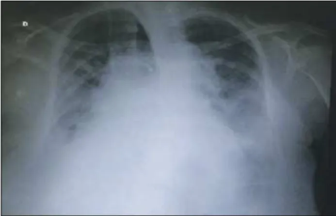
oxygen saturation (SpO2) and signiicant radiological worsening, with difuse iniltrates, signs of pulmonary congestion and opaciica-tion of the costophrenic angle of the breasts, as shown in Figure 1. She also presented oliguria and it was necessary to adminis-ter furosemide inadminis-termittently. Noninvasive mechanical ventilation (NIV) was used for an intermittent period, but with little improve-ment of the tachypnea.
Since there was no improvement of respiratory symptoms, even through using NIV, it was decided on August 2, 2015, to perform an intubation procedure and start continuous sedation for better respiratory management. Ater the clinical signs had become sta-ble, the weaning process was started.
At the minimum mechanical ventilation parameters, i.e. spon-taneous mode with FiO2 of 40%, positive end-expiratory pressure (PEEP) of 4 cmH20 and tidal volume of 5 ml/kg, the patient did not tolerate procedures to produce predictive indexes for with-drawal of mechanical ventilation. She showed signs suggestive of respiratory failure, according to the 2013 Brazilian guidelines for mechanical ventilation. A chest computed tomography (CT) scan was then performed, which revealed the presence of extensive bilat-eral pleural efusion at the bases of the lungs, and more evidently in the right lung. To relieve this, it was decided to perform thora-centesis, with removal of 1300 ml of citric luid.
To evaluate the lung mechanics, respiratory monitoring through a scanner by means of bioelectrical impedance analysis was chosen (Timpel Enlight 1800, São Paulo, Brazil). An EIT electrode belt with 16 electrodes was placed around the thorax at the level of the ith intercostal space, and one reference electrode was also placed on the patient. A tidal image was calculated as the diference between the EIT images at end-inspiration and end-expiration for one tidal breath, which represents the regional distribution of tidal volume (the tidal variation of impedance). hus, the predictive indexes for success or failure in withdrawing mechanical ventilation and the spontaneous breathing test (SBT) using a T piece were determined and could then be evaluated by means of tomography.
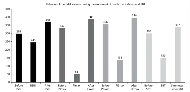
he results regarding the variation of heart rate (HR), respi-ratory frequency (RF), peripheral oxygen saturation (SpO2) and mean arterial pressure (MAP) are shown in Table 1. Any signii-cant variations in tidal volume and in the distribution of lung parenchyma could be checked through determining predictive indexes (RSBI, PImax and PEmax) from electrical bioimpedance tomography images and through determining spontaneous breath-ing. he behavior of changes in tidal volume at the time of deter-mining the predictive indices and SBT are illustrated in Graph 1. he changes in the distribution of air in the lung parenchyma are illustrated in Figures 2, 3, 4 and 5. hese were obtained from the scanner screen through bioimpedance at the following times: before, during and ater determining the predictive indices and before and 5 minutes ater completion of the spontaneous breathing test.
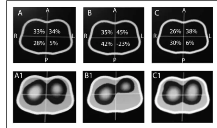
Importantly, the patient developed a high response to atrial ibrillation at the time of the spontaneous breathing test, with-out hemodynamic repercussions. Five minutes ater this test was started, the patient showed signs of respiratory failure. his requir-ing discontinuation of the test and return to mechanical ventila-tion in spontaneous mode, with the minimum parameters already described above. It was also observed that ater the predictive indexes had been determined, there was better distribution of air in the lung parenchyma. his was shown through the positive graphic percentages displayed on the scanner screen (Figures 2, 3, 4 and 5). he behavior of the air distribution within the lung paren-chyma before, during and ater determining the RSBI can be seen in the ventilation map distribution of 64%, 71% and 58% in the right lung and 36%, 28%, 43% in the let lung, as shown in Figure 2.
From these indings and because the weaning process failed for a second time, it was decided during a clinical visit and dis-cussion of the case to perform tracheostomy. his was done on
Figure 1. Chest X-ray in posterior-anterior view (April 24, 2015).
Table 1. Variation of heart rate (HR), respiratory frequency (RF),
peripheral oxygen saturation (SpO2) and mean arterial pressure
(MAP), before measuring the predictive indexes, during measurement of the predictive indexes and ive minutes after performing the spontaneous breathing test (SBT)
Times
Variables
HR (bpm) RF (rpm) SpO2 (%) MAP (mmHg)
Before tests 81 20 98 57
During RSBI 91 31 94 73
During PImax 82 29 100 65
During PEmax 84 36 100 70
During SBT 89 25 98 70
5 minutes after SBT 78 25 99 56
August 11, 2015. On the days following this, progressive reduction of ventilatory parameters was observed and the weaning process was again started, using intermittent mist in a T piece. he patient-progressed satisfactorily and continued to receive continuous 298
245
368
332
51
386
356
138
396
300
150
337
0 50 100 150 200 250 300 350 400 450
Before RSBI
RSBI After RSBI
Before PImax
PImax After PImax
Before PEmax
PEmax After PEmax
Before SBT
SBT 5 minutes after SBT Behavior of the tidal volume during measurement of predictive indexes and SBT
Graph 1. Behavior of the variation of tidal volume during measurement of the predictive indexes and spontaneous breathing test (SBT) by means of electrical bioimpedance tomography.
SBT = spontaneous breathing test; RSBI =rapid shallow breathing index; PImax = maximum inspiratory pressure; PEmax = maximum expiratory pressure. Before = before implementing the measurements; After = after the measurements.
A B C
A1 B1 C1
R A
36% 35% 42% 33% 39%
28% -3% 36% -14% 25% 4%
39%
P
A
P
A
P R
L L R L
Figure 2. Behavior of the air distribution in the lung parenchyma before, during and after measurement of the rapid shallow breathing index (RSBI).
A = air distribution in the lung parenchyma before measurement of RSBI; B = during measurement of RSBI; C = after measurement of RSBI; A1 = dynamic tomography image before RSBI; B1 = during RSBI; and C1 = after RSBI. Images represent a cross-section of the chest (electrode positioning level). Note: there was asymmetrical air distribution in the lung parenchyma, with smaller distribution of ventilation in the left lung and posterior lung ields during measurement of RSBI. There was better air distribution in the lung areas that were previously hypoventilated (left lung and posterior lung ields). R = right; L = left; A = anterior; P = posterior.
A B C
A1 B1 C1
R
A
32% 38%
78%
30% 41% 65%
25% 4% -100% 26% 3%
39%
P
A
P
A
P R
L L R L
Figure 3. Behavior of the air distribution in the lung parenchyma before, during and after measurement of the maximum inspiratory pressure (PImax).
nebulization. Ater a few days, the plastic tracheostomy tube was exchanged for a metal one.
DISCUSSION
Changes in lung function that occur subsequent to cardiac sur-gery using extracorporeal circulation are secondary to reactions to use of heparin and comprise protamine complex, edema, con-gestion, lung injury and microatelectasis. In most cases, mechan-ical ventilation is absent during extracorporeal circulation. his, together with the inlammatory response due to surgical trauma, leads to changes in respiratory function, consistent with those presented in cases of acute respiratory distress syndrome.7,8
Rodrigues et al. suggested that during and ater cardiac surgery, transient dysfunction of gas exchange (TDGE) is evident to varying degrees. Patients with preoperative hypertension and cardiogenic shock presented an association with occurrence of postoperative TDGE. During the postoperative period, presence of pneumonia, need for renal replacement therapy, need for blood therapy and pres-ence of cardiac arrhythmias were correlated with the appearance of a degree of TDGE, thus indicating a risk factor for reintubation.9
he case that Rodrigues et al. published consisted of a patient who was reintubated on the second day ater cardiac surgery.9 his patient presented a high response of atrial ibrillation while performing a SBT using a T piece without hemodynamic reper-cussions. Five minutes ater the SBT started, the patient showed signs of respiratory failure and the SBT had to be discontinued.
A B C
A1 B1 C1
R
A
33% 34%
28%
34% 36% 38%
27% 1% -0% 25% 6%
38%
P
A
P
A
P R
L L R L
Figure 4. Behavior of the air distribution in the lung parenchyma before, during and after measurement of the maximum expiratory pressure (PEmax).
A = air distribution in the lung parenchyma before measurement of PEmax; B = during measurement of PEmax; C = after measurement of PEmax; A1 = dynamic tomography image before PEmax; B1 = during PEmax; and C1 = after PEmax. Images represent a cross section of the chest (electrode positioning level). Note: there was asymmetrical distribution of air in the lung parenchyma, with smaller distribution of ventilation in the left lung and posterior lung ields during measurement of PEmax. There was better air distribution in the lung areas that were previously hypoventilated (left lung and posterior lung ields). R = right; L = left; A = anterior; P = posterior.
here was a second failure in withdrawing mechanical ventilation and conducting tracheostomy.
The images of Figures 2, 3, 4 and 5 were obtained in deter-mining rapid shallow breathing index superficial speed breath-ing index (RSBI), PImax, PEmax and SBT. They show variations in the distribution of air in the lung parenchyma. The behavior exhibited is compatible with the pendelluft effect.
Greenblatt et al. showed that presence of the pendellut efect may arise due to use of mechanical ventilation in spontaneous mode in cases in which spontaneous breathing eforts are detected, especially when the respiratory rate is high.10,11
Yoshida et al. conducted an experimental clinical study in which acute lung injury was induced in seven pigs, with the objec-tive of making comparisons with respiratory monitoring data from a clinical case of a male patient who underwent surgery for coro-nary revascularization. hey observed that when the seven pigs with acute lung injury were under mechanical ventilation without showing spontaneous ventilation efort, there was simultaneous inlation in diferent areas of their lungs. However, when the pigs showed spontaneous inspiratory eforts during mechanical ventila-tion, initial inlation of the dependent lung and simultaneous dela-tion could be observed. Despite these authors’ indings, it seems that the pendellut phenomenon can increase alveolar recruitment in areas of atelectasis that are selectively dependent on inlation, and that the degree of pendellut is proportional to the intensity of spontaneous efort on mechanical ventilation. hese indings corroborate what was found in the present case study.12
A B C
A1 B1 C1
R
A
33% 35%
42%
26% 38% 45%
28% 5% -23% 30% 6%
34%
P
A
P
A
P R
L L R L
Figure 5. Behavior of the air distribution in the lung parenchyma before, during and ive minutes after measurement of the spontaneous breathing test (SBT).
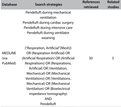
Table 2. Search of the literature in medical databases on October 17, 2016, for cases of pendelluft that have been diagnosed
Database Search strategies References retrieved
Related studies
MEDLINE (via PubMed)
Pendelluft during mechanical ventilation
Pendelluft during cardiac surgery Pendelluft during intensive care
Pendelluft during ventilator weaning
(“Respiration, Artiicial”[Mesh]) OR (Respiration Artiicial) OR (Artiicial Respiration) OR (Artiicial
Respirations) OR (Respirations, Artiicial) OR (Ventilation, Mechanical) OR (Mechanical Ventilations) OR (Ventilations,
Mechanical) OR (Mechanical Ventilation) OR (Bioelectrical impedance tomography)
AND Pendelluft
30 5
Greenblatt et al. considered that pendellut may be more evi-dent in diseases with a heterogeneous pattern of injury and major changes in respiratory mechanics, with changes in strength and in lung compliance. In the present case study, the chest X-ray sug-gested that the heterogeneous behavior described by these authors also occurred here.10
Recently, Yoshida et al. recommend that it would be important to balance muscle paralysis in relation to maintenance of sponta-neous breathing during mechanical ventilation for patients with acute respiratory distress syndrome network (ARDSnet). his would depend on the severity of ARDSnet, its evolution phase and its respiratory demands. In the early phase of severe ARDSnet, par-tial ventilatory support to promote spontaneous breathing should be avoided. Paralysis of the diaphragm muscle may be efective for preventing pendellut. In situations of less severity of ARDSnet, and ater a short period of diaphragm muscle paralysis in cases of severe ARDSnet, spontaneous breathing should be facilitated by means of partial mechanical ventilatory support. his would pre-vent large spontaneous respiratory eforts.13
he inding of pendellut can be determined through nonin-vasive monitoring conducted by means of electrical bioimpedance tomography, without exposing the patient to the invasive measures of other forms of monitoring. Furthermore, this monitoring can make a substantial contribution to research, thus facilitating implementa-tion of a series of studies that together can assist in understanding abnormal physiology. his would include investigating the regional heterogeneity of ventilation and factors associated with pendellut. A few studies have been conducted on pendellut diagnosed during weaning. he results from a systematic search in the main databases in the literature are presented in Table 2. Electrical bioimpedance tomography is simple to be performed at the bedside and has the potential to provide better understanding of the pathophysiology of a variety of lung disorders resulting from mechanical ventilation.14,15
CONCLUSION
Electrical bioimpedance tomography can contribute towards interpreting failures of withdrawal of ventilatory support and towards monitoring the side efects caused by mechanical ven-tilation, especially in patients with heterogeneous patterns of changes to respiratory mechanics. he indings from predictive indexes relating to withdrawal of mechanical ventilation might enable better distribution of air in the lung parenchyma and increased lung homogenization.
It should also be noted that ater the predictive indexes had been determined, the tidal volume did not return to its initial value. his efect was caused by alveolar recruitment through the increased respiratory efort during testing.
Electrical bioimpedance tomography can help in weaning patients of mechanical ventilation, as in the case presented here.
he limitation of pendellut while tests on weaning were being performed was deined.
Further studies should be conducted to investigate whether the alveolar recruitment achieved ater conducting spontaneous efort, at the time of determining the predictive indices, was maintained ater long reconnection to mechanical ventilation.
REFERENCES
1. Barbas CVS, Ísola AM, Farias AMC, et al. Recomendações brasileiras de ventilação mecânica 2013. Parte I [Brazilian recommendations of mechanical ventilation 2013. Part I]. Rev Bras Ter Intensiva. 2014;26(2):89-121. 2. Goldwasser R, Farias A, Freitas EE, et al. Desmame e interrupção da
ventilação mecânica. J Bras Pneumol. 2007;33(supl 2):128-136. 3. Fontoura IS, Pianezzola EM, Pacheco MTT. Tomograia por impedância
elétrica: uma alternativa para monitorização pulmonar continua em unidades de terapia intensiva. XII Encontro Latino Americano de Iniciação Cientíica e VIII Encontro Latino Americano de Pós-Graduação, Universidade do Vale do Paraíba; 2008. p. 1-4. Available from: http://www.inicepg.univap.br/cd/INIC_2008/anais/arquivosEPG/ EPG00320_02_A.pdf. Accessed in 2016 (Nov 17).
4. Frerichs I, Dargaville PA, Dudykevych T, Rimensberger PC. Electrical impedance tomography: a method for monitoring regional lung aeration and tidal volume distribution? Intensive Care Med. 2003;29(12):2312-6. 5. Muders T, Luepschen H, Putensen C. Impedance tomography as a new
monitoring technique. Curr Opin Crit Care. 2010;16(3):269-75. 6. Yoshida T, Torsani V, Gomes S, et al. Spontaneous efort causes occult
7. Morsch KT, Leguisamo CP, Coronel CC, Mattos W, Lima GG. Peril ventilatório dos pacientes submetidos a cirurgia de revascularização do miocárdio [Ventilatory proile of patients undergoing CABG surgery]. Rev Bras Cir Cardiovasc. 2009;24(2):180-7.
8. Szeles TF, Yoshinaga EM, Alencar W, et al. Hipoxemia após revascularização miocárdica: análise dos fatores de risco [Hypoxemia after myocardial revascularization: analysis of risk factors]. Rev Bras Anestesiol. 2008;58(2):124-36.
9. Rodrigues CD, Moreira MM, Lima NM, et al. Fatores de risco para disfunção transitória da troca gasosa após a cirurgia cardíaca [Risk factors for transient dysfunction of gas exchange after cardiac surgery]. Rev Bras Cir Cardiovasc. 2015;30(1):24-32.
10. Greenblatt EE, Butler JP, Venegas JG, Winkler T. Pendelluft in the bronchial tree. J Appl Physiol (1985). 2014;117(9):979-88.
11. Otis AB, McKerrow CB, Bartlett RA, et al. Mechanical factors in distribution of pulmonary ventilation. J Appl Physiol. 1956;8(4):427-43.
12. Yoshida T, Torsani V, Gomes S, et al. Spontaneous efort causes occult pendelluft during mechanical ventilation. Am J Respir Crit Care Med. 2013;188(12):1420-7.
13. Yoshida T, Uchiyama A, Fujino Y. The role of spontaneous efort during mechanical ventilation: normal lung versus injured lung. J Intensive Care. 2015;3:18.
14. Alzahrany M, Banerjee A. A biomechanical model of pendelluft induced lung injury. J Biomech. 2015;48(10):1804-10.
15. Vyshedskiy A, Murphy R. Pendelluft in chronic obstructive lung disease measured with lung sounds. Pulm Med. 2012;2012:139395.
Conlict of interest: None
Sources of funding: None
Date of irst submission: September 26, 2016
Last received: October 12, 2016
Accepted: October 14, 2016
Address for correspondence:
Ana Paula Ragonete dos Anjos Agostini
Faculdade de Ciências Médicas da Universidade Estadual de Campinas (FCM-UNICAMP)
Rua Tessália Vieira de Camargo, 126 Campinas (SP) — Brasil
CEP 13083-887 Tel. (+55 35) 3551-4097 Cel. (+55 35) 99201-5986