Arq Bras Cardiol volume 73, (nº 3), 1999
Lobo Fº et al. Total agenesis of the pericardium
3 0 7
Instituto do Coração e Pulmão–ICORP e Hospital Prontocárdio – Fortaleza Mailing address: José Glauco Lobo Fo - Rua Dr. José Lourenço, 625 - 60115-280 Fortaleza, CE - Brazil
J. Glauco Lobo Fº, Ricardo R. Maia Oliveira, Rafael Pontes de Siqueira, Rodrigo M. Landim, João Marcelo A .C. de Albuquerque, Carlos Bellini G. Gomes, Elita Borges, Maria Claúdia Leitão,
J. Nogueira Paes Jr
Fortaleza, CE - Brazil
Total Agenesis of the Left Pericardium
Case Report
This is the report of a 46-year-old patient with the preoperative diagnosis of an atrial septal defect (ASD) of the ostium secudum type. After sternectomy, partial agenesis of the left pericardium was diagnosed. It is our opinion that, if the radiographic picture is suggestive of this entity, a clinical search for cardiopulmonary anomalies should be performed, because the majority of these associated anomalies can and should be surgically corrected.
Total agenesis of the left pericardium is a rare conditi-on; no more then 200 cases have been reported in the medi-cal literature. There is a male predominance (3:1), and one third of the cases seem to be associated with other cardio-pulmonary anomalies, making the present case of the association of total absence of the left pericardium and ASD a very rare entity. Seventy-five percent of the conge-nital anomalies of the pericardium usually happen on the left side and are related to a premature atrophy of the left car-dinal vein (Cuvier’s duct) and compromise the circulation to the left pleuropericardial membrane.
Case Report
A 46-year-old male presented with fatigue on minor effort. No relevant clinical or surgical data were found in his medical history.
On physical examination he had signs of right heart failure, with enlargement of the jugular vein, palpable liver below the right costal border and edema of the legs. The examination of the thorax showed left deviation of the ictus
cordis . A 4/6 systolic murmur caused by an ASD was heard
on the left parasternal border. Another systolic murmur, caused by tricuspid regurgitation, was also detected at the fourth intercostal space in the left parasternal region. Car-diac rhythm was normal.
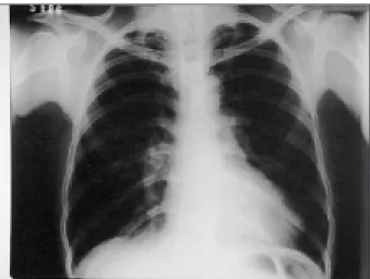
Routine laboratory examinations were normal. The chest X-ray showed global enlargement of the cardiac si-lhouette (2+/4+) and levoposition of the heart (fig. 1). Elec-trocardiogram showed normal sinus rhythm, with right
deviation of the axis, signs of enlargement of the right chambers and incomplete right bundle branch block. The echocardiogram showed situs solitus in levocardia, severe dilation of the right atrium, ASD of the ostium secundum type measuring 24 mm (fig. 2), dilation of the tricuspid ring, enlargement of the right ventricle and paradoxical motion of the ventricular septum. Because of the patient’s age and because of the fact that he was a heavy smoker and his pul-monary artery pressure needed to be measured, the patient underwent cardiac catheterizarion and coronary angiogra-phy in order for the angiogra-physicians to better plan surgery. In addition to the above-mentioned problems, a 90% obstruc-tion was detected at the proximal right coronary artery. The patient was then sent to surgery for correction of the ASD together with coronary artery bypass grafting.
After sternectomy, it was noted that the pericardium was absent (fig. 3). The heart was deviated to the left and there was no adherence between the left ventricle and the pleura. The right coronary artery was significantly calcified. The diaphragm was normal at inspection. Pericardial bor-ders were smooth and, in contiguity with the posterior left pericardium; fibrous-adipose tissue was found that was crossed over by the left phrenic nerve. Right pericardium and the right phrenic nerve were normally positioned. After extracorporeal circulation was started, with the heart still, the ASD was corrected and then revascularization of the right coronary artery was performed using a saphenous magna graft. No per- or postoperative complications occurred, and the patient was discharged seven days after surgery.
Discussion
Congenital pericardial defects are uncommon and
fre-quently asymptomatic, being diagnosed at post-mortem 2.
Congenital total absence of the left pericardium represent 57% of all congenital defects of the pericardium. Approxi-mately 78% of all of these defects occur on the left side and have been related to the premature atrophy of the left
common cardinal vein or left Cuvier’s duct 2,4.
The radiographic findings are levoposition of the
he-art and an elongated left border of the hehe-art 1. There is also
3 0 8
Lobo Fº et al.
Total agenesis of the pericardium
Arq Bras Cardiol volume 73, (nº 3), 1999
of the pulmonary tissue at this level. Retrospectively, these findings could be found in the present case.
In this case, the phrenic nerve was not located at its normal position but inside a fibrous-adipose tissue that was connected to the posterior border of the right peri-cardium. Because the phrenic nerve is usually mal-positioned in these cases of congenital anomalies of the pericardium, it is important to carefully identify it during surgery in order to avoid causing accidental lesions to it. In conclusion, we believe that, in the presence of radiographic suggestions of pericardial abnormalities, a clinical search for cardiopulmonary anomalies should be systematically performed, even if the patient is asymptomatic, because most associated findings can and should be corrected.
1. Chartrand-Lefebvre C, Filion R, Samson L, et al. Congenital complete absence of left pericardium. Can Assoc Radiol J 1997; 48: 353-6. 2. Samuels LE, Sharma S, Kaufman MS, et al. Absent pericardium during
coronary bypass. Arch Surg. 1997; 132: 318-9.
3. Higashikawa M, Nishio M, Kanamori T, et al. Traumatic tricuspid
References
gitation associated with congenital partial pericardial defect. Jpn Circ J 1997; 61: 358-60.
4. Hauser M, Berger MF. Congenital absence of left pericardium. JBR BTR, 1997; 80: 296.
5. Van Son JAM, Danielson GK, Schaff HV, et al. Congenital partial and complete absence of the pericardium. Mayo Clin Proc 1993; 68: 743-7. Fig. 1 - Chest X-ray in the antero-posterior position.
Fig. 2 - Enlarged heart and the ASD. Levoposition of the heart. AD- right atrium; AE- left atrium.