Article
0103 - 5053 $6.00+0.00
*e-mail: [email protected]
Synthesis, Characterization, EPR Spectroelectrochemistry Studies and
Theoretical Calculations of Manganese(II) Complexes with the
Ligands H
3bpeten and H
3bnbpeten
Stela Maris de M. Romanowski,a Sérgio P. Machado,b Geraldo R. Friedermann,a Antonio
S. Mangrich,a Monique de F. Hermann,a Hugo Oroino Limab and Shirley Nakagaki*,a
aDepartamento de Química, Universidade Federal do Paraná, 81531-990 Curitiba-PR, Brazil
bInstituto de Química, Universidade Federal do Rio de Janeiro, 21941-909 Rio de Janeiro-RJ, Brazil
Este trabalho relata a síntese e a caracterização dos complexos de manganês(II) [MnII(Hbpeten)] e [MnII(Hbnbpeten)], respectivamente [N,N’-bis-(2-hidroxibenzil)-N-(2-piridilmetil)-N ’-(2-hidroxietil)etano-1,2-diamine]manganês(II) e [N,N’-bis-(5-nitro-2-hidroxibenzil)-N’-(2-hidroxietil) etano-1,2-diamin]manganês(II), por análise elementar, voltametria cíclica, espectroscopias eletrônica (UV-Vis) e vibracional (FTIR), ressonância magnética nuclear de hidrogênio (1H NMR) e espectroeletroquímica de ressonância paramagnética eletrônica (EPR). Os dados de espectroeletroquímica de EPR foram consistentes com a redução de um grupo nitro, tanto no H3bnbpeten livre quanto no complexo de manganês. Além disso, os cálculos teóricos foram consistentes com os resultados experimentais, mostrando que somente um grupo nitro contribui para a formação dos orbitais LUMO e sugerindo que os resultados teóricos são adequados para explicar as propriedades eletrônicas dos complexos.
The synthesis and characterization of the manganese(II) complexes [MnII(Hbpeten)] and [MnII(Hbnbpeten)], where H
3bpeten and H3bnbpeten are respectively [N,N ’-bis-(2-hydroxybenzyl)-N-(2-pyridylmethyl)-N’-(2-hydroxyethyl)ethane-1,2-diamine] and [N,N ’-bis-(5-nitro-2-hydroxybenzyl)-N’-(2-hydroxyethyl)ethane-1,2-diamine], are reported. The characterization was carried out by elemental analyses, cyclic voltammetry, spectroscopic methods (UV-Vis, FTIR, 1H NMR), electronic paramagnetic resonance spectroelectrochemistry (EPR) and theoretical DFT calculations. The electrochemistry and EPR espectroelectrochemistry data were consistent with the reduction of one of the nitro groups in free H3bnbpeten and in the respective manganese(II) complex. These results were supported by DFT calculations, which showed that only one nitro group contributes to the LUMO. The theoretical data appear to be suitable to describe the electronic properties of the compounds.
Keywords: manganese complex, EPR, N,O-donor ligands, DFT, computational chemistry
Introduction
Reactions catalyzed by transition metal cations have attracted great interest in view of their applications in industrial processes.1 In fact, several M-salen complexes,
where M is manganese(III),2 chromium(III)3 or nickel(II),4
have been utilized as homogeneous catalysts for olein epoxidation.
The design of many manganese complexes and the investigation of their properties and applications (for example as catalysts) are bio-inspired. This is the case of
several manganese complexes whose preparation aims to model the structure, reactivity and spectroscopic properties of biomolecules that contain this transition metal in its various oxidation states. In this way, complexes with different ligand types and nuclearities have contributed substantially to the understanding ofthe role of manganese in biological systems such as SOD (superoxide dismutase), the oxygen-evolving complex (OEC) of photosystem II (PS II)5,6 and catalases,7 among others. Furthermore,
manganese complexes based on Schiff bases and other
N,O-donor ligands have been explored as model systems
the coordination geometry and electronic properties on the rich spectroscopic features of the biological Mn sites. In fact, such features and the chemical structures of the complexes were intensively studied in the past decades by different techniques, such as UV-Vis, FT-IR, EPR, XANES and EXAFS spectroscopies, X-ray crystallography, etc.8,9
In view of the development of molecular mechanics and quantum chemical calculations, geometry optimization, molecular orbital energy and vibrational frequency calculations of metallocomplexes by DFT methods have turned into valuable tools for the description of molecular properties. In recent years, this method has been extensively used in biomolecular chemistry involving coordination compounds of the first and second transition metal series.10-13 Recently, a detailed computational study of O-O
bond formation, catalyzed by monomeric and dimeric Mn-corrole complexes, was carried out by the density functional theory (DFT) approach, in the light of biomimetic models of OEC.14
Our interest in the chemistry of the H3bpeten and H3bnbpeten ligands led to the preparation and characterization (electrochemistry, vibrational and electronic spectroscopies) of the respective copper(II) complexes. The use of –NO2 electron-withdrawing groups in Hbnbpeten stabilizes the copper(II) oxidation state by several hundred millivolts, as conirmed by the anodic shift of the Epa value, when compared to the analogous H3bpeten complex which has no substituent in the phenolic moieties.15
In order to investigate the effect of the ligand structure in the coordination geometry and oxidation state of the manganese ion, complexes bearing the H3bpeten and H3bnbpeten ligands were synthesized and shown to present an interesting catalytic activity in oxidation reactions.16,17 They
were designed aiming the study of the effect of the –NO2 group on the physicochemical properties of the manganese complexes. The H3bnbpeten ligand is characterized by the presence of the electron-withdrawing nitro group, which should stabilize the manganese(II) species and make the synthesis of the complex much easier, as the greater acidity of H3bnbpeten15 facilitates the generation and coordination
of the deprotonated phenol group to the metal ion. Thus, this work reports the synthesis and characterization of the manganese-containing model compounds [MnII(Hbpeten)]
([N,N’-bis-(2-hydroxybenzyl)-N-(2-pyridylmethyl)-N
’-(2-hydroxyethyl)ethane-1,2-diamine]manganese(II)) and [MnII(Hbnbpeten)] ([N,N ’-bis-(5-nitro-2-hydroxybenzyl)-N’-(2-hydroxyethyl)ethane-1,2-diamine]manganese(II)) by
spectroscopic and electrochemical methods (UV-Vis, FTIR, cyclic voltammetry, EPR and EPR spectroelectrochemistry),18
as well as theoretical calculations envisaging the investigation
of the inluence of the frontier orbitals on the reactivity of the products.
Experimental
The following abbreviations were used throughout the text: H3bpten: N,N’-bis-(2-hydroxybenzyl)-N
-2-pyridylmethyl)-N’-(2-hydroxyethyl)ethane-1,2-diamine;
H3bnbpeten: N,N’-bis-(5-nitro-2-hydroxybenzyl)-N’
-(2-hydroxyethyl)ethane-1,2-diamine; Hpeten: N
-(2-pyridylmethyl)-N’-(2-hydroxyethyl)ethane-1,2-diamine;
Et3N: triethylamine; dmf: N,N-dimethylformamide;
DMF and [TBA][PF6]: tetra-n-butylammonium
hexaluorophosphate and Fc+/Fc: ferrocenium/ferrocene
redox pair.
Materials
2-pyridylcarboxyaldehyde, 2-bromomethylphenol, 2-hydroxy-5-nitrobenzaldehyde (5-nitrosalicylaldehyde),
N-(2-hydroxyethyl)ethylenediamine,
2-hydroxy-5-nitrobenzyl bromide (α-bromo-4-nitro-o-cresol),
manganese(II) acetate and [TBA][PF6] were obtained from Aldrich Chemical Co. For the electrochemical, spectroelectrochemical and spectroscopic studies, high-purity solvents were purchased from Merck and used as received. High-purity nitrogen gas was used to deoxygenate solutions. All other chemicals and solvents were reagent grade.
Syntheses
H3bpeten (L1):16,17 This unsymmetrical compound
was obtained in two steps, according to Scheme 1. The first step was a condensation reaction of N
-(2-hydroxyethyl)ethylenediamine (2.60 g; 25 mmol) with 2-pyridinecarboxyaldehyde (2.68 g; 25 mmol), followed by reduction with NaBH4 (1.90 g; 50 mmol) in methanol, producing N-(2-pyridylmethyl)-N’-(2-hydroxyethyl)
out with acetone, iltered out, washed with propan-2-ol and dried under vacuum. H3bpeten was obtained as a pale yellow solid. Yield: 3.20 g (78.5%), mp 157 °C. Anal.
Calc. for C24H29N3O3•2C
3H8O: C, 68.24; H, 7.01; N, 7.97%.
Found: C, 68.2; H, 7.1; N, 7.5%. IR ν
max/cm
−1: ν(O-H),
3188; ν(C=N, C=C), 1592, 1488, 1456 and 1438; d(O-H),
1368; ν(C-O), 1252. 1H NMR (DMSO-d
6) d 2.31 (4H, CH2
alcohol), 3.34 (4H, NCH2CH2N), 3.73 (2H, NCH2Py), 3.59 (4H, NCH2Ph), 7.50-6.72 (11H, PhH and PyH), 8.45 (1H, PyH). No signal was found for the -OH groups.
H3bnbpeten (L2):15 Also obtained according to Scheme
1, but using 2-hydroxy-5-nitrobenzyl bromide instead of 2-bromomethylphenol. H3bnbpeten was obtained as a yellow solid. Yield: 3.18 g (91.4%), mp 208 °C. Anal.
Calc. for C24H27N5O7: C, 57.95; H, 5.43; N, 14.08%. Found: C, 58.2; H, 5.3; N, 14.6%. IR ν
max/cm
−1: ν(O-H), 3478;
ν(C=N, C=C), 1.663, 1.597, 1.538 and 1.418; d(O-H),
1343; ν(C-O), 1280; d
assim(NO2), 1597; dsim (NO2), 1280 and
dC-N (ArNO2), 829. 1H NMR (DMSO-d6) d 2.21 (4H, CH2
alcohol), 2.82 (4H, NCH2CH2N), 3.77 (2H, NCH2Py), 3.72 (4H, NCH2Ph), 7.08-7.22-7.69-8.56 (4H, PyH), 6.79-7.83-8.05 (6H, PhH). No signal was found for the -OH groups.
[MnII(Hbpeten)] (1): This coordination compound was prepared by reluxing Mn(CH3COO)2 (0.173 g, 1 mmol) and H3bpeten (0.528 g, 1mmol) in methanol for 1 h, under magnetic stirring. Et3N (1 mL, 7.18 mmol) was added to the reaction mixture to yield a brown precipitate that was iltered out and recrystallized in acetonitrile at room temperature. The complex was obtained as a brown solid after work out (Yield: 0.24 g; 52.17 %). Anal. Calc. for C24H27N3O3Mn: C, 62.55; H, 5.86; N, 9.12%. Found: C, 62.63; H, 5.95; N, 9.16%.
[MnII(Hbnbpeten)] (2): The complex was prepared by refluxing Mn(CH3COO)2 (0.173 g, 1 mmol) and
H3bnbpeten (0.497 g, 1 mmol) in methanol for 1 h, under magnetic stirring. Et3N (1 mL, 7.18 mmol) was added to the reaction mixture to yield a brown precipitate, that was iltered out and recrystallized in acetonitrile at room temperature. The desired complex was obtained as a brown solid after work out (Yield: 0.32 g; 58.18%). Anal. Calc.
for C24H25N5O7Mn: C, 52.36; H, 4.55; N, 12.73%. Found: C, 52.53; H, 4.58; N, 12.79 %.
Physical measurements
IR spectra were recorded on a FTIR BOMEN-MICHELSON spectrophotometer, model MB, from KBr disks. Elemental analyses were performed on a Perkin Elmer model 2400 equipment. The 1H NMR spectra
were obtained with a Bruker AC-200F spectrometer in DMSO-d6. EPR spectra were measured at 298 K and
77 K on a Bruker ESP 300E spectrometer operating at a frequency of about 9.5 GHz (X-band), with a modulation frequency of 100 kHz, modulation amplitude of 10 G to 0.1 G, depending on the sample (see text), and microwave power of ca. 20 mW. Handling of EPR spectra was carried
out using Win-EPR® computer programs. The g-values
were determined using a Bruker weak pitch sample to calibrate the spectrometer. Molar conductivity was measured in dmf solution (10−3 mol L−1) at 298 ± 0.1 K
with a Digimed DM-31 conductivimeter. Visible and NIR spectra were recorded in dmf with a SHIMADZU model 2401 spectrophotometer. Cyclic voltammetry experiments were performed with a PAR 273 (Princeton Applied Research) equipment in dmf at room temperature and under nitrogen. A 0.1 mol L−1 solution of [TBA][PF
6] was
employed as supporting electrolyte. The voltammograms were obtained by using a standard three-component
system consisting of a carbon disk working electrode, a platinum wire auxiliary electrode, and an Ag/AgCl reference electrode. The redox potential of the Fc+/Fc
couple19 (found at +0.406 V vs. Ag/AgCl) was used to
calibrate the reference electrode. Spectropotentiostatic experiments were simultaneously performed using an optically transparent thin-layer cell constructed according to a procedure described in the literature.18,20 Potentials
were applied to the cell using a Microquímica (MQPG-01) potentiostat/galvanostat, and the spectra were collected on a Bruker ESP 300E spectrometer. The performance of the reference electrode was monitored by measuring the redox potential of the Fc+/Fc couple, which was
found at +0.518 V vs. Ag/AgCl, both before and after the
experiments. Spectra were registered for a selected series of applied potentials after the system attained equilibrium.
Theoretical calculations
The geometry optimization for H3bnbpeten was performed with the Gaussian 03W package, using the B3LYP hybrid functional and the 6-31G (d,p) and HF 6-31G (d,p) basis sets, on a personal computer equipped with a 3.0 GHz Intel Pentium D processor, 2 Gb of RAM memory and 160 Gb of hard disk space. Theoretical vibrational frequencies were calculated to check if the optimized structures presented true energy minima.
Results and Discussion
Syntheses
The syntheses of the unsymmetrical H3bpeten and H3bnbpeten molecules were conveniently carried out by reacting 2-bromomethylphenyl acetate and 2-hydroxy-5-nitrobenzyl bromide, respectively, with the diamine Hbpeten (Scheme 1). The characterization of H3bpeten and H3bnbpeten was unambiguously carried out by elemental analysis, IR and 1H NMR spectroscopies.
They were then allowed to react with Mn(CH3COO)2, in methanolic solution, to form the [MnII(Hbpeten)] (1) and
[MnII(Hbnbpeten)] (2)complexes, which are stable in air
and were isolated in good yields.
The IR spectra of 1 and 2 were similar to those of
the free H3bpeten and H3bnbpeten. They differed only in the following: (i) the appearance of well defined
band at 3424 cm−1 (1) and 3447 cm−1 (2), respectively,
attributed to the ν(O-H) stretching mode of the
non-coordinated 2-hydroxyethyl group and to the water molecule present in complex (3); (ii) the absence of in-plane
deformation bands of phenol groups, d (O-H), that appear at
1368 cm−1 (L1) and 1343 cm−1 (L2) in the free bases,
indicating coordination to the manganese(II) center as phenolate groups.
The molar conductivities of 1 and 2 in DMF solutions
at 298 K were insigniicant, being consistent with neutral coordination compounds.21
Electronic absorption
The electronic spectra of H3bpeten (L1), H3bnbpeten (L2)
and the respective manganese(II) complexes were collected in DMF, and the transitions (λ
max/nm (ε/L mol-1 cm-1))
are listed in Table 1. L1 exhibits absorption bands at 234
(5692), 260 (5731) and 282 nm (4360), while L2 showed
bands at 275 (21508) and 428 nm (34016). In contrast, the complex 1 exhibited a band at 267 nm (12218) while 2
showed bands at 264 (13024) and 389 nm (21803). The bands observed in L1 and L2 were attributed to
π → π* transitions in the pyridyl and nitrophenyl groups.
The presence of the electron withdrawing –NO2 substituent in the phenol ring of H3bnbpeten enhanced the molar absorptivity of the bands because it is in resonance with the aromatic ring. The absorption band observed for the [MnII(Hbpeten)] complex (1) was attributed to π → π∗
transitions of the pyridyl and phenyl groups. In the case of the [MnII(Hbnbpeten)] complex (2), the band at 264 nm
can be attributed to π → π∗ transitions of the pyridyl
group, while the 389 nm band was tentatively assigned to a charge transfer transition (LMCT), which overlapps with the π → π∗ transitions of the nitrophenyl groups of
the ligand, thus explaining its high molar absorptivity. No
d-d transition could be found in the UV-Vis spectra of
compounds 1 and 2, as expected for high spin d5 complexes.
Electron paramagnetic resonance
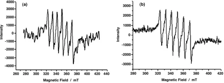
The EPR spectra of manganese complexes can provide valuable information regarding oxidation state, type of ligand and site symmetry. As expected, no signal was found in the X-band EPR spectra of free L1 and L2 in DMF solution
at room temperature, while six intense lines (Figure 1) were observed for compounds 1 and 2. These spectra are
characteristic of monomeric manganese(II) complexes with octahedral geometry and axial symmetry. The Hamiltonian parameters (Table 1) obtained from the analyses of the spectra were Aiso = 91.0 G and giso = 2.0059, and Aiso = 94.1 G and giso = 2.0081 for complexes 1 and 2 respectively.
Electrochemistry
manganese(II) complexes in dmf solution were investigated by cyclic voltammetry. The results are summarized in Table 1.
The electrochemical study of H3bpeten and H3bnbpeten was focused on the irst one-electron oxidation of the phenolic units and the effect of the nitro group on the redox potential. This may allow to predict the easiness of one-electron oxidation of the corresponding manganese(II) complexes because the oxidation potential of the protonated free proligand is similar to that of the corresponding manganese(II) complex (protonation parallels metallation).22
L1 exhibits an irreversible oxidation wave at Epa= 0.980 V
vs. Ag/AgCl (0.462 V vs. Fc+/Fc and 0.862 V vs. NHE) and
L2 a quasi-reversible wave at E1/2= 0.910 V vs. Ag/AgCl
(0.392 V vs. Fc+/Fc and 0.792 vs. NHE). These processes
correspond to the oxidation of the phenolic moieties leading
to unstable radical cations, undoubtedly of the phenoxyl type. Interestingly, L1 showed an anodic peak shifted by
0.070 V to more positive potentials as compared with L2,
in spite of the electron-withdrawing nitro group bound directly to the phenol group, probably due to the irreversible character of that wave. The cyclic voltammogram of L2
also presents a quasi-reversible redox couple at −1.781 V
vs. Ag/AgCl (−2.336 V vs. Fc+/Fc and −1.936 V vs. NHE),
that was tentatively attributed to the reduction of the nitro group present in the phenolic residues of H3bnbpeten.
The cyclic voltammograms of 1 and 2 in dmf at
100 mV s−1 showed a quasi-reversible redox couple,
respectively at 0.268 V and 0.165 V vs. Ag/AgCl, which can
be ascribed to the [MnII(HL)]/[MnIII(HL)]+ redox couple.
Compared with 1, the presence of an electron-withdrawing
group such as NO2 in the coordination environment of 2
Figure 1. X-band EPR spectra of 1 (a) and 2 (b) in DMF at 298 K. The Hamiltonian parameters are: Aiso = 91.0 G and giso = 2.0059 for 1; and Aiso = 94.1 G and giso = 2.0081 for 2.
Table 1. Spectroscopic and electrochemical data for the compounds H3bpeten (L1), H3bnbpeten (L2), [MnII(Hbpeten)] (1) and [MnII(Hbnbpeten)] (2)
Compounds UV-Vis dataa
λ
max/nm (ε / L mol−1 cm−1)
EPR parametersb Cyclic voltammetry datac
Aiso (G) giso Epa
V vs. Ag/AgCl (V vs. NHE)
E1/2
V vs. Ag/AgCl (V vs. NHE) H3bpeten (L1) 234 (5 692)
260 (5 731) 282 (4 360)
silent silent 0.980 (0.862) –
H3bnbpeten (L2) 275 (21 508)
428 (34 016) silent silent – 0.910 (0.792)
-1.781 (-1.936)
[MnII(Hbpeten)] (1) 267 (12 218) 91.0 2.0059 – 0.268 (0.150)
[MnII(Hbnbpeten)] (2) 264 (13 024) 389 (21 803)
94.1 2.0081
–
0.165 (0.040) -1.835 (-1.966)
a In DMF, 1 × 10⁻³ mol L−1 for L1 and 1 and 1 × 10⁻⁴ mol L−1 for L2 and 2; b In DMF solution, 1 × 10⁻³ mol L−1, at 298 K; c. In DMF solution, 1 × 10⁻³ mol L−1, under nitrogen, at 298 K, with 0.1 mol L−1 [TBA][PF
decreases E1/2 by about 0.100 V and is a consequence of the higher basicity of the H3bnbpeten ligand, which leads to a lower resistance to oxidation of complex 2. The cyclic
voltammogram of compound 2, similarly to the observed
for L2, presents a quasi-reversible redox couple at −1.835 V
vs. Ag/AgCl (−2.366 V vs. Fc+/Fc and −1.966 V vs. NHE),
that can be attributed to the reduction of the nitro group present in the phenolic residues of the H3bnbpeten ligand.
EPR spectroelectrochemistry
EPR spectroelectrochemistry was carried out in DMF to give support to the assignment of the redox process observed for free and coordinated L2 at the -1.800 V region.
The spectral changes are shown in Figures 2 and 3. The EPR spectroelectrochemistry studies (simultaneous EPR and electrochemistry measurements using an adapted EPR cell, see Experimental) of the [MnII(Hbnbpeten)]
complex (Figure 2a) showed no spectral change in the 0.0 to −0.5 V range, with the six characteristic manganese(II)
lines remaining unmodiied. However, when a potential of
−2.0 V (Figure 2b) was applied, there was a fast decrease
of the manganese(II) signals while a signal constituted by three sharp lines of similar intensity appeared, until inally only the new signal remained (Figure 2c).
The complete disappearance of the characteristic manganese(II) lines suggests that the metal center may be involved in the electrochemical process. In fact, the applied potential is suficient to promote reduction of the hydrated Mn2+ ions to Mn0 (Mn2+ + 2e → Mn0, E0 = −1.185 V),
leading to the deposition of the metal on the electrode and the disappearance of the EPR signal. The hypothesis of formation of manganese(III) complexes, that are EPR silent, can be ruled out because of the negative potential applied to the system.
The set of three signals can be assigned to a species with I = 1 and should not involve the metal center, as discussed above. In this case, some of the nitrogen atoms of the coordinated H3bnbpeten should be near one unpaired electron. It is improbable that the two saturated nitrogen atoms coordinated to the metal ion are involved, since a signal constituted by ive lines, in agreement with the 2n + 1 equation, is expected in this case. Accordingly, the unpaired electron should be localized on the nitro groups or on the pyridine nitrogen atoms.
The Hamiltonian parameters determined for the three-line signal are A = 13.7 G and g = 2.0048. However, a closer analysis showed that the signals are rather non-symmetric (Figure 3) and are probably constituted by a set of
non-Figure 2. EPR spectroelectrochemistry of the compound [MnII(Hbnbpeten)] (2), in DMF, 10−3 mol L−1 {0.1 mol L−1 [TBA][PF
6]}. (a) enhancement of the six line signal when the potential is stepped from 0.0 to −0.5 V; (b) decrease of the six line signal and appearance of a three line signal at
resolved hyperine lines. Thus, the acquisition parameters of the equipment were adjusted (sweep width from 20 to 5 ms, time constant from 40 to 5 ms, conversion time from 20 to 5 ms and modulation amplitude from 10 to 1 G) in order to try to improve the resolution of the spectra. The result is shown in Figure 3. Note that each line was resolved in three other lines separated by 3.0 G, denoting the hyperine interaction of one unpaired electron with two atoms with I = ½. Finally, a ine tuning of the acquisition parameters (sweep width to 0.64 ms, time constant to 0.64 ms, conversion time to 0.64 ms and modulation amplitude to 0.1 G) allowed a further increase in resolution, now splitting each one of the three lines in two lines separated by 0.7 G, indicating the superhyperine interaction with one atom of I = ½, that could be one hydrogen atom.
The EPR spectroelectrochemistry study described above was extended to H3bpeten (L1) and the [MnII(Hbpeten)]
complex (1). As expected from the cyclic voltammograms,
no spectral change could be observed in the –1.800 V region, suggesting that the above described redox process is associated with the nitro group in the phenolic moiety. In the case of L2,
as the molecule is unsymmetric, the nitro group at the same side of the pyridine group can probably be reduced before the one located at the side of the 2-hydroxyethyl residue.
Theoretical study
The analyses of the frontier orbitals of H3bnbpeten using DFT calculations allowed the determination of the energy of
the frontier orbitals at –0.337 (HOMO–1), −0.314 (HOMO), −0.207 (LUMO) and −0.205 hartree (LUMO+1), as well as
the mapping of the electronic density distribution of these orbitals in the molecule. The HOMO is mainly localized on the ethylenediamine and saturated substituents, while the LUMO is centered on the nitrophenol group in the pyridyl group side and the LUMO+1 is mainly localized on the nitrophenol group in the hydroxyethyl side (Figure 4). Thus, a strong participation of the nitro group and the phenolate ring is suggested, in agreement with the EPR spectroelectrochemistry data. This result reinforces the hypothesis that the unpaired electron in the reduced free H3bnbpeten and the respective manganese(II) complex is localized on the nitro group of the pyridyl side. Similar results were obtained using the HF method and the 6-31G (d,p) basis set, reinforcing the validity of our theoretical calculations.
The validity of the method was also conirmed by carrying out the theoretical calculation for the symmetric H2bbpen-NO2 ([N,N ’-bis-(2-hydroxy-5-nitro-benzyl)-N,N’-bis-(pyridine-2-methyl) ethylenediamine]) molecule
using the DFT method and the same basis set. As expected, the LUMO orbital had a similar contribution from both nitrophenol groups (Figure 5b), in contrast with the H3bnbpeten ligand, where the contribution was asymmetric, in agreement with EPR spectroelectrochemistry results. On the other hand, the HOMO orbital (Figure 5a) in both the symmetric and asymmetric molecules showed a similar electronic density distribution over the molecule, with no signiicant contribution from the nitrophenol groups.
Figure 3. EPR spectroelectrochemistry of H3bnbpeten in DMF, 1×10⁻³ mol L−1 {0.1 mol L−1 [TBA][PF
Figure 4. Graphic representation of the HOMO (a), LUMO (b) and LUMO+1 (c) calculated for H3bnbpeten using DFT.
Figure 5. Graphical representation of the HOMO (a) and LUMO (b) calculated for H2bbpen-NO2 using DFT.
Figure 6. Proposed structures of complexes 1 and 2.
Conclusions
Two Mn2+ complexes containing two polyfunctional
ligands, one symmetric bearing two phenol moieties (L1)
and one asymmetric, with electron-withdrawing –NO2 groups bound to the phenol rings (L2), were prepared and
characterized electrochemically and spectroscopically. The infrared, UV-Vis and EPR spectroscopy results, as well as the conductivimetry, elemental analysis and electrochemistry results were in agreement with the formation of manganese(II) complexes, whose proposed structures are presented in Figure 6.
The spectroelectrochemical studies of the free ligands and their respective manganese(II) complexes evidenced that the nitro group shifted the redox potentials by up to about 0.100 V, and was responsible for the appearance of a new quasi-reversible wave at −1.800 V, attributed to a
monoelectronic process involving the nitrophenol moiety. The localization of the electron on the nitro and nitrophenol groups in complex 2 and in L2,respectively, was conirmed
by EPR spectroelectrochemistry.
Thus, a potential of −2.0 V was applied to both
compounds at different intervals of time. After 30 s, a three-line signal that can be assigned to a free radical localized on the nitro group, appeared in the middle of the typical manganese(II) signal. In the same reducing conditions, the free L2 presented an eighteen-line
spectrum, due to the electronic spin interaction with the nuclear spins of the nitrogen atom of the nitro group, of two orto-hydrogen atoms and of one hydrogen atom
bound to the meta-carbon atom relative to the nitro
group. Accordingly, the electrochemistry and EPR espectroelectrochemistry data are consistent with the reduction of the nitro group bound to one of the phenol moieties of the free H3bnbpeten and the respective manganese(II) complex. This result was conirmed by DFT calculations that showed a major contribution of the nitrophenol group to the LUMO, indicating that the method can be used to predict some of the electronic properties of these compounds.
Supplementary Information
Acknowledgments
This work was supported by grants from CNPq, Fundação Araucária, UFPR/TN, PADCT, FAPERJ, Fundação Universitária José Bonifácio and Fundação José Pelúcio Ferreira.
References
1. Niu, S.; Hall, M. B.; Chem. Rev.2000, 100, 353.
2. Srinivasan, K.; Michaud, P.; Kochi, J. K.; J. Am. Chem. Soc.
1986, 108, 2309.
3. Samsel, E. G.; Srinivasan, K.; Kochi, J. K.; J. Am. Chem. Soc.
1985, 107, 7606.
4. Yoon, H.; Burrows, C. J.; J. Am. Chem. Soc. 1988, 110, 4087. 5. Manchanda, R.; Brudvig, G. W.; Crabtree, R. H.; Coord. Chem.
Rev.1995, 144, 1.
6. Su, J.-H.; Havelius, K. G. V.; Ho, F. M.; Han, G.; Mamedov, F.; Styring, S.; Biochemistry 2007,46, 10703.
7. Wieghardt, K.; Angew. Chem., Int. Ed. 1989, 28, 1153.
8. Dismukes, G. C.; Chem. Rev. 1996, 96, 2909.
9. Wu, A. J.; Penner-Hahn, J. E.; Pecoraro, V. L.; Chem. Rev. 2004,
104, 903.
10. Lanznaster, M.; Neves, A.; Bortoluzzi, A. J.; Assumpção, A. M. C.; Vencato, I.; Machado, S. P.; Drechsel, S. M.; Inorg. Chem.
2006, 45, 1005.
11. Scarpellini, M.; Casellato, A.; Bortoluzzi, A. J.; Vencato, I.;
Mangrich, A. S.; Neves, A.; Machado, S. P.; J. Braz. Chem. Soc. 2006, 17, 1617.
12. Paes, L. W.; Faria, R. B.; Machuca-Herrera, J. O.; Neves, A.; Machado, S. P.; Can. J. Chem. 2004,82, 1619.
13. Zhan, C. G.; Nichols, J. A.; Dixon, D. A.; J. Phys. Chem. A.
2003, 107, 4184.
14. Privalov, T.; Sun, L.; Åkermark, B.; Liu, J.; Gao, Y.;Wang, M.;
Inorg. Chem. 2007, 46, 7075.
15. Romanowski, S. M. M.; Tormena, F.; Santos, V. A.; Hermann, M. F.; Mangrich, A. S.; J. Braz. Chem. Soc.2004, 15, 897.
16. Neves, A.; Romanowski, S. M. de M.; Bortoluzzi, A. J.; Mangrich, A. S.; Inorg. Chim. Acta 2001, 313, 137.
17. Romanowski, S. M. de M.; Mangrich, A.S.; Neves, A.; Quim. Nova 2001, 24,592.
18. Friedermann, G. R.; Halma, M.; Castro, K. A. D. F.; Benedito, F. L.; Doro, F. G.; Drechsel, S. M.; Mangrich, A. S.; Assis, M. D.; Nakagaki, S.; Appl. Catal., A2006, 308, 172.
19. Gagné, R. R.; Koval, C. A.; Lisensky, G.C.; Inorg. Chem.1980,
19, 2854.
20. Poll, N.; J. Chem. Educ. 1974, 67, 970.
21. Geary, W. J.; Coord. Chem. Rev. 1971,7, 81.
22. Ménage, S.; Gellon, G.; Pierre, J-L.; Zurita, D.; Saint-Aman, E. ; Bull. Soc. Chim. Fr. 1997, 134, 785.
Received: February 25, 2009
Supplementary Information
*e-mail: [email protected]
Synthesis, Characterization, EPR Spectroelectrochemistry Studies and
Theoretical Calculations of Manganese(II) Complexes with the
Ligands H
3bpeten and H
3bnbpeten
Stela Maris de M. Romanowski,a Sérgio P. Machado,b Geraldo R. Friedermann,a Antonio
S. Mangrich,a Monique de F. Hermann,a Hugo Oroino Lima b and Shirley Nakagakia*
a Departamento de Química, Universidade Federal do Paraná, 81531-990 Curitiba-PR, Brazil
b Instituto de Química, Universidade Federal do Rio de Janeiro, 21941-909 Rio de Janeiro-RJ, Brazil
Figure S1. Molecular orbital energy levels for the frontier orbitals of the ligand H3bnbpeten.
Figure S2. Graphical representation of the HOMO (a) and LUMO (b) of the H3bnbpeten using Hartree-Fock.
![Figure 2. EPR spectroelectrochemistry of the compound [Mn II (Hbnbpeten)] (2), in DMF, 10 −3 mol L −1 {0.1 mol L −1 [TBA][PF 6 ]}](https://thumb-eu.123doks.com/thumbv2/123dok_br/18994412.461739/6.892.147.805.602.1050/figure-epr-spectroelectrochemistry-compound-mn-hbnbpeten-dmf-tba.webp)
![Figure 3. EPR spectroelectrochemistry of H 3 bnbpeten in DMF, 1×10 −3 mol L −1 {0.1 mol L −1 [TBA][PF 6 ]}](https://thumb-eu.123doks.com/thumbv2/123dok_br/18994412.461739/7.892.88.779.699.1074/figure-epr-spectroelectrochemistry-bnbpeten-dmf-mol-mol-tba.webp)


