UNIVERSIDADE DE LISBOA FACULDADE DE CIÊNCIAS
DEPARTAMENTO DE BIOLOGIA ANIMAL
Anticancer Potential of Azurin Interaction
with Lipid Rafts:
Deciphering the Role of Caveolae
Public Version
DISSERTAÇÃO
Mestrado em Biologia Humana e Ambiente
Ana Rita Cebola Garizo
Dissertação orientada por: Doutor Nuno Filipe Santos Bernardes Professora Deodália Maria Antunes Dias
II "One, remember to look up at the stars and not down at your feet. Two, never give up work. Work gives you meaning and purpose and life is empty without it. Three, if you are lucky enough to find love, remember it is there and do not throw it away." - Stephen Hawking -
III
ACKNOWLEDGEMENTS
During the realization of this MSc thesis I counted with important support and incentives, without them I would not be able to go through this step and to which I will always be eternally grateful.
First of all, I would like to thank my external supervisor Doctor Nuno Bernardes for all that he has taught me in the lab, for all the knowledge he has transmitted, for the patience, for the opinions and critics, for all the incentive words and being available… Thank you so much!
I would like to thank Professor Arsénio Fialho for the support and for the guidance throughout the development of this project. It was a huge pleasure to be able to help a bit more on this investigation.
A special thanks to both for believing in me and had given me this opportunity of working on Instituto Superior Técnico.
In addition, thanks to all Biological Sciences Research Group for the help in many occasions, especially to Dalila Mil-Homens, Joana Feliciano and Mónica Rato, and yet to my lab bench mates Bernardo Caniço, Marília Silva, Rui Martins, Soraia Guerreiro, Tiago Pita and Marcelo Ramires.
I would like to thank Sandra Pinto and Fábio Monteiro, who gave me support with the confocal microscopy and spectrofluorimeter, and shared ideas and advices about the work.
I also thank the financial support of the Institute for Biotechnology and Bioengineering (IBB).
I would also like to thank my internal supervisor, Professor Deodália Dias for all the determination to give me the best thesis opportunities, for being always available and for the affection shown.
Lastly, but not least, I would like to thank my family, specially my parents and my cousin Ana Lúcia, to my boyfriend and his parents, and to my best friends for always been there for me and never let me give up. To my grandmother, who will always be in my thought and that has given me her unconditional support.
IV
SUMÁRIO
O cancro é uma das principais doenças no mundo que pode levar à morte. Por ano, milhões de pessoas são diagnosticadas e mais de metade morre com esta doença.
Ao longo de várias décadas, diversos tratamentos alternativos à quimioterapia e radioterapia têm sido desenvolvidos no sentido de ultrapassar os problemas que estes tratamentos convencionais causam, nomeadamente os seus efeitos adversos associados com a sua elevada toxicidade sistémica.
Uma das possíveis alternativas que o campo da investigação tem vindo a seguir direcciona-se para factores, alguns solúveis, segregados por bactérias como enzimas, metabolitos secundários, toxinas, proteínas e péptidos derivados, que actuam especificamente nas células cancerígenas, sendo potenciais agentes anticancerígenos (Yamada et al., 2002a; Bernardes et al., 2010).
Um exemplo destes factores é uma pequena proteína solúvel em água, secretada por Pseudomonas aeruginosa, designada azurina com 128 aminoácidos e um peso molecular de 14 kDa (Yamada et al., 2005; Bernardes et al., 2013).
Existem muitos factores que suportam o potencial existente para a azurina poder actuar como um agente anticancerígeno. Um deles é que esta entra preferencialmente em células cancerígenas (Yamada et al., 2005). Para além disto, após a sua administração, não foram observados efeitos secundários em estudos in vivo (Choi et al., 2011; Warso et
al., 2013). Esta proteína bacteriana pode mediar interacções específicas de elevada
afinidade com várias proteínas das células humanas, conferindo-lhe a propriedade de proteína “molde”, que é provavelmente uma das suas características mais importantes (Fialho et al., 2007). Esta capacidade para actuar em múltiplos alvos é importante devido ao facto de poder ser mais difícil as células cancerígenas adquirirem resistência a este tratamento. Outra vantagem é que a azurina é uma molécula solúvel em água com um domínio hidrofóbico, que pode contribuir para a sua entrada nos tecidos e na eliminação para a corrente sanguínea (Kamp et al., 1990). Por último, esta proteína pode ser facilmente superexpressa em Escherichia coli, o que torna os processos de produção e purificação muito mais baratos (Bernardes et al., 2013).
O mecanismo de entrada da azurina nas células não é, no entanto, totalmente compreendido. Estudos sugerem que esta penetra a membrana plasmática por uma via endocítica mediada por caveolae, que são jangadas lípidicas não planares (Taylor et al., 2009). As jangadas lipídicas têm concentrações elevadas de ácidos gordos saturados, de esfingolípidos (incluindo esfingomielina, ceramida e gangliósidos como o GM1), e de
V colesterol (Quest et al., 2008; Martinez-Outschoorn et al., 2015). Estes microdomínios membranares são pequenos, dinâmicos, heterogéneos e conseguem recrutar certas classes de proteínas. Estão ainda implicados em vários processos celulares fisiológicos, tais como tráfico de proteínas pela membrana, transdução de sinal, transporte de colesterol, organização do citoesqueleto, motilidade, polaridade e endocitose (Simons and Toomre, 2000; Martinez-Outschoorn et al., 2015). Para além disto, sabe-se ainda que em melanomas e cancros da próstata e mama, as jangadas lipídicas estão em maior número, sugerindo que estas estruturas desempenham um papel funcional durante a tumorigénese (Irwin et al., 2011; Murai, 2015). Com isto, o estudo destas estruturas é importante para a prevenção e tratamento do cancro, uma vez que estas estão envolvidas na progressão da doença (Murai, 2015).
Existem dois tipos de jangadas lipídicas: jangadas lipídicas planares, que não têm características morfológicas específicas, e jangadas lipídicas não planares que, como foi referido anteriormente, são as caveolae. No primeiro caso, a proteína constituinte destes microdomínios é a flotilina (Martinez-Outschoorn et al., 2015), enquanto que no segundo caso, são as caveolinas e as cavinas (Parton et al., 2006; Parton and del Pozo, 2013).
Actualmente sabe-se que as caveolae tem um papel importante no cancro. Nestes microdomínios, a activação de cascatas de sinalização pode alterar a morfologia e o comportamento das células (Martinez-Outschoorn et al., 2015). A “hipótese de sinalização de caveolae” implica uma das suas proteínas constituintes obrigatórias, a caveolina-1, na integração de várias vias moleculares (Patani et al., 2012). A capacidade desta proteína em modular a sinalização intra-celular tem implicações importantes em vários estados patológicos e biológicos humanos, incluindo a tumorigénese. Na verdade, durante os últimos 20 anos, vários estudos foram feitos investigando o papel da caveolina-1 na iniciação e progressão do cancro, mostrando que esta proteína multifuncional regula diversos processos associados a esta doença, tais como transformação de células, crescimento de tumores, migração celular, invasão, resistência a múltiplas drogas e angiogénese (Senetta et al., 2013).
A compreensão do papel da caveolina-1 no desenvolvimento e progressão do cancro pode ser significativa para melhorar o prognóstico do paciente e prevenir o aparecimento desta doença.
Para além das vantagens da aplicação da azurina no tratamento do cancro descritas anteriormente, a utilização desta ou de péptidos seus derivados em combinação com fármacos quimioterapêuticos potencia o efeito anticancerígeno destes. Com isto, problemas como a aquisição de resistência ou toxicidade produzidos pela administração
VI sucessiva destes químicos podem ser ultrapassados, uma vez que passam a ser administrados em doses baixas (Bernardes et al., 2016; Yamada et al., 2016).
Neste projecto de investigação, foram utilizadas três linhas celulares cancerígenas humanas: MCF-7 que corresponde a uma linha celular cancerígena de mama, HT-29 que é uma linha cancerígena de cólon e A549 que são células cancerígenas de pulmão. O objectivo principal deste trabalho é esclarecer o potencial anticancerígeno resultante da interacção entre a azurina com as jangadas lipídicas, decifrando o papel de caveolae.
Neste estudo, demonstrámos que a azurina leva a um padrão de internalização das jangadas lipídicas, que consequentemente poderá remover receptores da superfície celular, que estão envolvidos na tumorigénese. Também verificámos que um dos primeiros passos de reconhecimento das células cancerígenas pela azurina dá-se ao nível do gangliósido GM1, que está localizado nas jangadas lipídicas. De seguida, observámos também que o silenciamento da expressão da caveolina-1 leva a uma diminuição, pelo menos em parte, da entrada da azurina nas células. Para além disso, foi possível verificar por técnicas de espectroscopia “in vitro”, que existe uma interacção física directa entra a azurina e um domínio funcional da caveolina-1, que é bastante importante na interacção com outras proteínas. O mesmo não se verifica para uma proteína mutante da azurina, onde foi operada a substituição de um aminoácido da sua estrutura nativa, identificando assim uma possível localização preferencial dentro da estrutura da azurina responsável pela interação desta com vários componentes dos microdomínios membranares.
Por último, administrámos azurina em conjunto com fármacos quimioterapêuticos (paclitaxel e doxorrubicina) nas células cancerígenas, e observámos que no geral, a acção terapêutica destes é beneficiada, levando a maiores níveis de morte celular.
Todos estes resultados elucidam sobre os mecanismos de entrada da azurina nas células cancerígenas, e mostram que esta proteína pode melhorar os efeitos de fármacos quimioterapêuticos que se encontram em uso clínico, e para os quais os doentes com cancro desenvolvem frequentemente resistência, dificultando a sua resposta terapêutica.
Palavras-Chave: Azurina, Potencial Anticancerígeno, Jangadas Lipídicas, Caveolae, Fármacos
VII
ABSTRACT
Azurin, a protein produced by Pseudomonas aeruginosa, acts as an anticancer agent. Studies suggest that this bacterial protein enters in cancer cells through the penetration of the plasma membrane via caveolae-mediated endocytic pathways.
Caveolae are non-planar lipid rafts characterized by an abundance of caveolin and cavin
proteins and it is known that the levels of lipid rafts are increased in melanomas, prostate, and breast cancers suggesting that these structures play a functional role during tumorigenesis.
In this project, three human cancer cell models have been used: the MCF-7 breast cancer cell line, the HT-29 colon cancer cell line and the A549 lung cancer cell line with the main objective to clarify the anticancer potential of azurin interaction with lipid rafts, deciphering the role of caveolae.
In this work, we demonstrate that azurin leads to a pattern of internalization of lipid rafts, through the staining of GM-1, a constituent of lipid rafts, with the Alexa488-labeled CtxB marker. We also show evidences that azurin recognizes cancer cells through the GM1 ganglioside which is located in lipid rafts, since its blockage with CtxB prevents the normal entry process of azurin in cancer cells. Then, we observed that silencing of Cav1 expression leads to a decrease at least in part, on the entry azurin in cells. In addition, it was verified by spectroscopic "in vitro" techniques, that there is a direct physical interaction between azurin and a functional domain of Cav1, which is very important in interacting with other proteins. The same is not true for an azurin mutant protein, which was operated at an amino acid substitution of its native structure, identifying a possible region within the sequence of azurin that may be of major importance for this mechanism.
Finally, we combined the azurin with chemotherapeutic drugs, such as paclitaxel and doxorubicin, and observed that in general, the therapeutic action of these is benefited, leading to higher levels of cell death than when the drugs are added alone.
In general, all these results elucidate on the azurin entry mechanisms in cancer cells, and show that azurin may be relevant as an adjuvant to improve the effects of other anticancer agents already in clinical use, to which patients often develop resistance hampering its full therapeutic response.
VIII
TABLE OF CONTENTS
ACKNOWLEDGEMENTS ... III SUMÁRIO ... IV ABSTRACT ... VII INDEX OF FIGURES ... X INDEX OF TABLES ... XIII LIST OF ABBREVIATIONS ... XIV1. INTRODUCTION ... 1
1.1. Bacterial protein azurin ... 2
1.1.1. Entry mechanism of azurin on human cells and subsequent effects ... 3
1.1.2. Azurin and cell surface receptors in cancer cells ... 4
1.2. Cholesterol effects in tumor progression ... 6
1.3. Lipid Rafts ... 7
1.3.1. Caveolae and Caveolins ... 8
1.3.2. Caveolin-1 Scaffolding Domain (CSD) ... 12
1.4. Azurin application in the treatment of cancer ... 14
1.4.1. Effects of azurin treatment in combination with drugs on human cancer cells ... 16
2. OBJECTIVES AND THESIS OUTLINE ... 18
3. MATERIALS AND METHODS ... 20
3.1. Human cancer cell lines and cell cultures ... 20
3.2. Bacteria growth, over-expression, extraction and purification of WT azurin or mutated protein ... 20
3.3. Pre-treatment with Cholera Toxin Subunit B (CTxB) ... 21
3.3.1. Protein extraction and Western blot ... 22
3.4. Confocal microscopy-Cholera Toxin Subunit B (CTxB) ... 23
3.5. Transfection of human cancer cells lines ... 24
IX
3.7. MTT cell viability assay ... 25
4. RESULTS / DISCUSSION / CONCLUSION AND FUTURE PERSPECTIVES ... 27
5. REFERENCES ... 28
CONFIDENTIAL APPENDIX ... 37
4. RESULTS ... 37
4.1. Blocking GM1 ganglioside reduces the penetration of azurin in cancer cells 37 4.2. WT azurin leads to an internalization of lipid rafts in the cancer cells ... 38
4.3. Silencing of CAV1 reduces the entry of WT azurin but not of mutated azurin ... 39
4.4. Azurin binds to the Caveolin-1 Scaffolding Domain (CSD) ... 41
4.5. The flotillin levels are not affected by the treatment of azurin in A549 and MCF-7 human cancer cell lines, but the same is not true in the HT-29 cell line ... 43
4.6. The WT azurin treatment in combination with drugs potentiates the anticancer effect of these agents in cancer cells ... 44
5. DISCUSSION ... 48
6. CONCLUSION AND FUTURE PERSPECTIVES ... 52
X
INDEX OF FIGURES
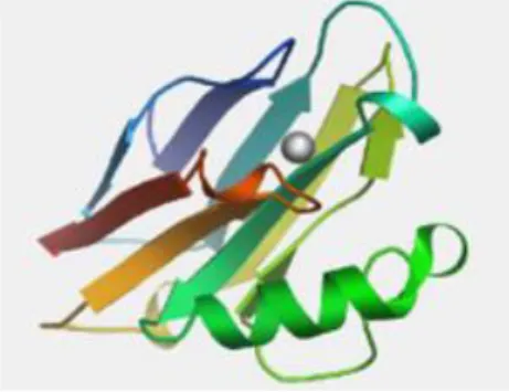
Figure 1: Structure of azurin (Adapted from Karpiński and Szkaradkiewicz, 2013). ... 2 Figure 2: Azurin can also bind avidly to the surface-exposed receptor tyrosine kinase EphB2, interfering in its binding with the ligand ephrinB2, and thereby preventing cell signaling that promotes cancer cell growth (Adapted from Bernardes et al., 2010). ... 5 Figure 3: Two types of lipid rafts: Planar lipid rafts membrane (a) contain high concentrations of flotillin proteins, which bind to cholesterol and sphingolipids. These microdomains are in the same plane as the non-raft membrane, hence the term planar lipid rafts. Invaginated lipid rafts (b; caveolae) are not in the same plane as the rest of the plasma membrane, hence the term non-planar lipid rafts. Caveolae require caveolin and cavin proteins for their formation. CAV1 is caveolin-1 (Adapted from Martinez-Outschoorn
et al., 2015). ... 8
Figure 4: Caveolae: Electron micrographs showing the ultrastructure of caveolae in fibroblasts (main panel and at high magnification upper left), and the complex arrangements of caveolae in cultured adipocytes (upper middle) and in skeletal muscle (top right). Scale bars represent 100 nm (Adapted from Parton and del Pozo, 2013). ... 9 Figure 5: Structure and general activities of caveolae/caveolin-1. Caveolae are flask-shaped invaginations in the cell membrane coated with multimers of caveolin scaffolding proteins. The N-termini and C-termini of caveolin proteins are in the cell cytoplasm, but a hairpin loop of the protein is inserted into the cell membrane. Various caveolae/caveolin-1 activities that have been reported in different cell types are depicted. BMP, bone morphogenetic protein; MLC, myosin light chain; PI-kinase, phosphatidylinositol 3-kinase; TGFβ, transforming growth factor beta (Baker and Tuan, 2013). ... 11 Figure 6: The topology of caveolin-1 (Cav1), depicted as a homo-dimer, permits anchorage to the plasma membrane through a central hydrophobic domain, flanked by hydrophilic N- and C-terminal cytosolic domains (Adapted from Patani et al., 2012). ... 12 Figure 7: Amino acid sequence of caveolin-1 (Cav1). The topology of this protein can be divided into domains: an oligomerization domain (residues 61-101; black and blue) with a caveolin scaffolding domain (CSD; residues 82-101, blue), a trans-membrane domain (residues 102-134; red) and bold residues indicate the C-terminal membrane attachment domain (CMAD; residues 135-178) and the N-terminal membrane attachment domain (NMAD; residues 1-60). The CSD also contains cholesterol recognition/interaction amino acid consensus (CRAC; Adapted from Hoop et al., 2012). ... 14
XI Figure 8: GM1 ganglioside blocking effect with CTxB in the penetration of azurin in cancer cells. MCF-7, A549 and HT-29 human cancer cell lines were grown overnight in 6-well plates with 5x10⁵ MCF-7 and HT-29 cells/well, and 2x10⁵ A549 cells/well. Next, these cells
were exposed to 1μg/mL of CTxB for 10 minutes. After this time, cells were treated with 50μM of WT azurin or mutated protein during 30 minutes. The controls were the cells exposed to bacterial proteins without CTxB. A decrease in WT azurin entry of 14%, 50% and 41% and a decrease in mutated azurin entry of 17%, 84% and 50% are observed in the MCF-7, A549 and HT-29 cells, respectively. The protein levels were normalized by the respective GAPDH level. ... 38 Figure 9: Effects of WT azurin and mutated azurin in the cell’s lipid raft organization. MCF-7 (left panel), A549 (central panel) e HT-29 (right panel) human cancer cell lines were grown overnight on a round glass coverslip in 24-well plates with a density of 5x10⁴
cells/well for the MCF-7 and HT-29 cell lines and 2x10⁴ cells/well for the A549 cell line.
After, these cells were exposed to 100μM of WT azurin or mutated protein for 24 hours. Untreated cells were the controls. The GM1 ganglioside of lipid rafts is marked with CTxB (green) and the nucleus of the cells is stained with DAPI (blue). ... 39 Figure 10: Effect of silencing of the Caveolin-1 siRNA in the entry of WT azurin and mutated azurin. MCF-7 and A549 human cancer cell lines were grown overnight in 6-well plates with a density of 5x10⁵ cells/well. In the next day, cells were transfected with 100nM
of Control siRNA and Caveolin-1 siRNA for 6-8 hours. After 24 hours, cells were treated with 50μM of WT azurin or mutated protein during 30 minutes. Control siRNAs with protein are the control condition. A decrease in WT azurin entry of 49% and 18% and a decrease in mutated azurin entry of 12% and 5% are observed in the MCF-7 and A549 cells, respectively. The protein levels were normalized by the respective actin level. ... 41 Figure 11: Interaction between the FITC-labeled Caveolin-1 Scaffolding Domain (CSD) and WT azurin or mutated azurin. Normalized Fluorescence Intensity for each protein concentration (0, 0.25, 0.5, 1, 5, 10 and 50µM). ... 42 Figure 12: Effect of WT azurin and mutated azurin in the flotillin levels. All human cancer cell lines used in this project were grown overnight in 6-well plates with a density of 5x10⁵
cells/well. Next, these cells were treated with 100μM of WT azurin or mutated protein during 48 hours. Untreated cells were the control condition. Any decrease in flotillin levels and a very small reduction of 11% when the cells were exposed to WT azurin and 5% when the cells were treated with mutated protein are observed in the MCF-7 A and A549 cells, respectively. In the case of HT-29 cells, a decrease in WT azurin entry of 30% and a
XII decrease in mutated azurin entry of 43% are observed. The protein levels were normalized by the respective GAPDH level. ... 44 Figure 13: Effects of WT azurin treatment in combination with drugs (paclitaxel black lines and doxorubicin blue lines) in MCF-7, A549 and HT-29 human cancer cell lines. All these cell lines were seeded overnight in 96-well plates (3 replicates) with a density of 2x10⁴,
5x10³ and 1x10⁴ cells/well, respectively. After this, these cells were treated with 25, 50 and
100μM of WT azurin together with 0.1, 1 and 10nM of paclitaxel or 0.1, 0.5 and 1µM of doxorubicin during 72 hours. Untreated cells were used as control. The cells only treated with protein (green lines) and only treated with drug serve to verify if the cell death caused by the combination of both was greater than the cell death caused by these components applied individually. ... 46 Figure S1: Effects of doxorubicin (blue line) and paclitaxel (black line) treatment MCF-7, A549 and HT-29 human cancer cell lines. All these cell lines were seeded overnight in 96-well plates (3 replicates) with a density of 2x10⁴, 5x10³ and 1x10⁴ cells/well, respectively.
After this, these cells were treated with 0.001, 0.01, 0.05, 0.1 and 1µM of doxorubicin or 0.0001, 0.001, 0.01, 0.1, 1 and 10nM of paclitaxel during 72 hours. In the case of HT-29, these cells were treated with 0.1, 1, 10, 100, 1.000, 10.000nM of paclitaxel. Untreated cells were used as control. ... 55
XIII
INDEX OF TABLES
Table 1: SDS-PAGE components. ... 22
Table 2: WT azurin Fluorescence Intensity. ... 42
Table 3: Mutated azurin Fluorescence Intensity. ... 42
Table S1: Polynomial adjust model relatively WT azurin Fluorescence Intensity. ... 54
XIV
LIST OF ABBREVIATIONS
CAV1 Caveolin-1 gene
CAV2 Caveolin-2 gene
CAV3 Caveolin-3 gene Cav1 Caveolin-1 Cav2 Caveolin-2 Cav3 Caveolin-3
CBM Caveolin-Binding Motif CDK2 Cyclin-Dependent Kinase 2 CLB Catenin Lysis Buffer
CMAD C-terminal Membrane Attachment Domain
CRAC Cholesterol Recognition/interaction Amino acid Consensus motif CSD Caveolin-1 Scaffolding Domain
CTxB Cholera Toxin B subunit
DMEM Dulbecco’s Modified Eagle Medium ECM Extracellular Matrix
EGFR Epidermal Growth Factor Receptor eNOS endothelial Nitric Oxide Synthase FBS Fetal Bovine Serum
FITC Fluorescein-5-IsoThioCyanate FOXM1 Forkhead box M1
HMG-CoA 3-Hydroxy-3-MethylGlutaryl-Coenzyme A reductase IPTG IsoPropyl-β-D-ThioGalactopyranoside
Kd Dissociation constant
LB medium Luria Broth medium LDL Low-Density Lipoprotein LXR Liver X Receptor
MAPK Mitogen-Activated Protein Kinase MTD Maximum Tolerated Dose
NMAD N-terminal Membrane Attachment Domain NOAEL No Observed Adverse Effect Level
PBS Phosphate Buffered Saline PED Protein Entry Domain SB medium Super Broth medium
XV SDS-PAGE Sodium Dodecyl Sulphate-PolyAcrylamide Gel Electrophoresis
TCR T Cell Receptor
TNF-α Tumor Necrosis Factor-α WT azurin Wild-Type azurin
1
1. INTRODUCTION
Cancer is a major disease in the world that can cause death. Each year, millions of people are diagnosed worldwide with cancer, and more than half of these patients die from this disease. Based on World Health Organization projections, in 2030, the number of people expected to die of cancer will be around 11.4 million. In 2012, the most diagnosed
types of cancer were lung (1.82 million), breast (1.67 million) and colorectal (1.36 million; Ferlay et al., 2014).
This disease is characterized by uncontrolled cell growth (benign tumors) and acquisition of metastatic properties (malignant cancers). Frequently, this occurs due to the activation of oncogenes and/or deactivation of tumor suppressor genes leading to uncontrolled cell cycle progression and inactivation of apoptotic events. Mechanisms such as mutations, chromosomal translocations or deletions, and dysregulated expression or activity of signaling pathways are involved in these genetic and cellular changes. Recent studies also suggest that epigenetic alterations can cause cancer due to its role in the generation of cancer progenitor cells and subsequent initiation of carcinogenesis (Sarkar
et al., 2013).
This rising problem is mostly due to a rapidly aging population, and demands a coordinated response from oncologists, public health professionals, policy-makers and researchers. Conventional cancer treatments, such as chemotherapy and radiotherapy, often fail to achieve a complete cancer remission and they are likely to cause side effects. This has been stimulating the development of many new approaches for the treatment of cancer, such as the use of live or attenuated bacteria (Bernardes et al., 2010).
The regression of cancer in humans and animals exposed to microbial pathogens agents has been verified more than 100 years ago (Yamada et al., 2002a). In 1909, William Coley used bacterial culture supernatants of Streptococcus pyogenes and Serratia
marcescens to treat patients with malignant cancer. This preparation was administrated in
approximately 1.200 patients leading to tumor regression in some cases, of which 30 healed completely. Nowadays, it is assumed that the central factor responsible for this therapeutic effect was increased Tumor Necrosis Factor-α (TNF-α) secretion in the body of the patient (Karpiński and Szkaradkiewicz, 2013).
Several reports have shown that microrganisms can replicate on the tumor locations, under hypoxic conditions (low concentration of oxygen), and also that microorganisms can stimulate the host’s immune system during the infections, blocking cancer progression (Yamada et al., 2002a).
2 Another example of a microbial pathogen strain that causes such effects is the
Mycobacterium bovis, which already in 1976 was widely used in the treatment of
superficial bladder cancer (Elkabani et al., 2000). Besides this, bacterial pathogens agents such as Listeria monocytogenes were tested as vaccine vectors for cancer prevention, since they induced the exposition of antigens on the cellular surface, leading to an immune response against cancer cells (Paglia et al., 1997). With this, it was believed that the infection with bacterial pathogen agents cause the activation of macrophages and lymphocytes, resulting in the production of cytotoxic agents with anticancer properties (Yamada et al., 2002a). However, the introduction of live bacteria on the human organism to treat cancer can produce significant side reactions, which may cause serious and eventually fatal infections that are presumed to be the resulted from the liberation of toxic bacterial products and limiting, that way, their use (Paglia et al., 1997; Dang et al., 2001).
1.1. Bacterial protein azurin
Currently, the investigation has been directed to segregated soluble factors by bacteria such as enzymes, secondary metabolites, proteins, or derived peptides and toxins, which may act specifically on cancer cells, being potential anticancer agents (Yamada et al., 2002a; Bernardes et al., 2010). An example of this factors is a small water-soluble protein secreted by Pseudomonas aeruginosa, called azurin (14 kDa; 128 amino acids), which is composed by one α-helix and eight β-sheets, forming a β-barrel motif and contains a hydrophobic patch (Figure 1). This protein is part of a group of type I redox proteins, which have an ion copper in its constitution, named cupredoxins (Kamp et al., 1990; Rienzo et al., 2000; Yamada et al., 2005; Fialho et al., 2012; Bernardes et al., 2013; Karpiński and Szkaradkiewicz, 2013). It is known that azurin is involved in the transport of electrons during the denitrification of these organisms (Yamada et al., 2009).
3 Azurin has structural similarity with variable domains of immunoglobulins and the ability to mediate specific high-affinity interactions with various unrelated mammalian proteins relevant in cancers, gives it the property of a natural scaffold protein (Fialho et al., 2007).
1.1.1. Entry mechanism of azurin on human cells and subsequent effects
The mechanism of entry for azurin is still not fully understood. The first hints suggested that azurin enters in mammal cells through the penetration of the plasma membrane via caveolae-mediated endocytic pathways and reach late endosomes, lysosomes, and the Golgi associated with caveolae (Taylor et al., 2009).
Currently, it is known that a peptide derived from azurin called p28 (50-77 amino acids) or Protein Entry Domain (PED) is per se, at least in part, responsible for mediating the entrance of the entire protein into cells. This peptide has an overall net negative charge, and forms an extended amphipathic α-helix with both hydrophobic amino acids (50-66) and hydrophilic amino acids (67-77). PED was further refined, by reducing the N-terminal to amino-acids 50-67 (p18) and it was found that this minimal fragment can be translocated to the inside of human cancer cells (Yamada et al., 2005; Taylor et al., 2009).
After the entrance of azurin to cancer cells, its derived peptide p28 is processed to the nucleus, it connects to a hydrophobic region inside of DNA-binding domain of tumor-suppressor protein p53 (21 kDa; 393 amino acids), forming a complex, and with this it inhibits the proteasomal degradation of p53 (Yamada et al., 2009). This protein is involved in innumerous cellular processes, including transcription, DNA repair, genomic stability and cell cycle control, being able to induce cellular death by apoptosis. In human cancers, p53 can suffer from inactivation by oncogenes and/or mutations (Martin et al., 2002; Apiyo and Wittung-Stafshede, 2005).
Experiments with isothermal calorimetry demonstrated that azurin binds to the NH2
-terminal domain of p53 with nanomolar affinity in a 4:1 stoichiometry, as well to the DNA-binding domain of this protein (Apiyo and Wittung-Stafshede, 2005).
A few studies, supported by site-directed mutagenesis, suggest that a specific region of azurin has been implicated in this complex formation. This region consists in amino acids Met-44, Met-64 located in a hydrophobic patch, which have been shown to be important for the interactions with p53, and their substitutions resulted in altered complex formation (Yamada et al., 2002b). Thus, with the inhibition of proteasomal degradation of
4 p53 occurs a raise of the cytoplasmic and nuclear levels of this protein, and consequently, increased DNA binding activity. The cyclin-dependent kinase inhibitors p21 and p27 levels also increase, which in turn reduces the intracellular levels of Cyclin-Dependent Kinase 2 (CDK2) and Cyclin A1, essential proteins in the mitotic process, as well as Forkhead box M1 (FOXM1), a transcription factor for G2/M progression. Since these components are
involved in controlling the cell cycle, the reduction in their levels interrupts this process at G2/M phase, thus leading to apoptosis (Yamada et al., 2009). With this, it was possible to
understand that the use of the p28 segment of azurin can be a good therapeutic option for the regression of tumors (Warso et al., 2013). It will then act like a cytostatic and cytotoxic agent, having yet been suggested that COOH-terminal of p28, with 10 to 12 amino acids, is responsible for its antiproliferative activity (Taylor et al., 2009).
Additionally, it is documented that the azurin penetration rate into cancer cells decreases after the elimination of cholesterol on the plasma membranes using methyl-β-cyclodextrin and after treatments with nocodazole or with monensin, which disrupt membrane caveolar by disruption of the microtubules and inhibit the activity of endosomes and lysosomes, respectively (Yamada et al., 2009). This suggests that this protein penetrates the plasma membrane via caveolae-mediated endocytic pathways. It is also known that this process is not dependent on membrane bound glycosaminoglycans neither on clathrins. This suggested that p28 and p18 penetrate the plasma membrane via a nonclathrin-caveolae-mediated process. In addition to all this, it is possible that N-glycosylated proteins may have a role at least in the initial steps of recognition (Taylor et
al., 2009).
Beyond this, it is important to note that azurin shows a preferred internalization to the cancer cells rather than the normal ones. That way, the application of this bacterial protein on cancer therapy will bring a new way to fight this disease (Yamada et al., 2005).
1.1.2. Azurin and cell surface receptors in cancer cells
In 2014, Bernardes et al. revealed trough microarray analyses that in MCF-7 breast cancer cells treated with azurin occurred an up-regulation of genes associated with cellular processes, such as vesicle transport and pathways associated with lysosomes, as well as an increased expression of genes associated with endocytosis, membrane organization and endosome transport. Also, azurin caused a reduction in the expression of an important number of genes coding for cell surface receptors, as it was previously said, resulting in a down-regulating of their downstream signaling, which usually sustains cell proliferation and
5 aberrant constitutive signaling (Bernardes et al., 2014). It is known that cancer cells have the capability of grow, even in the absence of external growth stimulatory signals, frequently by overexpressing growth factor receptor tyrosine kinases (Hanahan and Weinberg, 2011). Some of these receptors, for example Epidermal Growth Factor Receptor (EGFR), when activated, stimulate signaling pathways involved in cell growth, survival and migration. EGFR is located normally on the plasma membrane, namely in discrete heterogeneous microdomains, denominated by lipid rafts, which are less fluid than the surrounding bulk plasma membrane, and enriched in cholesterol, sphingolipids and certain types of proteins, acting as platforms for cellular signaling. These microdomains are divided into two types: planar lipid rafts and non-planar lipid rafts. Levels of lipid rafts are increased in melanomas, prostate, and breast cancers, which suggests that these structures may play a functional role during tumorigenesis (Quest et al., 2008; Irwin et al., 2011; Martinez-Outschoorn et al., 2015; Murai, 2015). The tyrosine kinase receptors can become extremely active by genomic amplification, overexpression or by mechanisms that inhibit their degradation upon their endocytosis. That way, this deregulation can lead to an excessive accumulation of these receptors on the surface of cancer cells (Abella and Park, 2009).
Azurin can also binds to several Eph receptor tyrosine kinases, a family of extracellular receptor proteins known to be upregulated in many tumors. This protein binds to the EphB2 receptor, interfering with its phosphorylation at the tyrosine residue, which in turn interferes with the binding to the ligand ephrinB2, resulting in the inhibition of cell signaling and cancer growth. It was suggested that such events occurred due to structural similarities between azurin and the ligand ephrinB2 (Figure 2; Chaudhari et al., 2007).
In cancer cells, the removal of functional receptors from cell surface and their targeting to lysosome was proven to be an important mechanism by which their permanent activation and consequent tumorigenesisis are prevented (Abella and Park, 2009).
Figure 2: Azurin can also bind avidly to the surface-exposed receptor tyrosine kinase EphB2, interfering in
its binding with the ligand ephrinB2, and thereby preventing cell signaling that promotes cancer cell growth (Adapted from Bernardes et al., 2010).
6
1.2. Cholesterol effects in tumor progression
Cholesterol is required for the assembly and maintenance of cell membranes and modulates membrane fluidity and function, including transmembrane signaling and cell adhesion to the extracellular matrix but various evidences also suggest that this steroid may play a critical role in cancer progression (Murai, 2015).
One of the first observations linking cholesterol and cancer was made in 1909 in a study, which noted the presence of crystals of a ‘fatty nature’ in tumor sections (White, 1909). Nevertheless, over 100 years later the cause and effect relationships between cholesterol and increased cancer risk remain unknown (Nelson et al., 2014).
It was first noted in the early 1950s that obesity and elevated total cholesterol increase tumor incidence in mouse models of breast cancer. To clarify this issue, the impact of elevated cholesterol on breast tumor pathogenesis was evaluated in a mouse model, and thus found that a diet high in cholesterol but normal in fat content significantly decreased tumor latency and increased tumor growth, supporting the hypothesis that cholesterol itself can impact upon tumor pathophysiology (Nelson et al., 2014).
Studies still demonstrated that malignant breast cells have the propensity to accumulate intracellular cholesterol, potentially seek cholesterol by invasion when their needs are not being met in their current environment. This may have implications for the control of progression and metastasis by regulation of dietary cholesterol (Martin and Golen, 2012).
The maintenance of cholesterol homeostasis is a fundamental requirement for the normal growth of eukaryotic cells (Murai, 2015). Free cholesterol in most cells is maintained at a constant level by a series of homeostatic processes that regulate it: partitioning into the plasma and endoplasmic membranes; efflux, uptake, and de novo synthesis; esterification by acyl-CoA: cholesterol acyltransferase (Das et al., 2014).
Given the complexity and redundancy of the mechanisms that regulate intracellular cholesterol homeostasis, it has been difficult to understand how an increase in circulating cholesterol can influence cancer pathogenesis. However, it is clear that under conditions of high cholesterol demand, as occurs during rapid proliferation, the cells should be able to overcome the processes that function to maintain cholesterol homeostasis. In particular, it has been demonstrated that activation of the T Cell Receptor (TCR) results in increased expression of SULT2B1, an enzyme that sulfates and inactivates the oxysterol ligands of Liver X Receptor (LXR). Consequently, the loss of LXRs activity, which is involved in maintaining intracellular cholesterol homeostasis, enables the cells to accumulate the
7 cholesterol needed for new membrane synthesis (Bensinger et al., 2008). It will be interesting to see whether cancer cells have adopted a similar mechanism to accumulate the cholesterol needed for cell proliferation (Nelson et al., 2014).
Thus, cholesterol synthesis is tightly regulated in normal cells, but dysregulated cholesterol synthesis and sterol-dependent proliferation are frequently found in various cancer cell types, and may lead to cancer progression. In addition, proliferating cancer cells exhibit increased 3-Hydroxy-3-MethylGlutaryl-Coenzyme A reductase (HMG-CoA) and Low-Density Lipoprotein (LDL) receptor activities, resulting in increased cholesterol levels and higher cholesterol consumption compared to normal proliferating cells (Nelson
et al., 2014).
There are data suggesting that increased cholesterol content alters the biophysical properties of membranes, facilitating the formation of lipid rafts and increasing the activity of signaling events that initiate at the membrane (Nelson et al., 2014).
As mentioned above, the levels of lipid rafts are increased in melanomas, prostate, and breast cancers suggesting that these structures play a functional role during tumorigenesis (Irwin et al., 2011; Murai, 2015). With this, the study of lipid rafts is important for the prevention and treatment of cancer, since these structures are involved in the progression of this disease (Murai, 2015).
1.3. Lipid Rafts
Lipid rafts (10-200 nm) have high concentrations of saturated fatty acids and sphingolipids (including sphingomyelin, ceramide and gangliosides like GM1), which are self-aggregate with cholesterol via interactions between their saturated hydrocarbon chains and the sterol ring of cholesterol. This specific composition results in a higher degree of organization of the lipid constituents in these membrane microdomains, known as the liquid ordered state (Quest et al., 2008; Martinez-Outschoorn et al., 2015).
The most important properties of lipid rafts are that they are small, dynamic, heterogeneous, and can selectively recruit certain classes of proteins. These are implicated in various physiological cellular processes, such as protein membrane trafficking, signal transduction, cholesterol transport, cytoskeletal organization, motility, polarity and endocytosis (Simons and Toomre, 2000; Martinez-Outschoorn et al., 2015).
As mentioned above, the gangliosides are characteristic components of the plasma membrane of eukaryotic cells, specifically located in lipid rafts (Margheri et al., 2015). GM1 ganglioside has a special interest, since it is involved in the cellular signaling. Through
8
Figure 3: Two types of lipid rafts: Planar lipid rafts membrane (a) contain high concentrations of flotillin
proteins, which bind to cholesterol and sphingolipids. These microdomains are in the same plane as the non-raft membrane, hence the term planar lipid rafts. Invaginated lipid rafts (b; caveolae) are not in the same plane as the rest of the plasma membrane, hence the term non-planar lipid rafts. Caveolae require caveolin and cavin proteins for their formation. CAV1 is caveolin-1 (Adapted from Martinez-Outschoorn et al., 2015).
interaction with this ganglioside, some biomolecules are endocytosed, triggering cellular functions such as microdomain regulation, ion transport modulation, neuronal differentiation, immune cell reactivity and neurotrophin signaling. The five glycosyl units forming the oligosaccharide chain of GM1 constitute a coding configuration that promotes selective interactions with other glycoconjugates as well as specific peptide sequences. The ceramide unit of this amphipathic molecule is also essential, because it maintains appropriate hydrophobic associations between GM1 and the lipid bilayer. Thereby, GM1 has acquired the status of raft marker owing to its enrichment in lipid rafts and facile detection by ligands such as Cholera Toxin B subunit (CTxB) and anti-GM1 antibodies (Gonatas et al., 1983; Ledeen and Wu, 2015).
There are two main types of lipid rafts: planar lipid rafts (Figure 3a) lack specific morphological features, as opposed to caveolae, which are non-planar lipid rafts (Figure 3b). In the case of planar lipid rafts, these are constituted by flotillin proteins (Martinez-Outschoorn et al., 2015). On the other hand, caveolae are characterized by an abundance of caveolin and cavin proteins (Parton and del Pozo, 2013).
1.3.1. Caveolae and Caveolins
Invaginated lipid rafts called caveolae have important roles in cancer. The activation of signaling cascades in this microdomain can change cell morphology and behavior (Martinez-Outschoorn et al., 2015).
9
Caveolae (from the Latin word for ‘little cavities’) were first described in the 1950s
as 50-100 nm non-clathrin, flask-shaped invaginations of the plasma membrane, being rich in cholesterol, sphingomyelin and glycosphingolipids (Figure 4; Yamada, 1955; Senetta et al., 2013; Yang et al., 2015).
The biological functions associated with caveolae are diverse. These include endocytosis, transcytosis, cell adhesion, cell migration, lipid regulation, compartmentalization of signaling pathways, calcium signaling and tumorigenesis (Razani and Lisanti, 2002; Parton and del Pozo, 2013; Anwar et al., 2015). Furthermore, caveolae can flatten in response to membrane stretch, providing a way to prevent rupture of the membrane. In addition, mechanosensing by this structure might induce protective downstream signaling responses, thereby regulating the composition of the Extracellular Matrix (ECM; Parton and del Pozo, 2013). Thus, caveolae interact with the actin cytoskeleton and microtubule network (Mundy, 2002).
These non-planar lipid rafts can exist as invaginations of the plasma membrane, as completely enclosed vesicles or as aggregates of several vesicles. This led to the conclusion that these structures are conduits for the endocytosis of macromolecules (Razani and Lisanti, 2002). Interestingly, several studies have also shown that caveolae-mediated uptake of materials is not limited to these molecules. In certain cell types, viruses and even entire bacteria are engulfed and transferred to intracellular compartments in a
caveolae-dependent fashion (Anderson et al., 1996; Razani and Lisanti, 2002). Thereby, caveolae represent one of the multiple raft endocytic pathways. Furthermore, these
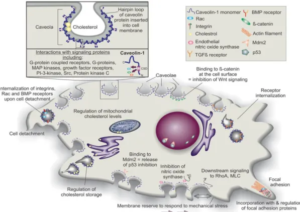
structures contain some signaling molecules, such as G-proteins, non-receptor tyrosine kinases and endothelial Nitric Oxide Synthase (eNOS). These also function as organizing centers that concentrate key signaling transducers (Figure 5; Senetta et al., 2013).
Figure 4: Caveolae: Electron micrographs showing the ultrastructure of caveolae in fibroblasts (main panel
and at high magnification upper left), and the complex arrangements of caveolae in cultured adipocytes (upper middle) and in skeletal muscle (top right). Scale bars represent 100 nm (Adapted from Parton and del Pozo, 2013).
10 Recently, the fundamentals of caveolae biogenesis are beginning to be discovered (Parton and del Pozo, 2013). Caveolins, tightly bound to cholesterol and sphingolipids, are essential for caveola formation and they are the main integral proteins of this structure, in which they work together with another group of proteins termed cavins (Parton and del Pozo, 2013; Martinez-Outschoorn et al., 2015). Each caveolae contains approximately 100 to 200 caveolin molecules formed by three principle members: Caveolin-1 (Cav1), Caveolin-2 (Cav2) and Caveolin-3 (Cav3; Fujimoto et al., 2000). The caveolin gene family is highly conserved with inter-species sequence homology (Patani et al., 2012) and includes CAV1, CAV2 and CAV3 genes. CAV1 is widely expressed in various tissues such as epithelial and endothelial cells, fibroblasts, adipocytes, and type I pneumocytes, and
CAV2 shares a similar expression distribution to CAV1 as it requires CAV1 for
stabilization. By contrast, CAV3 is specific to glia cells, skeletal and cardiac muscle cells (Scherer et al., 1994; Scherer et al., 1996; Tang et al., 1996; Martinez-Outschoorn et al., 2015). The exception is smooth muscle cells, where all three proteins are produced (Tang
et al., 1996).
With the ability to form homo- and hetero-oligomers, caveolins directly interact with numerous proteins in plasma membrane and are involved in various signaling pathways (Anwar et al., 2015).
The ‘caveolae signaling hypothesis’ implicates Cav1 in the integration of numerous molecular pathways (Patani et al., 2012). Cav1 in endothelial cells regulates angiogenesis, microvascular permeability and vascular remodeling (Hehlgans and Cordes, 2011). This protein facilitates transport of fatty acid and cholesterol in a lipoprotein chaperone complex as well as mediates transport of albumin and LDL through transcytosis pathway. Secretion of insulin is also mediated by Cav1 via ATP dependent-potassium channel and interaction with G-protein coupled receptor located at caveolae (Anwar et al., 2015). This protein also interacts with glycosyl-phosphatidylinositol-linked proteins (Patani et al., 2012), estrogen receptor (ER; Razandi et al., 2002), p85 regulatory subunit of PI3K and eNOS (Garcia-Cardena et al., 1997; Ju et al., 1997). Furthermore, Cav1 has been reported to bind to several proteins involved in cell proliferation such as EGFR, Src-family tyrosine kinases, H-Ras, protein kinase C, components of the Mitogen-Activated Protein Kinase (MAPK) cascade and HER2/Neu (Figure 5; Zhang et al., 2013; Patani et al., 2012). The ability of Cav1 to modulate intracellular signaling has important implications in numerous human biological and pathological conditions, including tumorigenesis. Actually, during the past 20 years, studies have investigated the role of Cav1 in cancer initiation and progression, showing that this multifunctional protein regulates many cancer-associated processes,
11
Figure 5: Structure and general activities of caveolae/caveolin-1. Caveolae are flask-shaped invaginations
in the cell membrane coated with multimers of caveolin scaffolding proteins. The N-termini and C-termini of caveolin proteins are in the cell cytoplasm, but a hairpin loop of the protein is inserted into the cell membrane. Various caveolae/caveolin-1 activities that have been reported in different cell types are depicted. BMP, bone morphogenetic protein; MLC, myosin light chain; PI-kinase, phosphatidylinositol 3-kinase; TGFβ, transforming growth factor beta (Baker and Tuan, 2013).
such as cell transformation, tumor growth, cell migration, invasion, multidrug resistance and angiogenesis (Senetta et al., 2013). However, the relationship between Cav1 and tumorigenesis remains contentious (Patani et al., 2012). The observed expression profiles indicated that the role of Cav1 varied according to tumor types (Felicetti et al., 2009). Downregulation appears in ovarian cancer (Wiechen et al., 2001a), colon cancer (Bender
et al., 2000) and mesenchymal sarcomas (Wiechen et al., 2001b). On the contrary,
upregulation is associated with lung (Ho et al., 2002), bladder (Rajjayabun et al., 2001), breast (Anwar et al., 2015), esophageal (Kato et al., 2002), thyroid (Janković et al., 2012) and prostate cancers (Yang et al., 1999). Hence, inferences drawn from one cancer type may not be generalizable (Patani et al., 2012), because Cav1 apparently possesses mutually exclusive functions, as tumor suppressor or tumor promoting gene, depending on tumor type/stage, cell context and the deriving availability of Cav1 interacting partners (Felicetti et al., 2009).
Cav1 is an integral membrane 178-amino acid protein of 21–22 kDa that was first identified in 1953 (Senetta et al., 2013). This protein is synthesized in the endoplasmic reticulum (ER), is shipped to the Golgi and finally, is transported to the cell surface to form
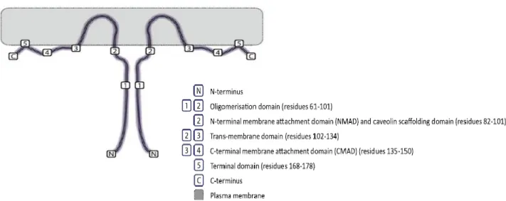
12 region q31.1 at the D7S522 locus, which is close to a known fragile site (FRA7G) frequently deleted in cancer (Senetta et al., 2013). The molecular structure of Cav1 resembles as a hairpin and the topology of this protein can be divided into domains (Figure 6): a N-terminal Membrane Attachment Domain (NMAD; residues 1-60), an oligomerisation domain (residues 61-101) with a Caveolin-1 Scaffolding Domain (CSD; residues 82-101), a trans-membrane domain (residues 102-134) and a C-terminal Membrane Attachment Domain (CMAD; residues 135-178). Both the C- and N-terminal face the cytoplasm (Patani et al., 2012).
Due to alternative splicing or initiation, Cav1 exists in two isoforms, α (residues 1-178) or β (residues 32-1-178). Cav1β is distinct in that it has a 31 amino acid residue deletion at the amino terminus (Quest et al., 2008; Wang et al., 2015).
Elucidation of Cav1 in cancer development and progression may be significant for improving patient prognosis and preventing tumor onset.
1.3.2. Caveolin-1 Scaffolding Domain (CSD)
The most prominent domain of Cav1 is the CSD (residues 82-101). Mutational studies indicate that this segment is necessary and sufficient for membrane binding (Schlegel et al., 1999). Nevertheless, these residues are also critical for oligomerization, protein interactions, and cholesterol recognition (Hoop et al., 2012).
In an active form, Cav1 is frequently phosphorylated on tyrosine-14 and/or serine-80 leading to activation of CSD (Anwar et al., 2015).
Figure 6: The topology of caveolin-1 (Cav1), depicted as a homo-dimer, permits anchorage to the plasma
membrane through a central hydrophobic domain, flanked by hydrophilic N- and C-terminal cytosolic domains (Adapted from Patani et al., 2012).
13 Residues within the CSD are required for oligomerization of Cav1 monomers into homo-oligomers of 14-16 proteins, which themselves assemble into higher-order oligomers during the formation of caveolae (Hoop et al., 2012).
The F92TVT95 segment within the CSD is important for signaling, as it is required for
interaction with other proteins, such as Src-family tyrosine kinases, H-Ras, HER2, estrogen receptor, MAPK and G protein-coupled receptors (Hoop et al., 2012; Wang et al., 2015). This binding event involves a consensus motif in the partner protein with high aromatic content, occasionally referred to as a Caveolin-Binding Motif (CBM; Hoop et al., 2012). The original definition of the CBM arises from the work of Couet et al., who obtained random peptides binding to the CSD by phage display. The peptides obtained were statistically enriched in tryptophan or other aromatic amino acids. Noting that certain separations of aromatic residues were particularly common, the investigators identified a 16-residue portion of the bovine Gi2α subunit (the GP peptide) which bounds to CSDs from Cav1 and Cav3 and much less so to Cav2. Interestingly, when all four aromatic residues were simultaneously mutated to Alanine or Glycine, the interaction was lost. Based on this finding, three CBM variants were defined, each containing three or four aromatic residues separated by unspecified amino acids, and shown to occur in known or possible caveolin binding proteins. The aromatic residues of the defined CBMs are largely hydrophobic, especially phenylalanine (Couet et al., 1997). However, some studies suggest that the CBM, despite its prevalence in the caveolin literature, is not necessary for all caveolin interactions being only implicated in a small minority of events (Byrne et al., 2012).
Finally, the CSD facilitates direct interaction with cholesterol regulating raft organization and cholesterol trafficking (Tagawa et al., 2005). More precisely, formation of
caveolae strictly requires tight binding of Cav1 to cholesterol (Murata et al., 1995). This
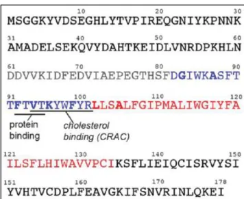
functionality is localized to a Cholesterol Recognition/interaction Amino acid Consensus motif (CRAC; residues 94-101) in residues V94TKYWFYR101 (Figure 7; Epand et al., 2005).
Thus, despite its short length (20-residue segment), the CSD appears to incorporate an array of critical functionalities (Hoop et al., 2012).
14 In summary, future studies are needed to unravel the relationship between lipid rafts and the adhesion and migration capacity of cancer cells, and to clarify the anticancer potential of azurin interaction with lipid rafts, deciphering the role of caveolae. Studies on the regulation of cholesterol are also important to understand the mechanisms related to cancer progression. With this, new targets may be developed for the treatment and prevention of cancer.
1.4. Azurin application in the treatment of cancer
There are many reasons which support the theory that azurin have the potential to act as an anticancer agent. Besides the preferential entry in cancer cells, no adverse side effects were observed in vivo studies (Yamada et al., 2005; Choi et al., 2011; Warso et al., 2013). As mentioned above, this protein also can mediate specific high-affinity interactions with various unrelated mammalian proteins relevant in cancer, conferring on it the property of natural scaffold protein, which is probably the most important characteristic of this protein (Fialho et al., 2007). This ability to act on multiple targets is important due to the fact that might be harder to trigger resistance by the cells. Another advantage of this bacterial protein is that azurin is a water-soluble molecule with a hydrophobic patch, and this might help in its tissue penetration and clearance from the blood stream (Kamp et al., 1990). In addition to all this, azurin can be easily hyper-expressed in Escherichia coli, which makes the process of production very cheap (Bernardes et al., 2013), and being a
Figure 7: Amino acid sequence of caveolin-1 (Cav1). The topology of this protein can be divided into
domains: an oligomerization domain (residues 61-101; black and blue) with a caveolin scaffolding domain (CSD; residues 82-101, blue), a trans-membrane domain (residues 102-134; red) and bold residues indicate the C-terminal membrane attachment domain (CMAD; residues 135-178) and the N-terminal membrane attachment domain (NMAD; residues 1-60). The CSD also contains cholesterol recognition/interaction amino acid consensus (CRAC; Adapted from Hoop et al., 2012).
15 small protein it can be hypothesized that it expression may occur in different vectors, including some human cell types. All these reasons make azurin an attractive molecule to be used in cancer therapy.
Preclinical pharmacological studies recurring to the use of p28 provided significant evidences that there is no apparent toxicity or immune response in the patients with solid tumors p53+/+, on which No Observed Adverse Effect Level (NOAEL) and Maximum Tolerated Dose (MTD) were established (Warso et al., 2013). With these results, it can be concluded that azurin has low immunogenicity, being a non-antibody recognized protein and for that, it is not susceptible to immune attack, even though it is a bacterial protein.
p28, as a lead compound supported by CDG Therapeutics, has finished Phase I clinical trial, which defined it as an anticancer agent under an investigational new drug application (IND 77.754) approved by the Food and Drug Administration (Bernardes et al., 2013). Recent studies have also shown that p28 is safe and well tolerated in children with progressive CNS malignancies (Lulla et al., 2016). Subsequent studies will focus on the establishment of an adequate dose for Phase II clinical trial, in obtaining a pharmacokinetic profile, determining potential immunogenicity and if possible assessing preliminary antitumor activity (Warso et al., 2013).
However, there are other domains in azurin with anticancer property (Chaudhari et
al., 2007) that should provide better efficacy and will likely make azurin less susceptible to
resistance development provided lack of toxicity of azurin in animals and cancer patients can be demonstrated, as has been done for p28 (Fialho and Chakrabarty, 2012).
Given azurin’s propensity for both therapeutic and cancer preventive activity, a weekly or bi-weekly injection of azurin in vulnerable people, for example women with family history of breast or ovarian cancers and with diagnosed BRCA1/BRCA2 mutations, may be one way to prevent, or greatly reduce, the onset of cancer in such people. Other pathways of administration azurin for cancer treatment, such as oral are currently being investigated (Chakrabarty et al., 2014).
The p28 segment of azurin or the entire protein can be combined with drugs, resulting in the transport of these to the interior of cancer cells. For example, p28 can be combined with cargo proteins, which cannot enter by themselves in eukaryotic cells (Yamada et al., 2005), or nanoparticle-loaded drugs can be surface coated with azurin to improve its therapeutic efficiency. In addition, azurin or its derived peptides can be fluorescently labeled, providing good diagnostic markers to locate tumors inside the body, since it preferentially moves toward cancer cells (Chakrabarty et al., 2014).
16
1.4.1. Effects of azurin treatment in combination with drugs on human cancer cells
Nowadays, chemotherapeutics include DNA-damaging and antimitotic agents. The first intercalate with DNA, inducing double strand breaks that induce ataxia-telangiectasia mutated (ATM)-dependent nuclear accumulation of p53 (Kurz et al., 2004). In addition, the apoptotic pathway via Bcl-2/Bax and the caspase cascade, as well as the necrotic pathway through Toll-like receptors are targets for DNA-damaging agents (Yamada et al., 2016). On the other hand, antimitotic agents bind to the β-tubulin subunits of microtubules. This interaction leads to a prolonged activation of the mitotic spindle checkpoint and mitotic arrest followed by mitotic slippage and induction of apoptosis. This agents, also called taxanes, still induce post-transcriptional acetylation and phosphorylation of p53, which leads to its intracellular increase, upregulating p21 protein, inhibiting the cell cycle, and also leading to apoptosis (Kim et al., 2013).
Unfortunately, with the consecutive application of these agents, cancer cells acquire resistance. Beyond this, these drugs can also lead to significant toxicity that may force treatment to become dose-limiting (Yamada et al., 2016). With this, new therapeutic strategies, more effective in killing cancer cells but also more selective, are needed in order to increase the efficiency and decrease the toxic side effects associated to administration of drugs (Bernardes et al., 2016).
One of these strategies is based on simultaneous use of the p28 peptide derived from azurin or the protein with drugs.
Recent studies have shown that p28 in combination with lower concentrations of DNA-damaging drugs like doxorubicin, dacarbazine, temozolamide, and antimitotic agents such as paclitaxel and docetaxel, increased their cytotoxicity by activating tumor-suppressor protein p53, which subsequently induced the endogenous cyclin-dependent kinase inhibitor p21, reducing levels of CDK2, resulting in cell cycle inhibition at G2/M
phase and leading to apoptosis. Thus, the enhanced activity of these anticancer agents in combination with p28 was facilitated through the p53/p21/CDK2 pathway. Taken together, these results highlight a new approach to maximize the efficacy of chemotherapeutic agents while reducing dose-related toxicity (Yamada et al., 2016).
In addition, a recent study also assessed the potential synergy of a co-treatment with azurin. The drugs used were gefitinib or erlotinib, both EGFR inhibitors, in low concentrations. These combined treatment demonstrated an increase in cell death when
17 compared to the sum of each agent alone, i.e., a synergistic effect occurred in comparison to the single treatments (Bernardes et al., 2016).
In the same study, it was demonstrated by Atomic Force Microscopy that azurin administration leads to changes in biophysical properties of the plasma membrane of cancer cells, thereby causing changes in signaling pathways that mediate drug resistance. These effects may be of particular interest in drug resistant cancers, where the more rigid nature of the membrane was associated to increased resistance to the accumulation of anticancer drugs. Therefore, since azurin may disrupt lipid rafts, the effects of co-administrated drugs are enhanced (Bernardes et al., 2016).
Another study demonstrating the mentioned above was performed by Choi et al., 2011. In this study, azurin-treated oral squamous carcinoma cells showed decreased cell viability accompanied by apoptotic phenotypes including morphological change, DNA breakage, and increases in p53 and cyclin B1 protein levels. In these cancer cells, with combined treatment of azurin and anticancer agents (5-fluorouracil and etopside), they discovered that this protein increased the sensitivity of oral squamous carcinoma cells to these anticancer drugs (Choi et al., 2011).
In conclusion, azurin has a strong enhancer anticancer effect on cancer cells when it is used along with anticancer drugs.
18
2. OBJECTIVES AND THESIS OUTLINE
Several studies have shown that caveolae, a non-planar lipid raft, have an important role in cancer. These can exist as invaginations of the plasma membrane, as completely enclosed vesicles or as aggregates of several vesicles. This led to the conclusion that these structures are conduits for the endocytosis of macromolecules (Razani and Lisanti, 2002). In addition, it is known that the essential components for the formation of caveolae are caveolins (Cav1, Cav2 and Cav3), which are tightly bound to cholesterol and sphingolipids (Parton and del Pozo, 2013; Martinez-Outschoorn et al., 2015). In the case of Cav1, the ability of this protein to modulate intracellular signaling has important implications in numerous human biological and pathological conditions, including tumorigenesis (Senetta et al., 2013).
Besides this, previous studies suggest that azurin enters in mammal cells through penetration the plasma membrane via caveolae-mediated endocytic pathway (Taylor et al., 2009).
With all this, this research project aims to clarify the anticancer potential of azurin interaction with lipid rafts, deciphering the role of caveolae. Moreover, recent studies from our group suggested that the link between the Cav1 and azurin involves a consensus motif rich in aromatic amino acids in azurin, occasionally referred to as a CBM (unpublished data). This led to the hypothesis that this hot-spot of aromatic amino acids are critical to the first recognition steps between azurin and cancer cells. To understand the importance of these aromatic residues in azurin interaction with cancer cells, a mutation in this region was made and the interaction of this mutated azurin with cancer cells was studied. It was shown that mutated azurin has a reduced entry capacity in cancer cells. Furthermore, the levels of Cav1 in cancer cells upon treatment with the mutated azurin were also studied, showing that the mutated protein cannot decrease Cav1 content like the WT azurin. By invasion and MTT assays, it was shown that this mutated protein cannot decrease significantly invasion and cell viability like the WT azurin. These data showed that this mutation in one particular residue of azurin sequence plays an especially important role in the entry process of azurin, since a mutation in that residue affected the ability of this protein to enter and exert his cytotoxic effects in cancer cells (unpublished data). Due to all these results, the mutated azurin was also used in the development of this work.
We started to observe if human cancer cells treated with azurin alter their lipid raft staining profile through immunostaining with a fluorescent-labeled for CTxB.


