R E S E A R C H
Open Access
Antibacterial potential of a basic phospholipase
A
2
(VRV-PL-VIIIa) from
Daboia russelii pulchella
(Russell
’
s viper) venom
Shivalingaiah Sudharshan
1and Bhadrapura Lakkappa Dhananjaya
1,2*Abstract
Background:Microbial/bacterial resistance against antibiotics poses a serious threat to public health. Furthermore, the side effects of these antibiotics have stimulated tremendous interest in developing new molecules from diverse organisms as therapeutic agents. This study evaluates the antibacterial potential of a basic protein,Vipera russellii venom phospholipase A2fraction VIIIa (VRV-PL-VIIIa), fromDaboia russelii pulchellavenom against gram-positive and gram-negative bacteria.
Methods:The antibacterial potential of VRV-PL-VIIIa in the presence and absence of an inhibitor (p-bromophenacyl bromide) was tested against gram-positive and gram-negative bacteria and the minimum inhibitory concentration was determined by microdilution tests.
Results:VRV-PL-VIIIa demonstrated potent antibacterial activities against all the human pathogenic strains tested. It more effectively inhibited such gram-positive bacteria asStaphylococcus aureusandBacillus subtilis,when compared to the gram-negative bacteriaEscherichia coli, Vibrio cholerae, Klebsiella pneumoniaeandSalmonella paratyphi.It inhibited bacterial growth at minimum inhibitory concentration values ranging from 11.1 to 19.2μg/mL. The anti-bacterial potential of VRV-PL-VIIIa was comparable to the standards gentamycin, chlorophenicol and streptomycin. The PLA2’s hemolytic and antibacterial activities were strongly correlated. Furthermore, even in the presence of p-bromophenacyl bromide, intense antibacterial activity was observed, suggesting a dissociation or partial overlapping of the bactericidal/antimicrobial domains.
Conclusion:VRV-PL-VIIIa demonstrated potent antibacterial activities against all the human pathogenic strains tested. The study shows that despite a strong correlation between enzymatic and antimicrobial activities of VRV-PL-VIIIa, it may possess additional properties that mimic the bactericidal/membrane permeability-increasing protein. This study encourages further in-depth studies on the molecular mechanisms of antibacterial properties of VRV-PL-VIIIa, which would thereby facilitate development of this protein into a possible therapeutic lead molecule for treating bacterial infections.
Keywords:Snake venom, Bactericidal, Antibiotics, Drug, Human pathogenic bacteria
* Correspondence:[email protected]
1Toxinology Group, Adichunchanagiri Biotechnology and Cancer Research
Institute (ABCRI), Balagangadharanatha Nagara, Mandya District, Karnataka, India
2Toxinology/Toxicology and Drug Discovery Unit, Center for Emerging
Technologies (CET), Jain University, Jakksandra Post, Ramanagara 562112, India
Background
Microbial/bacterial resistance against antibiotics consti-tutes a therapeutic problem that poses a significant threat to public health [1–4]. The prevalence of bacterial resist-ance to conventional antibiotics has prompted an intense search for new therapeutic agents from diverse animal ori-gins [5]. Proteins/peptides with potent antimicrobial activ-ities are found in many secretions of organisms, including snakes [5, 6]. Venoms from snakes, particularly Crotalidae, are known to be a rich natural source for the discovery and development of novel antimicrobial agents [6, 7]. Among the various components of snake venom, phospo-holipase A2 (svPLA2) enzyme, apart from its catalytic
activity of hydrolyzing the sn-2 ester bond of glyceropho-spholipids, shows other important toxic/pharmacologic effects that include myonecrosis, neurotoxicity, cardio-toxicity, as well as hemolytic, hemorrhagic, hypertensive, anticoagulant, platelet-aggregation-inhibiting and edema-inducing activities [8, 9]. This array of biological actions may be either dependent or independent of catalytic activ-ities. The svPLA2s are also reported to act as antimicrobial
agents and are emphasized for their potential as thera-peutic lead molecules [6, 7]. Crotapotin, a secretory phospholipase A2 isolated from the venom of Crotalus
durissus terrificus, has been demonstrated to exert anti-bacterial activity as well as antiviral activity against the human immunodeficiency virus [10–12]. Acidic PLA2s,
both Asp49 and Lys49 PLA2homologues, have previously
been shown to perform bactericidal activity [13–15]. A cationic protein isolated from venom of the inland taipan (Oxyuranus microlepidotus) has been demonstrated to selectively and dose-dependently kill the gram-positive bacteria through membrane disruption [16]. Recently, a phospholipase A2from the venom of the saw-scaled viper
with novel bactericidal and membrane-damaging activities was characterized [17]. These molecules are shown to be highly attractive due to their biochemical diversity, and broad spectrum of activity against enveloped bacteria, fungi, viruses, protozoa, and parasites [6, 7, 18].
Despite the therapeutic potential of svPLA2s as
anti-microbial agents, very few with bactericidal/antimicro-bial activities have been characterized [6, 18, 19]. Found in India, Russell’s viper (Daboia russelii) is a widely dis-tributed snake responsible for potent toxic and lethal ef-fects [20–23]. Despite several reports on its various biological effects, relatively little is known regarding its antimicrobial activity [20–25]. Furthermore, there are no reports available in relation to the antimicrobial activity exhibited by PLA2s from the venom of Daboia russelii
pulchella. A basic PLA2, namely Vipera russellii venom
phospholipase A2fraction VIIIa (VRV-PL-VIIIa), isolated
fromDaboia russelii pulchellavenom is reported to pro-voke various biological effects such as edema, platelet aggregation, pulmonary hemorrhage etc. [26, 27]. In the
present work, we evaluate the antibacterial potential of VRV-PL-VIIIa and investigate its possible biochemical mechanism of action. Additionally, this study exemplifies the therapeutic utility of VRV-PL-VIIIa as an antimicro-bial agent.
Methods
Venom of Daboia russelii pulchella (Southern region) was purchased from the Irula Co-operative Society Ltd. (India). Agar, beef extract, yeast extract and peptone were bought from Hi Media Private Ltd. (India). The p-Bromophenacyl bromide (p-BPB) and other chemicals used were all of analytical grade and purchased from Sigma Chemicals Ltd. (USA). Authentic pure clinical isolated cultures of human pathogenic bacteria – Staphylococcus aureus, Bacillus subtilis, Escherichia coli, Salmonella typhi, Vibrio cholerae, Klebsiella pneumoniae and Salmonella paratyphi – were obtained from the Department of Microbiology, Adichunchanagiri Institute of Medical Sciences (AIMS), B.G. Nagara, Karnataka, India. These are all human pathogens that have developed some resistance to common antibiotics particularly in the clinical environment. Bacteria were multiplied in nutrient agar at 36 ± 2 °C. After 2 days, cultures were harvested and prepared at a final concentration of 1 × 10⁸ cfu/mL and used for thein vitroinhibition assay.
Isolation of VRV-PL-VIIIa and chemical modification by p-Bromophenacyl bromide
VRV-PL-VIIIa from the venom ofDaboia ruselii pulchella was purified until homogeneous as described previously by the method of Kasturi and Gowda [26], with modifica-tions by Srinivasan [27]. The protein concentration was estimated by Lowry’s method. Chemical modification of PLA2by p-Bromophenacyl bromide (p-BPB) was carried
out as described by Condreaet al.[28]. Briefly, 100μL of 40 mM p-BPB in acetone was added to 3 mL of PLA2
solution (0.5 mg/mL, in 0.05 M Tris–HCl buffer, pH 7.5). The reaction was allowed to proceed for 40 min, and then acidified with glacial acetic acid to pH 4.0 to stop the reac-tion. Excess of reagent was removed by dialyzing against 0.05 M Tris–HCl buffer pH 7.5.
Phospholipase A2activity
The phospholipase A2 assay was carried out according
to the method described by Bhat and Gowda [29]. Phos-phatidyl choline (PC) was diluted with petroleum ether (60–80 °C) to obtain a concentration of 1000 nmoles/ 50 mL. The reaction mixture containing VRV-PL-VIIIa (5 μg) was augmented to 680 μL with water. To the reaction mixture, 200 μL of ether, 100 μL of Tris–HCl buffer (0.05 M, pH 7.5) and 20μL of CaCl2(0.4 M) were
(isopropanol:pet ether:1NH2SO4, 40:10:1) was added,
mixed and centrifuged at 1000 rpm for 3 min. To the organic phase, 0.5 mL of CHCl3:pet ether (1:5) was
added, mixed and centrifuged at 1000 rpm for 3 min. To the upper phase, cobalt reagent [1.35 vol. of trietha-nolamine increased to 10 mL with solution A (6 g of CO(NO3)2.6H2O + 0.8 mL glacial acetic acid) and 7 mL
of solution B (saturated Na2SO4)] was added, mixed
and centrifuged 1000 rpm for 3 min. The upper organic phase was carefully transferred and 0.75 mL ofα -nitroso-β-naphthol reagent (0.4 % α-nitroso-β-naphthol in 96 % ethanol) was added. The intensity of the orange coloration is directly proportional to the amount of cobalt present. After 30 min, 2 mL of ethanol was added to dilute the contents and absorbance was read at 540 nm. The amount of free fatty acid released was estimated using the standard linolenic acid curve. The enzyme activity was expressed as nmoles of fatty acid released/minute/mg of protein.
For inhibition studies, VRV-PL-VIIIa (5 μg) was pre-incubated with or without a different concentration of p-Bromophenacyl bromide (1–6 μm) at 37 °C for 15 min. Appropriate controls were set up and further experiments were performed as described above. The inhibition is expressed as a percentage, considering the activity of venom alone as 100 %.
Hemolytic activity assay
Hemolytic (direct/indirect) activity of isolated VRV-PL-VIIIa was determined according to the method of Boman and Kaletta [30], using packed human erythro-cytes (blood group A). The human erythroerythro-cytes used for the study were sourced from previously published work, which had ethical approval from the ethics committee of the University of Mysore (UOM) for the withdrawal of blood [23]. The direct and indirect hemolytic assays were carried out using washed erythrocytes. For the dir-ect hemolytic assay, the packed erythrocytes (1 mL) were suspended in nine volumes of phosphate-buffered saline (PBS), which formed the stock. The stock (1 mL) was incubated with various concentrations of isolated VRV-PL-VIIIa (0–5 μg) for 30 min at 37 °C. For the indirect hemolytic assay, stock was prepared by mixing packed erythrocytes (1 mL), egg yolk (1 mL) and phosphate-buffered saline (8 mL). One milliliter of suspension from stock was incubated with various concentrations of iso-lated VRV-PL-VIIIa (0–6 μg) for 30 min at 37 °C. The reaction was terminated by adding 10 mL of ice-cold PBS and then centrifuged at 4 °C and 800 g. The amount of hemoglobin released in the supernatant was measured at 540 nm. One milliliter of stock erythrocytes with 10 mL ice-cold PBS alone was defined as constituting 0 % lysis.
For inhibition studies, VRV-PL-VIIIa (6 μg) was pre-incubated with or without a different concentration of p-Bromophenacyl bromide (1–6 μM) at 37 °C for
15 min. Appropriate controls were set up and further experiments were accomplished as described above. The inhibition is expressed as a percentage, considering the activity of venom alone as 100 %.
Bactericidal activity of VRV-PL-VIIIa
Bactericidal activity was evaluated by the well diffusion method on nutrient agar medium [31]. This was con-firmed by the inhibitory effect on bacterial growth as reflected by the inhibition zone, compared to that of known antibiotics such as gentamicin (G), chloram-phenicol (Cp) and streptomycin (Sm) at 30 μg/mL. The sterile nutrient agar medium (20 mL) in petri dishes was uniformly smeared using sterile cotton swabs with pure test cultures of the human pathogenic bacteria S. aureus, B. subtilis, E. coli, S. typhi, V. cholerae, K. pneumoniae andS. paratyphi. The nutrient agar media was prepared by dissolving 0.3 % beef extract, 0.3 % yeast extract, 0.5 % peptone, 0.5 % NaCl and 1.5 % agar in 1:l of distilled water and 0.2 % methanol (v/v). The wells of 5 mm diameter were made using a sterile cork borer in each petri dish and the isolated VRV-PL-VIIIa (0-12 μg) pre-incubated independently with or without p-Bromophenacyl bromide (15μM) was added; a blank well loaded without test compound was considered the control. For each treatment, ten replicates were prepared. The plates were incubated at 37 °C for 24 h and the result-ing zone of inhibition was measured by comparresult-ing the control and the standard antibiotics.
For inhibition studies, VRV-PL-VIIIa (12 μg) was pre-incubated with or without a different concentration of p-Bromophenacyl bromide (1–6 μM) at 37 °C for 15 min and antimicrobial activity was assessed as de-scribed above with appropriate controls. The inhibition is expressed as a percentage, considering the activity of venom alone as 100 %.
Determination of minimum inhibitory concentration (MIC)
The minimum inhibitory concentration of the isolated VRV-PL-VIIIa and the antibiotics used were determined by serial dilution in the nutrient agar, with concentra-tions ranging from 2–20μg/mL. The inoculum was pre-pared overnight from fresh broth culture in nutrient broth and plates were incubated for 24 h at 37 °C. MIC was recorded as the lowest VRV-PL-VIIIa at which the antibiotic concentrations demonstrated no visible growth in the broth [32].
Statistical analysis
Results and discussion
The snake venom PLA2s (svPLA2s), apart from their
well known toxic and lethal effects, are also known to be of therapeutic utility, particularly as bactericidal/ antibacterial agents [6, 19]. In a previous study, it was shown that the crude Russell’s viper venom from India exhibited strong antibacterial actions [25]. However, the principal component responsible for it was unexplored. In the present work, we evaluate the basic PLA2
(VRV-PL-VIIIa) of Daboia russelii pulchella venom for its antibacterial activity on different human pathogenic bacteria [26, 27].
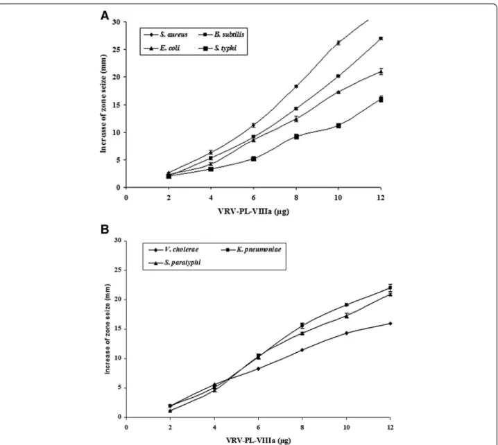
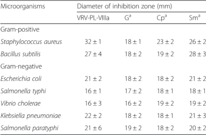
When tested, VRV-PL-VIIIa (0–12 μg/mL) showed a broad spectrum of highly significant antibacterial activities by producing a clear zone of inhibition that was dose-dependent in the range of 17–30 mm (Fig. 1– a and b) (Table 1). Interestingly, it showed more significant inhib-ition of gram-positive bacteria including S. aureus and B. subtilis (in the range of 27–32 mm) in relation to such gram-negative species as E. coli, S. typhi, V. cholerae, K. pneumoniae andS. paratyphi(in the range of 16–22 mm) (Table 1). Furthermore, it was interesting to note that VRV-PL-VIIIa exhibited similar or greater antibacterial activities than those of the
standards gentamicin, chlorophenicol and streptomycin (which was in the range of 16–20 mm) (Table 1). When VRV-PL-VIIIa was tested via the agar dilution assay for determining minimum inhibitory concentration (MIC), it significantly inhibited the bacterial growth with MIC values ranging from 11.2 to 20μg/mL, when compared to standard antibiotics whose range was between 18 and 24 μg/mL (Table 2). Thus, VRV-PL-VIIIa was as potent as standard antibiotics in inhibiting the growth of bacterial strains.
A strong correlation is usually found between the PLA2’s hemolytic and antibacterial activities [6, 19, 25].
The VRV-PL-VIIIa also showed a potent hemolytic (indirect) activity that is usually associated with svPLA2s.
It was found that VRV-PL-VIIIa produces a dose-dependent hemolysis of blood cells and at 5μg concentra-tion provoked 100 % hemolysis (Fig. 2). From these data it may be concluded that the antibacterial effects of VRV-PL-VIIIa are dependent upon catalytic activity, i.e., an
enzymatic membrane degradation effect that is usually observed in sPLA2s [6, 33]. The correlation between
PLA2, hemolytic and antibacterial activities indicates that
the catalytically activity of PLA2is principally involved in
bactericidal/antibacterial activities [6, 19, 25]. However, despite the existence of another mechanism, namely svPLA2isolated fromBothrops asper(also classified within
group IIA) venom, which was shown to directly kill both gram-positive and gram-negative bacteria [13].
Furthermore, a toxin from B. asper (myotoxin II, a catalytically inactive Lys49 PLA2) exhibited a bactericidal
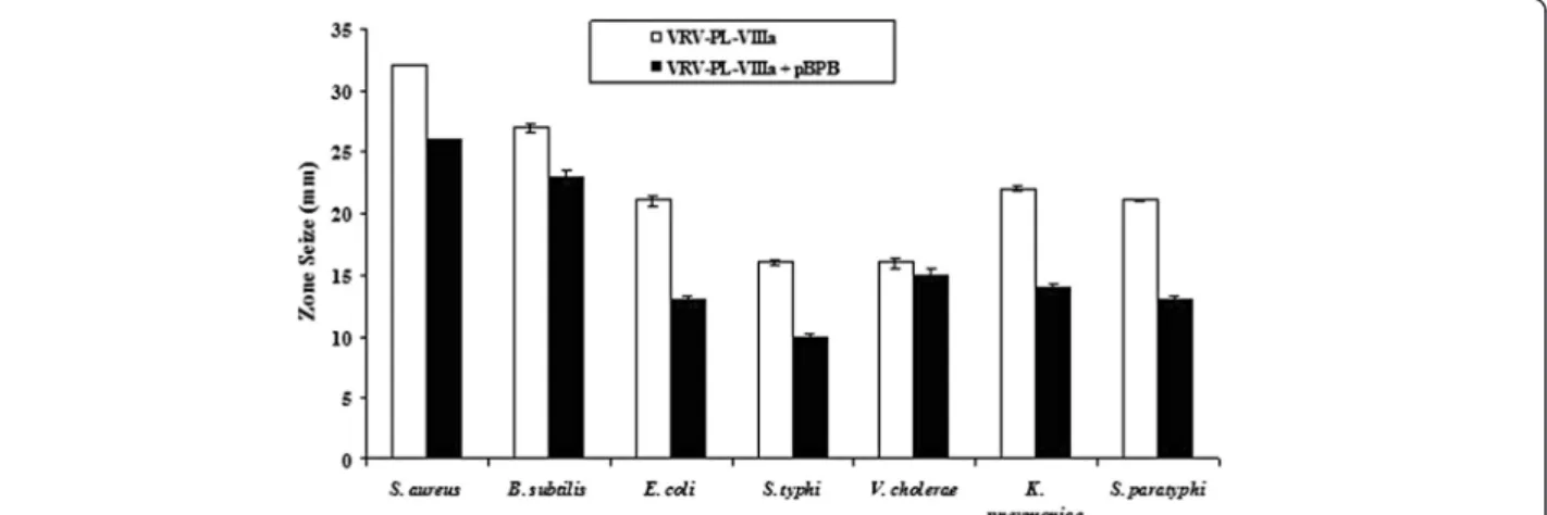
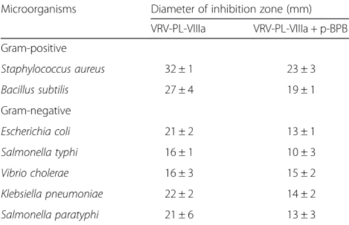
mechanism that was independent of catalytic activity [13, 6, 19]. Additional in-depth studies showed that a short sequence of the protein, corresponding to residues 115–129 of its cytolytic C-terminal region, was respon-sible for its bactericidal activity, emphasizing the fact that bactericidal activity is not associated with enzymatic activities [6, 13, 19]. Similarly, the study found that when VRV-PL-VIIIa was pre-incubated with p-BPB, an inhibitor of svPLA2enzymatic activity, both enzymatic and
antibac-terial activities were inhibited (Fig. 3 and Table 3) [34]. However, it should be noted that there was incomplete abolition of antibacterial activity (Table 3), even though the enzymatic activity was completely abolished (Table 4). This suggests a dissociation between enzymatic activity and bactericidal/antibacterial activity of VRV-PL-VIIIa. It is usually observed that the potent bactericidal activity of sPLA2s is accomplished by binding to anionic surfaces
along with the enzymatic degradation of phospholipids in the target membranes, i.e., preferentially in the case of gram-positive species. However, the bactericidal activity against gram-negatives is known to require a synergistic action of bactericidal/permeability-increasing protein (BPI), and is also equally dependent on enzymatic-based membrane degradation [13, 6, 17, 19].
It has been demonstrated that, in addition to cationic properties of PLA2molecules, the polyanionic properties
of lipoteichoic acids from bacterial cell wall promote the attack of membrane phospholipids by svPLA2s.
There-fore, the action mode of svPLAs depends on the type of bacteria species involved (gram-positive or negative). Recently, an association was shown between antibacter-ial and enzymatic activity of an antibacterantibacter-ial PLA2
(EcTx-I) purified fromEchis carinatus venom [6]. How-ever, the present study found that the bactericidal activity of VRV-PL-VIIIa is partially independent of catalytic activ-ity and antibacterial activities (Tables 3 and 4), which is supported by the homogenous nature of the protein with no other associated enzymatic activities (such as L-amino-oxidase, proteases etc.) in the preparation (data not shown). Therefore, the VRV-PL-VIIIa bacteri-cidal mechanism may include “fatal depolarization” of the bacterial membrane, creation of physical holes in the membrane, scrambling of normal distribution of
Table 2Minimum inhibitory concentration (MIC) of VRV-PL-VIIIa and standard antibiotics by serial dilution method
Microorganisms MIC (μg/mL)
VRV-PL-VIIIa G Cp Sm
Gram-positive
Staphylococcus aureus 15.3 ± 1 20.8 ± 1 14.4 ± 2 13.6 ± 1
Bacillus subtilis 11.1 ± 3 20.8 ± 3 14.4 ± 1 16.6 ± 1
Gram-negative
Escherichia coli 16.3 ± 2 23.8 ± 1 14.4 ± 2 14.6 ± 1
Salmonella typhi 17.2 ± 2 18.8 ± 1 17.4 ± 2 13.6 ± 1
Vibrio cholerae 17.3 ± 2 19.8 ± 3 14.4 ± 1 19.6 ± 1
Klebsiella pneumoniae 19.2 ± 1 20.8 ± 1 14.4 ± 2 13.6 ± 1
Salmonella paratyphi 17.3 ± 2 23.8 ± 1 14.4 ± 2 14.6 ± 1
The results are mean ± SD (n= 6)
Ggentamicin,Cpchloramphenicol,Smstreptomycin
Table 1Antibacterial activity of VRV-PL-VIIIa and standard antibiotics
Microorganisms Diameter of inhibition zone (mm)
VRV-PL-VIIIa Ga Cpa Sma
Gram-positive
Staphylococcus aureus 32 ± 1 18 ± 1 23 ± 2 26 ± 2
Bacillus subtilis 27 ± 4 18 ± 2 19 ± 2 28 ± 3
Gram-negative
Escherichia coli 21 ± 2 18 ± 2 18 ± 2 21 ± 2
Salmonella typhi 16 ± 1 17 ± 2 18 ± 1 18 ± 1
Vibrio cholerae 16 ± 3 16 ± 2 19 ± 2 19 ± 2
Klebsiella pneumoniae 22 ± 2 18 ± 2 18 ± 1 21 ± 3
Salmonella paratyphi 21 ± 6 19 ± 2 18 ± 2 20 ± 2
The results are mean ± SD (n= 6)
lipids between the bilayer leaflets, damage of critical intracellular targets after internalization of the peptide, and also inhibition of macromolecular biosynthesis as observed in many svPLA(2)s and/or interaction with
specific vital components inside the bacteria [6, 35]. It was reported that PLA2s purified from Agkistrodon
piscivorus piscivorusrely on a membrane-permeabilizing mechanism to exert their bactericidal effects [36]. A recent study showed the presence of a large number of PLA2-sensitive phospholipid domains/composition,
rather than only the phosphatidylcholine (PC) content of a particular membrane that determines the extent of membrane damage by a particular venom PLA2enzyme
[37]. This might be one of the reasons for the differen-tial inhibitory potency of VRV-PL-VIIIa on various bacterial species. Therefore, it may be concluded that the isolated VRV-PL-VIIIa phospholipase A2, will
mani-fest its antimicrobial activity not only by acting upon the membrane through its enzymatic activity, but also by other mechanisms as discussed above, independently
Fig. 3Bactericidal activity against different human pathogenic strains of VRV-PL-VIIIa pre-incubated with or without p-Bromophenacyl bromide. VRV-PL-VIIIa (12μg/mL) was pre-incubated with or without a different concentration of p-Bromophenacyl bromide (6μM) at 37 °C for 15 min and
bactericidal activity was estimated in agar diffusion assay. The diameter of the clear zone was measured and plotted after subtracting the diameter of the well (5 mm). Results are expressed as mean ± SD for three independent assays, each performed in triplicate
of its catalytic activities. Further in-depth studies on molecular mechanism of action of VRV-PL-VIIIa anti-bacterial activity would be of interest to develop this as a therapeutic lead molecule for application.
Conclusion
This study shows that VRV-PL-VIIIa – a PLA2 from
Daboia russelii pulchellavenom–presents potent anti-bacterial activity. Significant antianti-bacterial activity is observed, even in presence of an inhibitor of PLA2
enzymatic activity (p-BPB), suggesting a dissociation or partial overlapping of the bactericidal/antimicrobial do-mains of the enzyme. This study demonstrates that although there is a strong correlation between enzym-atic and antimicrobial activities of VRV-PL-VIIIa, it may also possess other properties that mimic the bac-tericidal/membrane permeability-increasing protein. These results should encourage further in-depth studies on molecular mechanisms of anti-bacterial properties of VRV-PL-VIIIa, which would thereby facilitate its development into a therapeutic lead molecule for treat-ing bacterial infections.
Ethics committee approval
The use of human erythrocytes was approved by the Ethics Committee of the University of Mysore (UOM).
Competing interests
The authors declare that they have no competing interests.
Authors’contributions
DBL contributed to the conception and design of the study, acquisition of data, analysis and interpretation of data, and drafting of the manuscript. SS analyzed and interpreted data. Both authors read and approved the final manuscript.
Acknowledgments
DBL thanks Jain University for the constant encouragement to the progress of research. SS and DBL acknowledges the Adichunchanagiri Mahasamstana Mutt and Shikshana Trust for providing facilities in the Adichunchanagiri Biotechnology and Cancer Research Institute (ABCRI). We thank Balagangadharanatha Swamiji Institute for Technology (BGS-IT) and Sri Adichunchangiri College of Pharmacy for the support to carry out the research.
Received: 28 October 2014 Accepted: 15 May 2015
References
1. Norrby SR, Nord CE, Finch R, European Society of Clinical Microbiology and Infectious Diseases. Lack of development of new antimicrobial drugs: a potential serious threat to public health. Lancet Infect Dis. 2005;52:115–9. 2. Choudhury R, Panda S, Singh DV. Emergence and dissemination of
antibiotic resistance: a global problem. Indian J Med Microbiol. 2012;30(4):384–90.
3. Echols RM. A long and winding road; evolution of antimicrobial drug development–crisis management. Expert Rev Anti Infect Ther. 2012;10(11):1311–9.
4. Ghafur A. The Chennai declaration: a solution to the antimicrobial resistance problem in the Indian subcontinent. Clin Infect Dis. 2013;56(8):1190. 5. Zasloff M. Antimicrobial peptides of multicellular organisms. Nature.
2002;415:389–95.
6. Samy RP, Gopalakrishnakone P, Stiles BG, Girish KS, Swamy SN, Hemshekhar M, et al. Snake venom phospholipases A(2): a novel tool against bacterial diseases. Curr Med Chem. 2012;19(36):6150–62.
7. Perumal Samy R, Pachiappan A, Gopalakrishnakone P, Thwin MM, Hian YE, Chow VTK, et al.In vitroantimicrobial activity of natural toxins and animal venoms tested againstBurkholderia pseudomallei. BMC Infect Dis. 2006;6:1–16. 8. Kini RM. Phospholipase A2- a complex multifunctional protein puzzle. In:
Wiley J, editor. Venom phospholipase A2enzymes: structure, function and mechanism. Chichester, England: John Wiley & Sons; 1997. p. 1228. 9. Gutiérrez JM, Lomonte B. Phospholipases A2: unveiling the secrets of a
functionally versatile group of snake venom toxins. Toxicon. 2013;62:27–39. 10. Soares AM, Mancin AC, Cecchini AL, Arantes EC, Franca SC, Gutierrez JM,
et al. Effects of chemical modifications of croprotein B, the phospholipase A2subunit of croprotein fromCrotalus durissus terrificussnake venom, on its enzymatic and pharmacological activities. Int J Biochem Cell Biol. 2001;33(9):877–88.
11. Toyama MH, de Oliveira DG, Beriam LOS, Novello JC, Rodrigues-Simioni L, Marangoni S. Structural, enzymatic and biological properties of new PLA2 isoform fromCrotalus durissus terrificusvenom. Toxicon. 2003;41(8):1033–8.
12. Sampaio SC, Brigatte P, Sousa-e-Silva MC, dos Santos EC, Rangel-Santos AC, Curi R, et al. Contribution of crotoxin for the inhibitory effect ofCrotalus durissus terrificussnake venom on macrophage function. Toxicon. 2003;41(7):899–907.
13. Paramo L, Lomonte B, Pizarro-Cerda J, Bengoechea JA, Gorvel JP, Moreno E. Bactericidal activity of Lys49 and Asp49 myotoxic phospholipases A2from
Bothrops aspersnake venom: synthetic Lys49 myotoxin II-(115–129)-peptide identifies its bactericidal region. Eur J Biochem. 1998;253:452–61. 14. Vargas LJ, Londoño M, Quintana JC, Rua C, Segura C, Lomonte B, et al. An
acidic phospholipase A2with antibacterial activity fromPorthidium nasutum
snake venom. Comp Biochem Physiol B Biochem Mol Biol. 2012;161(4):341–7. 15. Soares AM, Andrião-Escarso SH, Bortoleto RK, Rodrigues-Simioni L, Arni RK,
Ward RJ. Dissociation of enzymatic and pharmacological properties of piratoxins-I and -III, two myotoxic phospholipases A2fromBothrops pirajai
snake venom. Arch Biochem Biophys. 2001;387:188–96. Table 3Antibacterial activity of VRV-PL-VIIIa with or without
p-Bromophenacyl bromide (p-BPB)
Microorganisms Diameter of inhibition zone (mm)
VRV-PL-VIIIa VRV-PL-VIIIa + p-BPB
Gram-positive
Staphylococcus aureus 32 ± 1 23 ± 3
Bacillus subtilis 27 ± 4 19 ± 1
Gram-negative
Escherichia coli 21 ± 2 13 ± 1
Salmonella typhi 16 ± 1 10 ± 3
Vibrio cholerae 16 ± 3 15 ± 2
Klebsiella pneumoniae 22 ± 2 14 ± 2
Salmonella paratyphi 21 ± 6 13 ± 3
The results are expressed as mean ± SD (n= 6)
Table 4Phospholipase A2activity of VRV-PL-VIIIa with or without
p-Bromophenacyl bromide (p-BPB)
Specific activity
Enzymatic activity VRV-PL-VIIIa VRV-PL-VIIIa + p-BPB
PLA2a 93.4 ± 3.1 3.2 ± 0.8
Values are presented as mean ± SD (n= 6)
aSpecific activity is expressed in terms of fatty acid released in nmoles/minute/
16. Nair DG, Fry BG, Alewood P, Kumar PP, Kini RM. Antimicrobial activity of omwaprin, a new member of the waprin family of snake venom proteins. Biochem J. 2007;402(1):93–104.
17. Perumal Samy R, Gopalakrishnakone P, Bow H, Puspharaj PN, Chow VT. Identification and characterization of a phospholipase A2from the venom of the Saw-scaled viper: Novel bactericidal and membrane damaging activities. Biochimie. 2010;92(12):1854–66.
18. Pereira HA. Novel therapies based on cationic antimicrobial peptides. Curr Pharm Biotechnol. 2006;7(4):292–34.
19. de Oliveira Junior NG. e Silva Cardoso MH, Franco OL. Snake venoms: attractive antimicrobial proteinaceous compounds for therapeutic purposes. Cell Mol Life Sci. 2013;70(24):4645–58.
20. Jayanthi GP, Gowda TV. Geographical variation in India in the composition and lethal potency of Russell’s viper (Vipera russelli) venom. Toxicon. 1988;26(3):257–64.
21. Woodhams BJ, Wilson SE, Xin BC, Hutton RA. Differences between the venoms of two sub-species of Russell’s viper:Vipera russelli pulchellaand
Vipera russelli siamensis. Toxicon. 1990;28(4):427–33.
22. Prasad NB, Uma B, Bhatt SK, Gowda VT. Comparative characterisation of Russell’s viper (Daboia/Vipera russelli) venoms from different regions of the Indian peninsula. Biochim Biophys Acta. 1999;1428(2–4):121–36. 23. Dhananjaya BL, Zameer F, Girish KS, D’Souza CJ. Anti-venom potential of
aqueous extract of stem bark ofMangifera indicaL. againstDaboia russelii
(Russell’s viper) venom. Indian J Biochem Biophys. 2011;48(3):175–83. 24. Venkatesh M, Prasad N, Sing T, Gowda TV. Purification, characterization, and
chemical modification of neurotoxic peptide fromDaboia russeliisnake venom of India. J Biochem Mol Toxicol. 2013;27(6):295–304.
25. Perumal Samy R, Gopalakrishnakone P, Thwin MM, Chow TK, Bow H, Yap EH, et al. Antibacterial activity of snake, scorpion and bee venoms: a comparison with purified venom phospholipase A2enzymes. J Appl Microbiol. 2007;102(3):650–9.
26. Kasturi S, Gowda TV. Purification and characterization of a major phospholipase A2from Russell’s viper (Vipera russelli) venom. Toxicon. 1989;27(2):229–37.
27. Srinivasan S. Mechanism of action of snake venom toxic phospholipases, Thesis. India: University of Mysore; 2004.
28. Condrea E, Fletcher JE, Rapuano BE, Yang CC, Rosenberg P. Effect of modification of one histidine residue on the enzymatic and
pharmacological properties of a toxic phospholipase A2fromNaja nigricollis
snake venom and less toxic phospholipases A2fromHemachatus haemachatusandNaja naja atrasnake venoms. Toxicon. 1981;19(1):61–71. 29. Bhat MK, Gowda TV. Purification and characterization of a myotoxic
phospholipase A2 from Indian cobra (Naja naja naja) venom. Toxicon. 1989;27(8):861–73.
30. Boman HG, Kaletta U. Chromatography of rattlesnake venom, a separation of three phosphodiesterases. Biochim Biophys Acta. 1957;24(3):619–31. 31. Forbes BA, Sahm DF, Weissfeld AS, Trevino EA. Methods for testing
antimicrobial effectiveness. In: Baron EJ, Petrson LR, Finegold SM, editors. Bailey and Scott’s diagnostics microbiology. St. Louis, Missouri: Mosby Co; 1990. p. 171–94.
32. Prescott LM, Harley JP, Klein DA. Introduction to microbiology. 5th ed. San Francisco: Benjamin-Cummings Publishing Co Inc; 1996. p. 681–4. 33. Buckland AG, Wilton DC. The antibacterial properties of secreted
phospholipases A2. Biochim Biophys Acta. 2000;1488:71–82. 34. Rudrammaji LM, Machiah KD, Kantha TP, Gowda TV. Role of catalytic
function in the antiplatelet activity of phospholipase A2cobra (Naja naja naja) venom. Mol Cell Biochem. 2001;219(1–2):39–44.
35. Park CB, Kim HS, Kim SC. Mechanism of action of the antimicrobial peptide buforin II: buforin II kills microorganisms by penetrating the cell membrane and inhibiting cellular functions. Biochem Biophys Res Commun. 1998;244(1):253–7.
36. Shen Z, Cho W. Highly efficient immobilization of phospholipase A2and its biomedical applications. J Lipid Res. 1995;36(5):1147–51.
37. Saikia D, Bordoloi NK, Chattopadhyay P, Choklingam S, Ghosh SS, Mukherjee AK. Differential mode of attack on membrane phospholipids by an acidic phospholipase A2(RVVA-PLA2-I) fromDaboia russellivenom. Biochim Biophys Acta. 2012;1818(12):3149–57.
Submit your next manuscript to BioMed Central and take full advantage of:
• Convenient online submission
• Thorough peer review
• No space constraints or color figure charges
• Immediate publication on acceptance
• Inclusion in PubMed, CAS, Scopus and Google Scholar
• Research which is freely available for redistribution