Vol.50, n. 2 : pp.249-258, March 2007
ISSN 1516-8913 Printed in Brazil BRAZILIAN ARCHIVES OF
BIOLOGY AND TECHNOLOGY
A N I N T E R N A T I O N A L J O U R N A L
Characterization of Enterocins Produced by
Enterococcus
mundtii
Isolated from Humans Feces
Alessandra Einsfeld Ferreira, Natália Canal, Daiana Morales, Daiane Bopp Fuentefria and Gertrudes Corção*
Departamento de Microbiologia; Instituto de Ciências Básicas da Saúde; Universidade Federal Rio Grande do Sul; corcao@ufrgs.br; 90050-170; Porto Alegre - RS - Brasil
ABSTRACT
The aim of this study was to characterize bacteriocins produced by 70 strains of Enterococcus mundtii.Four strains exhibited antibiotic activity towards Listeria innocua, L. monocytogenes, Lactobacillus plantarum, and Salmonella Enteritidis. They remained active under temperatures of up to 121oC for 20 min, and under pH treatments that varied from 2.0 to 10.0. Antimicrobial activity was maintained during the storage test for 60 days under freezing. The kinetics of production revealed the peak activity of 1600 AU /mL during the logarithmic growth phase and the molecular weight found was approximately 3.0 kDa. The characterization of the products with antimicrobial activity indicated their proteic nature, presenting a typical kinetics of primary metabolite and a molecular weight similar to many purified enterocins.
Key words: Enterococcus mundtii, enterocins, antimicrobial activity, biopreservative
*
Author for correspondence INTRODUCTION
Bacteriocins are ribosomally synthesized peptides and proteins which may inhibit the growth or eliminate certain bacterial species, affecting the permeability of the membrane or even interfering with essential roles played by the cell, such as DNA replication and translation (Bennick et al., 1998). The study of bacteriocins produced by lactic acid bacteria (LAB) has gained much attention recently. The potential use of LAB as non-toxic biopreservation agents in the industrial processing of human food and animal feeds is chiefly due to the fact that they inhibit the growth of pathogenic and degradative bacteria (Cintas et al., 2000). Bacteriocins are subdivided into four classes, in terms of their biochemical and genetic characteristics. Class I bacteriocins comprises the
enterocins classified to date, including mundticin, the only enterocin produced by E. mundtii so far characterized. Class IIb comprises the bacteriocins whose activity is dependent on the complementary activity of two different peptides, and class IIc is formed by non-lantibiotic bacteriocins that failed to be ranked under classes IIa and IIb (Casaus et al., 1997; Franz et al., 1999). Class III bacteriocins are large size (above 30 kDa), heat-labile proteins. Class IV is formed by complex proteins that require association with lipids and carbohydrates in order to exhibit antimicrobial activity (Lauková et al., 1998).
Since E. mundtii enterocins have been scarcely studied, this study was designed to characterize the bacteriocins produced by E. mundtii for their antimicrobial activity spectrum, evaluating their antimicrobial activity measured in Arbitrary Units (AU /mL), their sensitivity to heat, pH, storage conditions and proteolytic enzymes. Molecular size and the kinetics production curve were also determined.
MATERIALS AND METHODS
Bacterial strains
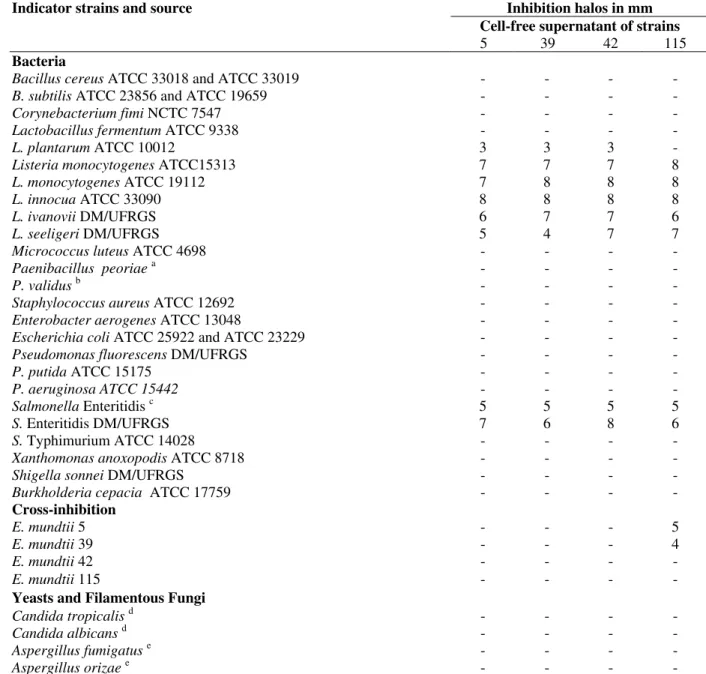
This study used 70 E. mundtii strains, from the Department of Microbiology Collection (Instituto de Ciências Básicas da Saúde, Universidade Federal do Rio Grande do Sul, Brazil). Strains were from animal and clinical and nonclinical human isolates. In order to establish the antimicrobial activity spectrum of enterocins, 16 Gram-positive and 12 Gram-negative bacterial strains, related to food poisoning; two yeasts, isolated from clinical samples, and two filamentous fungi, isolated from rice, were used (Table 1).
Screening of bacteriocin activity
Initially, a triage procedure was carried out for the 70 strains using five indicator bacteria: Listeria innocua ATCC 33090, L. monocytogenes ATCC 15313, L. welshimeri DMIC/UFRGS,
Corynebacterium fimi NCTC 7547 and
Micrococcus luteus ATCC 4698. The double layer test (Tagg et al., 1971) was used. The E. mundtii
isolates were spotted onto a Petri dish containing Trypticasein soya agar (TSA), and then incubated at 35o C for 24 h. Subsequently, each dish was overlaid with semi-solid TSA agar previously
inoculated with a 106 cell/mL solution of each indicator bacterium. After another incubation period at 35o C for 24 h, inhibition halos were observed. All strains that inhibited the growth of at least one of the five indicator bacteria, underwent hydrogen peroxide test, using double layer method in anaerobic conditions, and the bacteriophage assay described by Lewus et al. (1991). These procedures were adopted to rule out hydrogen peroxide and bacteriophage action as inhibitors of bacterial growth.
Preparation of the cell-free culture supernatant
The strains that presented antimicrobial activity but with a negative result for hydrogen peroxide and bacteriophage tests were used to prepare the cell-free culture supernatant. The strains were incubated in MRS broth (Accumedia) at 35o C for 18 h under agitation. Subsequently, the pH of the culture was adjusted to 6.2 with NaOH 1M. The culture was then centrifuged at 3000g for 15 min, and sterilized by filtration through a 0.22-µm-pore size membrane (Schleicher and Schuell).
Quantitative analysis of the antimicrobial activity of the cell-free culture supernatant
The quantitative analysis of the antimicrobial activity was measured in Arbitrary Units per milliliter (AU/mL). Antimicrobial activity was evaluated using the agar well diffusion method, with L. innocua ATCC 33090 as indicator strain at a concentration of 106 cell /mL.Serial 1:1 dilutions in phosphate buffer (PBS) of the supernatant were carried out. Aliquots of 80 µL from these dilutions were inoculated into the wells. Aliquots of the crude supernatant underwent the same procedure. After 10 minutes at 4ºC for diffusion, the agar plates were incubated at 35o C for 18 h, after which inhibition halos were measured. All the experiments were carried out in triplicate, with three different supernatants prepared as described before. An Arbitrary Unit (AU/mL) was defined as the reciprocal of the highest dilution that produced an inhibition halo larger than 2 mm in diameter (Kawamoto et al., 2002).
Determination of the antimicrobial activity spectrum of the cell-free culture supernatant
with 80 µL of the cell-free culture supernatant and 106 cells/mL of the indicator bacteria cell culture. After incubation at 35oC for 18 h, inhibition halos were measured. A cross-inhibition test was carried out, in which cell-free supernatants were tested against their own bacteriocin-producing strains.
Sensitivity to heat, pH variation, storage conditions and proteolytic enzymes
Aliquots of the cell-free supernatant were exposed to heat treatments of 40, 50, 60, 70, 80, 90 and 100o C for 30 min, and at 121o C for 10 and 20 min. Aliquots of the supernatants after treatment as well as a control aliquot (supernatant without treatment) were inoculated using the agar well diffusion method. In a separate experiment, cell-free culture supernatants were adjusted to values between pH 2 and pH 10 using buffers. The agar plates were incubated at 35oC for another 24h. Controls were prepared adding PBS instead of the buffer with predefined pH. In order to confirm the proteic nature of the bacteriocin, sensitivity to proteolytic enzymes trypsin, proteinase K and lysozyme at 0.1 and 1 mg /mL were assessed in a concomitant experiment. The agar well diffusion method was used to measure the activity of the supernatants after treatments. The bacteriocin stability under storage was assessed using two sets of aliquots of the cell-free supernatants: one set was kept refrigerated at 4o C, and the other kept frozen at –20o C for 60 days. The antimicrobial action was determined every 15 days, and expressed in AU/mL using the agar well diffusion method. All tests used 106 cells/mL preparations of
L. innocua ATCC 33090 as indicator strain. Plates were incubated at 35o C for 18 h and inhibition halos were measured in mm. All tests were carried out in duplicate using three different supernatants.
Bacteriocin Production kinetics
Bacteriocin production kinetics was determined for E. mundtii strains 39 and 115, which were chosen in the light of the results of cross-inhibition analyses. The antimicrobial activity, expressed in AU/mL, and sensitivity to storage conditions were also carried out. Strains were grown in MRS broth (Accumedia) at 35o C for 18 h, under constant agitation. Aliquots of 100 µL were retrieved every 1h. Antimicrobial activity (AU/mL) was evaluated using the agar well diffusion method, and L.
innocua was the indicator microorganism used. Bacterial growth was followed by 620 nm absorbance analysis, the pH of the culture was also determined.
Molecular weight analysis
The molecular weight of bacteriocin was determined by 16% SDS-PAGE of the cell-free culture supernatant (Ausubel et al., 1987). Polyacrylamide gels were loaded with two aliquots of each cell-free supernatant. After electrophoresis gels were cut in halves, one half was stained with Coomassie blue to observe molecular size, and the other half was thoroughly washed in sterile distilled water and plated onto a thin TSA agar layer and overlaid with a layer of TSA 0.7% agar, previously inoculated with indicator bacterium L. innocua. The Petri dishes were incubated at 35o C for 18 h for further inspection of inhibition halos.
Statistical analysis
The results of sensitivity to heat, pH variations and storage conditions were subjected to analysis of variance (Mixed Hierarchical ANOVA) at a probability of p <0.05.
RESULTS
Screening of the bacteriocin-producing strains
Table 1: Indicators strains used and results of the antimicrobial activity test for the cell free supernatants of E. mundtii strains.
Indicator strains and source Inhibition halos in mm
Cell-free supernatant of strains
5 39 42 115
Bacteria
Bacillus cereus ATCC 33018 and ATCC 33019 - - - -
B. subtilis ATCC 23856 and ATCC 19659 - - - -
Corynebacterium fimi NCTC 7547 - - - -
Lactobacillus fermentum ATCC 9338 - - - -
L. plantarum ATCC 10012 3 3 3 -
Listeria monocytogenes ATCC15313 7 7 7 8
L. monocytogenes ATCC 19112 7 8 8 8
L. innocua ATCC 33090 8 8 8 8
L. ivanovii DM/UFRGS 6 7 7 6
L. seeligeri DM/UFRGS 5 4 7 7
Micrococcus luteus ATCC 4698 - - - -
Paenibacillus peoriaea - - - -
P. validusb - - - -
Staphylococcus aureus ATCC 12692 - - - -
Enterobacter aerogenes ATCC 13048 - - - -
Escherichia coli ATCC 25922 and ATCC 23229 - - - -
Pseudomonas fluorescens DM/UFRGS - - - -
P. putida ATCC 15175 - - - -
P. aeruginosa ATCC 15442 - - - -
Salmonella Enteritidis c 5 5 5 5
S. Enteritidis DM/UFRGS 7 6 8 6
S. Typhimurium ATCC 14028 - - - -
Xanthomonas anoxopodis ATCC 8718 - - - -
Shigella sonnei DM/UFRGS - - - -
Burkholderia cepacia ATCC 17759 - - - -
Cross-inhibition
E. mundtii 5 - - - 5
E. mundtii 39 - - - 4
E. mundtii 42 - - - -
E. mundtii 115 - - - -
Yeasts and Filamentous Fungi
Candida tropicalisd - - - -
Candida albicans d - - - -
Aspergillus fumigatuse - - - -
Aspergillus orizaee - - - -
Strains a to d are from Instituto de Ciências Básicas da Saúde, Universidade Federal do Rio Grande do Sul a - strains isolated from soil; b - Strains isolated from water; c - Strains isolated from cheese; d - Strains isolated from clinical samples provided by HIV+ patients; e - Strains isolated from rice, Instituto de Ciência e Tecnologia dos Alimentos, Universidade Federal do Rio Grande do Sul.
Antimicrobial activity of the cell-free supernatant
All cell-free culture supernatants obtained exhibited the same antimicrobial activity spectrum, inhibiting L. innocua ATCC33090, L. monocytogenes ATCC 15313, L. ivanovii, L. seeligeri, Lactobacillus plantarum ATCC 10012 and 2 Salmonella Enteritidis strains, isolated from cheese (Table 1). The cross-inhibition test carried out against the antimicrobial strains themselves,
revealed supernatant 115 to inhibit E. mundtii
strains 5 and 39, yet without inhibiting its own strain or strain 42 (Table 1).
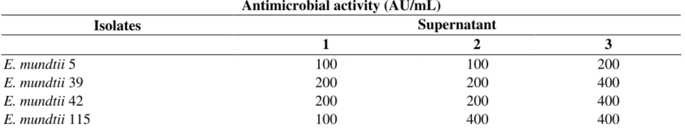
Quantitative analysis of antimicrobial activity of cell-free supernatants expressed in AU /mL
observed for the three supernatants obtained for the same strain. This was observed especially for
E. mundtii 115, which produced 100 AU/mL for the first supernatant, and remarkably, an
antimicrobial activity that was 4 times higher for the other 2 supernatants. E. mundtii 5 presents the lowest AU results and E. mundtii 115 presented the highest ones (Table 2).
Table 2 - Antimicrobial activity results (AU/mL) for the three supernatants prepared with the four E. mundtii
strains, using L. innocua ATCC 33090 as indicator strain
Antimicrobial activity (AU/mL)
Isolates Supernatant
1 2 3
E. mundtii 5 100 100 200
E. mundtii 39 200 200 400
E. mundtii 42 200 200 400
E. mundtii 115 100 400 400
Sensitivity to heat, pH, storage conditions and proteolytic enzymes
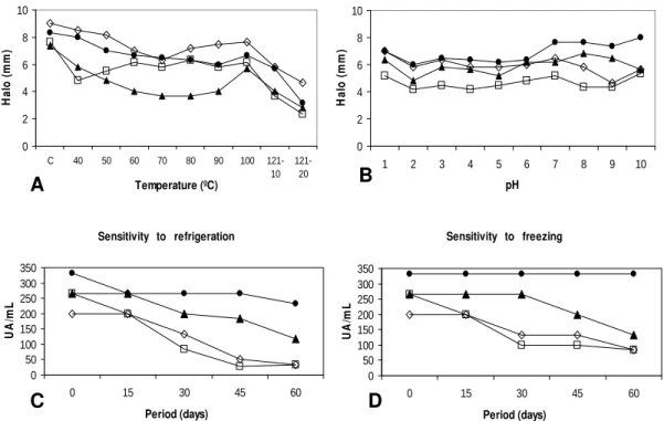
The test carried out to establish the sensitivity of bacteriocins to heat revealed that supernatants maintained antimicrobial activity at all temperatures adopted. The supernatant produced by E. mundtii strain 39 was an exception since it lost all antimicrobial activity when kept at 121o C for 20 min (Fig. 1A). Antimicrobial activity was more affected at 121o C for all supernatants. The pH sensitivity test showed that antimicrobial activity was not entirely lost along the pH range adopted (Fig. 1B). Yet, the greatest decrement in activity was observed at pH 2 for the four supernatants tested. Supernatants 5 and 42 suffered some degree of antimicrobial activity loss at alkaline pHs, whereas supernatants 39 and 115 produced but a narrow increase in inhibition halos. Antimicrobial activity was the same or even higher at neutral pH when compared to the control experiment. The increased inhibition halos under alkaline pH conditions could be due to alterations in peptide diffusion in such conditions.
As regards the storage test, no supernatant lost completely the antimicrobial activity initially exerted during the 60 day refrigeration and freezing experiments. All supernatants were proved to be more stable when frozen (-20o C), exhibiting little antimicrobial activity loss during the 60 day period adopted (Fig. 1D). The same supernatants kept under refrigeration (4o C) for the same period exhibited a more considerable antimicrobial activity loss (Fig. 1C). The greatest detriment was observed for supernatant 39, either under refrigeration or freezing. Supernatant 115
was considered the most stable among the four tested under both storage conditions. The test to measure sensitivity to proteolytic enzymes showed that all supernatants were rendered inactive by trypsin and proteinase K at the two concentrations tested. Lysozyme did not completely inactivate any culture supernatant.
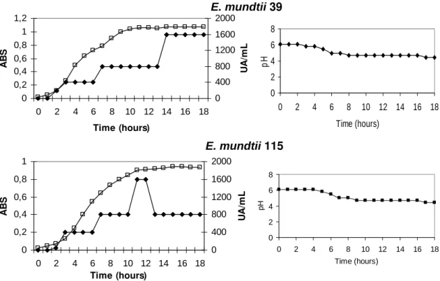
Production kinetics and molecular size of E. mundtii 39 and E. mundtii 115 bacteriocin
After a 1 h incubation period, it was possible to observe an increase in absorbance for both E. mundtii strains 39 and 115. The production of bacteriocins was observed only after a 2 h incubation period. Stationary phase began at approximately 11 h after incubation for strain 39, and roughly 14 h after incubation for strain 115 (Fig. 2). Both strains began to produce bacteriocins during the logarithmic growth phase. Strain 115 presented a peak during the production with 1.600 AU /mL at the end of the logarithmic phase, and partly lost antimicrobial activity during the stationary phase. Strain 39 showed maximum antimicrobial activity of 1.600 AU /mLfor up to 18h incubation. Initial pH of cultures was 6.1, and after 18h incubation, it decreased to 4.4.
The molecular weight estimated for bacteriocins produced by E. mundtii 39 and 115 was between 2.3 and 3.4 kDa (Fig. 3).
Figure 1 - Results of enterocin sensitivity test of cell-free supernatants produced by E. mundtii
strains under (A) heat, (B) pH. Results are presented as halo sizes (mm). Temperatures between 40o C and 100o C were applied for 30 min. The temperature of 121o C was applied for 10 and 20 min. Results of enterocin sensitivity test of cell-free supernatants produced by E. mundtii strains for (C) refrigeration at 4o C and (D) freezing at –20o C. Results are presented as Arbitrary Units (AU mL-1). The data presented in theses graphics are the means of three supernatants for which diferences were not significant (p<0.05). (C=untreated control, = E. mundtii 42, = E. mundtii 39, ◊= E. mundtii 5, = E. mundtii 115).
DISCUSSION
Enterococcus spp. play an important role in the maturation of some kinds of cheese and in the preparation of some probiotic compositions (Franz et al., 1999). Apart from this, they produce bacteriocins, specifically called enterocins, which inhibit the growth of pathogenic and food-deteriorating bacteria, including the genera
Listeria and Clostridium (Giraffa, 1995).
Nevertheless, it is necessary that a strain used commercially was its biological safety duly ascertained. The use of E. faecalis and E. faecium
as biopreservers would be questionable, as both
species infected human hosts deleteriously (De Vuyst et al., 2002).
Forthis reason and due to the paucity of published data on other enterococci species, the E. mundtii
strains were chosen to characterize the bacteriocins the species produces. During the triage experiment carried out in this study, only a low number (four in a total of 70 strains) of bacteriocin-producing strains was found. This was in accordance with the results of other studies. Du Toit et al. (2000) carried out a study with 92 isolates, of which only seven were enterocin producers (7.6%), and Jennes et al. (2000) found only one enterocin-producing strain among 77 LAB tested.
Sensitivity to refrigeration
0 50 100 150 200 250 300 350
0 15 30 45 60
Period (days)
U
A
/m
L
0 2 4 6 8 10
C 40 50 60 70 80 90 100
121-10
121-20 Temperature (ºC)
H
a
lo
(
m
m
)
Sensitivity to freezing
0 50 100 150 200 250 300 350
0 15 30 45 60
Period (days)
U
A
/m
L
0 2 4 6 8 10
1 2 3 4 5 6 7 8 9 10
pH
H
a
lo
(
m
m
)
A B
Figure 2 – Growth kinetics of E. mundtii 39 and E. mundtii 115. Results obtained for antimicrobial activity in AU/mL using the agar well diffusion method and L. innocua as the indicator (), microbial growth in absorbance (620 nm) ( ) and pH
(39 and115) (■), in MRS broth at 35ºC for 18 h.
Figure 3 - Characterization of molecular weight of enterocins produced by E. mundtii strains39 and 115. Comassie Blue gel (A) and antimicrobial activity gel (B) using the indicator strain L. innocua. The arrows indicate the molecular weight at gel A and the inhibition halo at the gel B.
The enterocins produced by Enterococcus sp. typically inhibited Gram-positive bacteria, including the Listeria and Lactobacillus genera; and this was observed even for the Enterococcus
genus itself, as reported in the present study. There are only few enterocins that can inhibit Gram-negative bacteria. To our knowledge, the majority of the studies carried out on enterocins have not
tested the Salmonella genus. The culture supernatants obtained from E. mundtii isolates inhibited two Salmonella Enteriditis strains. In studies that characterized enterocins as producing antimicrobial activity against Salmonella spp., a similar result was found for enterocin 012 produced by Enterococcus gallinarum, which inhibited the growth of Salmonella Typhimurium
0 2 4 6 8
0 2 4 6 8 10 12 14 16 18
Time (hours)
p
H
0 0,2 0,4 0,6 0,8 1
0 2 4 6 8 10 12 14 16 18
Time (hours)
A
B
S
0 400 800 1200 1600
U
A
/m
L
0 0,2 0,4 0,6 0,8 1
0 2 4 6 8 10 12 14 16 18
Time (hours)
A
B
S
0 400 800 1200 1600 2000
U
A
/m
L
E. mundtii 115
0 2 4 6 8
0 2 4 6 8 10 12 14 16 18
Time (hours)
p
H
2,3 3,4 6,2
A B
(Jennes et al., 2000; Lauková et al., 1998; 2000; 2004). Lactobacillus fermentum was not inhibited by any of the culture supernatants tested, but L. plantarum was weakly inhibited. These could be interesting results, as the species mentioned were commonly used in bacteria associations developed as additives to food fermentation processes and probiotic combinations. Todate, only two studies have characterized enterocins produced by E. mundtii, and only one of these enterocins was identified and characterized as mundticin (Bennik et al., 1998; Kawamoto et al., 2002). Which inhibited L. monocytogenes, L. plantarum, and
Enterococcus strains (Kawamoto et al., 2002). The estimated molecular size of mundticin was 3.4 kDa, which was in accordance with the results found in the present study.
The cross inhibition test revealed that strains 5 and 39 were inhibited by culture supernatant 115, which suggested that this supernatant produced a different enterocin. Yet, in the light of the fact that strain 115 was not inhibited by supernatants 5 and 39, it was possible to conclude that strain 115 was resistant to the supernatants mentioned, and could be producing more than one enterocin.
Enterocins were shown to be consistently stable under high temperatures, different pH ranges, and long storage periods. These characteristics were typical of various enterocins already characterized, such as enterocin A (Aymerich et al., 1996), enterocin B (Casaus et al., 1997), enterocin P (Cintas et al., 1997) produced by E. faecium, and enterocin 1071A and 1071B, produced by E. faecalis (Balla et al., 2000). The stability exhibited by enterocins under these conditions is a very important feature, since the main use of these peptides lies in biopreservation of fermented foods. The losses in antimicrobial activity after the treatment with proteolytic enzymes reveal the proteic nature of enterocins.
The tests carried out to establish virulence factors and resistance to antimicrobial agents are valuable tools when choosing a safer strain to be used as biopreservative. The four E. mundtii strains presented a capsule and produced negative results for hemolysin, gelatinase and adhesin production tests. The resistance profile exhibited by the strains involved between 3 and 5 antibiotics, no strain was resistant to vancomycin. (data not shown).
The kinetics of production showed primary metabolite kinetics, as the production of enterocins started as of the logarithmic growth phase. Such a
finding had previously been reported in studies on enterocins (Torri Tarelli et al., 1994; Franz et al., 1996; Mareková et al., 2003). The decrease in antimicrobial activity observed for strain 115 after a longer incubation time could be due to the degradation of the bacteriocin by proteolytic enzymes present in the medium, or else by the low pH (Torri Tarelli et al., 1994). Another aspect to be considered would be the re-adsorption of enterocin by the cell through its membrane in low pH medium (Franz et al., 1996). Similar results have been found by Herranz et al. (2001) for E. faecium P21, in which the activity peak was observed after a 12 h incubation period, as of the early stationary phase and which likewise had a final pH of 4.4. Mareková et al. (2003) analyzed the production of enterocins in E. faecium strain EK13 at 37oC without pH adjustments in a fermentation vat. The production started within 1 h incubation period, and the peak activity was at the 7th h of incubation (25,600 AU/mL), with the beginning of the stationary phase at 6 h of incubation. The final pH was 4.4 after a 25 h incubation period. The kinetics of the production of E. faecium FAIR – E198 revealed a peak activity of 800 AU/mL after a 5 h fermentation period in MRS broth supplemented with 2% glucose at 37oC and pH kept at 6.5. Under the same conditions, but with pH kept at 5.5, a lower activity was determined (200 AU/mL) after a 10 h incubation period (Sarantinopoulos et al., 2002). Among the enterocins characterized todate, the majority have been ranked under class IIa, with molecular weight under 10 kDa (Eijsink et al., 2002). Most of the enterocins showed their molecular weight between 2 and 5 kDa. Enterocins A and B are those that have been best characterized and are produced by E. faecium
strains, with molecular weight of 4.8 and 5.4 kDa, respectively. Other enterocins, such as enterocin 32 (produced by E. faecium) has a molecular weight of 5 kDa and enterocin P has 4.5 kDa (Ennahar et al., 2000). All these molecular weight figures are over the molecular weight observed at the present study. However, enterocin O12, produced by E. galinarum presented a molecular size of 3.4 kDa (Jennes et al., 2000). Similarly, enterocin ON-157, produced by E. faecium strain NIAI 157, showed one of the lowest molecular weight characterized (2.5 kDa, Ohmomo et al., 2000).
biopreservation agents exhibited by these strains and the enterocins they produced. This was observed in the light of the fact that the enterocins and the strains analyzed showed little sensitivity to high temperatures and pH variations. Also, the capacity to inhibit Listeria monocytogenes and
Salmonella Enteritidis — two very important pathogens associated to food poisoning — reinforced the use of E. mundtii in food preservation strategies.
ACKNOWLEDGEMENTS
We are grateful to Prof. Dr Patricia Valente who kindly supplied the yeast isolates used as indicators. This study was supported by a grant from CNPq (National Council for Research Development) and CAPES.
RESUMO
O objetivo do presente estudo foi caracterizar bacteriocinas produzidas por 70 cepas de
Enterococcus mundtii. Estas foram caracterizadas quanto a sua atividade antimicrobiana, sensibilidade ao aquecimento, pH, armazenamento e enzimas proteolíticas. Foi também determinada sua cinética de produção e peso molecular. Entre as 70 cepas analisadas, quatro apresentaram atividade antibiótica contra Listeria innocua, L.
monocytogenes, Lactobacillus plantarum, e
Salmonella Enteritidis. Esta atividade foi mantida em temperaturas até 121ºC por 20 minutos, e sob condições de pH entre 2,0 e 10,0. A atividade antimicrobiana foi mantida nos testes de armazenamento a -20°C, por 60 dias. A cinética de produção revelou picos de atividade de 1600 AU/mL durante a fase logarítmica de crescimento e o peso molecular foi de aproximadamente 3,0 kDa. A caracterização dos produtos com atividade antimicrobiana revelaram suas naturezas protéicas, cinéticas de metabólito primário e peso molecular semelhante aos das enterocinas já purificadas.
REFERENCES
Aymerich, T.; Holo, H.; Havarstein, L. S.; Hugas, M.; Garrida, M. and Nes, I. (1996), Biochemical and genetic characterization of enterocin A from
Enterococcus faecium, a new antilisterial bacteriocin in the pediocin family of bacteriocins. Appl. and Environ. Microbiol.,62, 1676-1682.
Ausubel, F.; Brent, R.; Kingston, R.E.; Moore, D.D.; Seidman, J.G.; Smith, J.A. and Struhl, K. (1987) Current protocols in molecular biology vol. 2 New York John Wiley and Sons.
Balla, E.; Dicks, L. M. T.; Du Toit, M.; Van Der Merwe, M. J. and Holzapfel, W. H. (2000), Characterization and cloning of the genes encoding enterocin 1071A and enterocin 1071B, two antimicrobial peptides produced by Enterococcus faecalis BFE 1071. Appl. and Environ. Microbiol.,
66, 1298-1304.
Bennik, M. H. J.; Vanloo, B.; Brasseur, R.; Gorris, L. G. M. and Smid, E. J. (1998), A novel bacteriocin with a YGNGV motif from vegetable-associated
Enterococcus mundtii: full characterization and interaction with target organisms. Biochim et Biophys. Acta,1373, 47-58.
Casaus, P.; Nilsen, T.; Cintas, L.M.; Nes, I. F.; Hernández, P. E. and Holo, H. (1997), Enterocin B, a new bacteriocin from Enterococcus faecium T136 which can act synergistically with enterocin A.
Microbiology,143, 2287-2294.
Cintas, L.; Casaus, P.; Havarstein, L. S.; Hernández, P. E. and Nes, I. F. (1997), Biochemical and genetic characterization of enterocin P, a novel sec-dependent bacteriocin from Enterococcus faecium P13 with a broad antimicrobial spectrum. Appl and Environ. Microbiol., 63, 4321-4330.
Cintas, L.; Casaus, P.; Herranz, C.; Havarstein, L. S.; Holo, H.; Hernández, P. E. and Nes, I. F. (2000), Biochemical and genetic evidence that Enterococcus faecium L50 produces enterocins L50A and L50B, the sec-dependent enterocin P, and a novel bacteriocin secreted without an N-terminal extension termed enterocin Q. J. of Bacteriol.,182, 6806-6814. De Vuyst, L.; Foulquié Moreno, M. and Revets, H.
(2002), Screening for enterocins and detection of hemolysin and vancomycin resistance in enterococci of different origins. Intern. J. of Food Microbiol.,
2635, 1-20.
Du Toit, M.; Franz, C. M. A. P.; Dicks, L. M. T. and Holzapfel, W. H. (2000), Preliminary characterization of bacteriocins produced by Enterococcus faecium
Eijsink, V.; Axelsson, L.; Diep, D. B.; Havarstein, L. S.; Holo, H. and Nes, I. F. (2002), Production of class II bacteriocins by lactic acid bacteria; an example of biological warfare and communication. Antonie van Leeuwenhoek, 81, 639-654.
Ennahar, S.; Sashihara, T.; Sonomoto, K. and Ishizaki, A. (2000), Class IIa bacteriocins: biosynthesis, structure and activity. FEMS Microbiol. Reviews,24, 85-106.
Franz, C. M. A. P.; Schillinger, U. and Holzapfel, W. H. (1996), Production and characterization of enterocin 900, a bacteriocin produced by
Enterococcus faecium BFE 900 from black olives. Intern.J. of Food Microbiol.,29, 255-270.
Franz, C. M. A. P.; Holzapfel, W. H. and Stiles, M. E. (1999), Enterococci at the crossroads of food safety?
Intern. J. of Food Microbiol., 47, 1-24.
Giraffa, G. (1995), Enterococcal bacteriocins: their potential use as anti-Listeria factors in dairy technology. Food Microbiology,12, 551-556. Herranz, C.; Casaus, P.; Mukhopadhyay, S.; Martínez,
J. M.; Rodríguez, J. M.; Nes, I. F.; Hernández, P. E. and Cintas, L. M. (2001), Enterococcus faecium P21: a strain occurring naturally in dry-fermented sausages producing the class II bacteriocins enterocin A and enterocin B. Food Microbiology, 18, 115-131. Jennes, W.; Dicks, L. M. T. and Verwoerd, D. J.
(2000), Enterocin 012, a bacteriocin produced by
Enterococcus gallinarum isolated from the intestinal tract of ostrich. J. of Appl. Microbiol., 88, 349-357. Kawamoto, S.; Shima, J.; Sato, R.; Eguchi, T.;
Ohmomo, S.; Shibato, J.; Horikoshi, N.; Takeshita, K. and Sameshima, T. (2002), Biochemical and genetic characterization of mundticin KS, an antilisterial peptide produced by Enterococcus mundtii NFRI 7393. Appl. and Environ. Microbiol.,
68, 3830-3840.
Lauková, A.; Czokková, S.; Vasilková, Z.; Juris, P. and Mareková, M. (1998), Occurrence of bacteriocin production among environmental enterococci. Letters in Appl. Microbiol.,27, 178-182.
Lauková, A.; Juris, P.; Vasilková, Z. and Papajová, I. (2000), Treatment of sanitary-important bacteria by bacteriocin substance V24 in cattle dung water.
Letters in Appl. Microbiol., 30, 402-405.
Lauková, A.; Guba, P.; Nemcova, R. and Mareková, M. (2004), Inhibition of Samonella enterica serovar
Dusseldorf by enterocin A in gnotobiotic Japanese quails. Vet. Med.-Czech.,49, 47-51.
Lewus, C.; Kaiser, A. and Montville, T. (1991), Inhibition of food-borne bacterial pathogens by bacteriocins from lactic acid bacteria isolated from meat. Appl. and Environ. Microbiol., 57, 1683-1688. Mareková, M.; Lauková, A.; De Vuyst, L.; Skaugen, M.
and Nes, I. F. (2003), Partial characterization of bacteriocins produced by environmental strain
Enterococcus faecium EK13. J. of Appl. Microbiol.,
94, 523-530.
Sarantinopoulos, P.; Leroy, F.; Leontopoulou, E.; Georgalaki, M. D.; Kalantzopoulos, G.; Tsakalidou, E. and De Vuyst, L. (2002), Bacteriocin production by Enterococcus faecium FAIR-E 198 in view of its application as adjunct starter in Greek Feta cheese making. Intern. J.l of Food Microbiol., 72, 125-136. Ohmomo, S.; Murata, S.; Katayama, N.; Nitisinprasart,
S.; Kobayashi, M.; Nakajima, T.; Yajima, M. and Nakanishi, K. (2000), Purification and some characteristics of enterocin ON-157, a bacteriocin produced by Enterococcus faecium NIAI 157. J. of Appl. Microbiol., 88, 81-89.
Tagg , J. R. and Mcgiven, A. R. (1971), Assay system for bacteriocins. Applied Microbiol.,21, 943.
Torri Tarelli, G.; Carminati, D. and Giraffa, G. (1994), Production of bacteriocins active against Listeria monocytogenes and Listeria innocua from dairy enterococci. Food Microbiology, 11, 243-252.