UNIVERSIDADE DA BEIRA INTERIOR
Ciências
Role of STEP in dopaminergic synapses
Emika Calado Teixeira
Dissertação para obtenção do Grau de Mestre em
Bioquímica
(2º ciclo de estudos)
Orientadora: Prof
a. Doutora Graça Maria Fernandes Baltazar
iii
“Bad times have a scientific value. These are occasions a good learner would not miss.”v
Acknowledgements
Aos meus pais por sempre me terem ensinado que para tudo na vida é preciso esforço, a não desistir, a ter discernimento, a respeitar o tempo apropriado dos acontecimentos, a ser justa e que mais valiosas que as vitórias são os que nos sustentam nas derrotas;
Aos meus irmãos Anderson e Sayuri pelo amor que me transmitem em cada abraço e por nunca pouparem esforços para colocar um sorriso no meu rosto;
À minha tia Maria José, que sempre me acolheu, por ser esse grande porto seguro na minha vida sem o qual a conclusão dessa tese não seria possível;
Ao Ivo, meu maior suporte, quem enxugou cada lágrima e me fez acreditar que tudo nunca deixou de ser possível, que sempre esteve lá independente do meu egoísmo e da dificuldade. Por tudo que me tem ensinado, por me fazer ser melhor e por me fazer feliz sempre;
À toda minha família por sempre me acolher na “volta a casa” e me mostrar que sou e sempre serei amada;
À Débora, a DJ, irmã de tese e PQT, que com as suas loucuras sempre deu melodia e muitas gargalhadas às longas noites de trabalho e que mesmo nos momentos mais angustiantes nunca deixou faltar um abraço apertado e um xêru carinhoso de irmã, e a certeza de que tudo ficaria bem;
À Julieta, que prontamente me acolheu e a quem devo toda a aprendizagem laboratorial, agradeço todo o empenho, por me ter acompanhado sempre e mais que tudo pela amizade que sempre levarei comigo;
Às minhas amigas Patrícia, pela imensa fortaleza que é e pela força que sempre me transmitiu, Daniela, por ser um apoio e pela amizade inesquecível, Nádia, por todas as longas conversas e confissões, e Elisabete, por toda a tranquilidade transmitida e por ser sempre compreensiva. A todas pelos momentos incríveis que passámos, por sempre ouvirem as minhas angústias, por estarem presentes quando mais precisei, e independente do caminho que se segue estarão sempre comigo;
Às minhas de sempre, Aysa, por tanto que nem tenho como agradecer, Trifas e PT, pela amizade incrível, Diva e Inocente, por sempre acreditarem no meu potencial. Levar-vos-ei sempre comigo;
Ao Ricardo e a Alice, Miguel e a Carina, Jorge e Bianca, Olga e Henrique, por todo o apoio incondicional. A todos os amigos dançantes, bem-haja!
Aos meus amigos e companheiros de sala, Joel por todo o apoio nos momentos difíceis e por toda a compreensão, Sandra e Helena por todos os conselhos, seja no laboratório ou na vida, pelas conversas e pelo ambiente acolhedor que sempre houve, e Júnior e Ana, por fazerem estes últimos meses mais divertidos, muito obrigada;
À Catarina, Marta Esteves, Marta Pereira, André e Jéssica, pelas risadas nos corredores deste centro, pelas conversas de laboratório, pelo apoio e por toda a compreensão;
vi
À Professora Graça Baltazar, por toda a paciência e preocupação, por toda a aprendizagem, por todo o encorajamento quando foi preciso, por todo o apoio, por ter sido mãe em muitos momentos e por não me ter deixado desistir quando tudo parecia impossível. Muito obrigada!
vii
Resumo
A proteína fosfatase de resíduos de tirosina enriquecida no estriado (STEP) é um dos membros da família PTP, e existe maioritariamente em duas isoformas, a STEP61 e a STEP46 sendo
diferencialmente expressas durante o desenvolvimento. A STEP é conhecida por se opor ao fortalecimento sináptico através da desfosforilação de moléculas chave envolvidas na sinalização neuronal. A maioria dos trabalhos publicados sobre a atividade e regulação da STEP têm foco no seu papel pós-sináptico, embora recentemente tenha sido confirmada a sua presença pré-sinapticamente em neurónios glutamatérgicos, onde contribui para a regulação dos níveis de cálcio. Em relação aos neurónios dopaminérgicos, foi reportado que os níveis de STEP estão aumentados em pacientes com a doença de Parkinson que tenham a mutação da parkina e em murganhos modelo da doença. A mutação na parkina pode levar a uma acumulação de STEP, que pode estar envolvida na morte neuronal associada a essa patologia. A doença de Parkinson é idiopática e caracteriza-se pela disfunção motora devido a perda progressiva de neurónios dopaminérgicos que ocorre na substantia nigra pars compacta. Estudos revelaram elevados níveis de tirosina hidroxilase (TH), fosforilação da THSer31 e elevados níveis de dopamina no estriado de murganhos STEP KO, o que sugere que a STEP está a agir como depressora da transmissão dopaminérgica. Além disso, murganhos STEP KO ou com fenótipo selvagem tratados com o inibidor da STEP, mostraram elevada resistência a lesão dopaminérgica exercida pelo MPTP. Este trabalho tem como objetivo verificar se a STEP pré-sináptica afeta a formação e a função das sinapses dopaminérgicas. Os nossos dados mostraram que os neurónios dopaminérgicos expressam STEP nos terminais pré-sinápticos e que a inibição da STEP não alterou significativamente os níveis dos marcadores sinápticos em culturas de neurónios do mesencéfalo ventral de murganhos. A avaliação específica dos terminais dopaminérgicos mostrou que a inibição da STEP também não alterou a intensidade dos punctas nem de sinaptofisina nem de sinapsina. Os dados recolhidos da avaliação morfológica sugerem um efeito de compensação na condição controlo do MPP+. A redução no número de neurónios
dopaminérgicos pode estar a levar ao aumento do número de neurites para compensar a carência na transmissão dopaminérgica. Mais estudos serão necessários para clarificar o papel desempenhado pela STEP nos neurónios dopaminérgicos e além disso ajudar a perceber se a STEP pré-sináptica contribui para a morte retrógrada dos neurónios dopaminérgicos observada na doença de Parkinson.
Palavras-chave
STEP; neurónios dopaminérgicos, sinapses dopaminérgicas, Doença de Parkinson; neurodegeneração; células gliais
ix
Resumo alargado
A família de proteínas fosfatases de tirosina (PTP) desempenha um papel fundamental desfosforilando proteínas que afetam processos de sobrevivência e morte celular. A proteína fosfatase de resíduos de tirosina enriquecida no estriado (STEP), membro da família PTP, é conhecida por contrariar o fortalecimento sináptico ao desfosforilar proteínas chave do processo de sinalização neuronal. O estudo dos substratos da STEP é fundamental para a compreensão da sua atividade e dos mecanismos de regulação. A maior parte dos trabalhos publicados sobre a STEP, centram-se no seu papel ao nível pós-sináptico, pouco se sabendo sobre o papel exercido pré-sinapticamente. Estudos reportaram que os níveis de STEP estão aumentados tanto no estriado de doentes com a doença de Parkinson como no modelo da doença induzido pelo MPTP em murganhos. Além disso, murganhos STEP KO mostraram ser mais resistentes ao insulto realizado com a toxina dopaminérgica MPTP, o mesmo ocorrendo com murganhos com fenótipo selvagem tratados com o inibidor específico de STEP, TC-2153. A DP é uma desordem do movimento crónica e progressiva causada pela perda de neurónios dopaminérgicos da substantia nigra pars compacta que projetam para o estriado. Atualmente, os fatores envolvidos na origem deste processo neurodegenerativo não estão bem estabelecidos. No entanto, vários estudos indicam que a neurodegeneração associada à DP dá-se através de um processo de morte retrógrada iniciado nos terminais estriatais. Dados revelaram que o estriado de murganhos STEP KO apresenta níveis mais elevados de tirosina hidroxilase (TH), maior fosforilação de THSer31, bem como níveis aumentados de dopamina, o que sugere que a STEP pode atuar como inibidor da transmissão dopaminérgica. Pouco se sabe acerca do papel pré-sináptico dessa fosfatase, todavia recentemente foi reportado que a STEP está localizada também na região pré-sináptica de sinapses glutamatérgicas e está implicada na regulação dos níveis de cálcio nos terminais destes neurónios. Uma vez que a STEP está de facto presente ao nível pré-sináptico e havendo dados que sugerem a sua influência depressora na transmissão sináptica dopaminérgica, este trabalho tem como intuito explorar o impacto da STEP pré-sináptica na formação e atividade das sinapses dopaminérgicas. Este trabalho possibilitou localizar pela primeira vez a STEP ao nível pré-sináptico em terminais dopaminérgicos. Em culturas enriquecidas em neurónios da região do mesencéfalo ventral foi possível verificar uma tendência para aumento dos níveis dos marcadores pré-sinápticos sinaptofisina, sinapsina I e VMAT 2 quando a STEP foi inibida. Na presença de células gliais o nível do marcador sinaptofisina reduziu significativamente quando a STEP foi inibida. Quando avaliados especificamente os terminais dopaminérgicos em culturas de neurónios do mesencéfalo ventral, verificou-se que na situação de exposição a MPP+ o número de células
dopaminérgicas diminuiu 35% em relação ao controlo, validando o modelo proposto para o estudo. A avaliação dos puncta realizada nos terminais destes neurónios revelou uma redução consistente do marcador sinaptofisina aquando da administração da toxina dopaminérgica, e quando a STEP foi inibida os níveis não sofreram alteração. Portanto, a modulação da STEP foi
x
capaz de prevenir a redução dos níveis de sinaptofisina que ocorreu na situação de lesão. Por outro lado, os resultados da análise morfológica da arborização neuronal revelaram que a inibição da STEP alterou significativamente o comprimento máximo dos neurites, mas não alterou o comprimento médio nem o número de neurites por neurónio. Já na condição de exposição a MPP+ ocorreu um aumento do número de neurites embora se verifique que o
comprimento destes é menor. Os dados apresentados podem sugerir um efeito de compensação pois embora a quantidade de células dopaminérgicas seja menor elas estão mais ramificadas, de forma a compensar essa redução.
Este trabalho permitiu localizar a STEP ao nível pré-sináptico nos neurónios dopaminérgicos. Embora sejam necessários mais os dados obtidos neste trabalho, em conjunto com o descrito na literatura, sugerem que a STEP pré-sináptica pode estar envolvida na morte neuronal associada à doença de Parkinson, sendo essa fosfatase um potencial alvo terapêutico nesta patologia.
xi
Abstract
Striatal-enriched protein tyrosine phosphatase (STEP) is a member of the PTP family, and exists in two major isoforms, STEP61 and STEP46, being differentially expressed during development.
STEP is known to oppose synaptic strengthening by the dephosphorylation of key molecules involved in neuronal signaling. Most of the published works on STEP activity and regulation focus on its postsynaptic role, but recently STEP has also been located in glutamatergic presynaptic terminals where it contributes to the regulation of calcium levels. Concerning dopaminergic systems, it was reported that STEP levels are increased in Parkinson’ disease (PD) mice model and in parkin-mutated PD patients. A parkin mutation may lead to STEP accumulation, which may contribute to PD-associated dopaminergic neuronal death. PD is an idiopathic pathology characterized by motor dysfunction due to the progressive loss of dopaminergic neurons in the substantia nigra pars compacta. Reported data show high levels of tyrosine hydroxylase (TH), THSer31 phosphorylation and increased dopamine levels in striatum of STEP KO mice, suggesting that STEP acts as a repressor of dopaminergic transmission. Additionally, STEP KO mice or wild type (WT) mice treated with a specific STEP inhibitor shown increased resistance to the dopaminergic toxin MPTP. This work aims to explore if presynaptic STEP affects the formation and function of dopaminergic synapses. Our data showed that dopaminergic neurons express STEP at the presynaptic terminals and STEP inhibition did not significantly increase the levels of presynaptic markers in neuron cultures from mice midbrain. The specific evaluation of dopaminergic neurons showed that STEP inhibition does not affect synaptophysin levels. The morphological data suggest a compensation effect when the dopaminergic toxin was administrated. The reduction of dopaminergic cells may lead to an increase in the number of neurites to compensate the lack of dopaminergic transmission. Taking together, more studies are necessary to clarify the role played by presynaptic STEP in dopaminergic neurons and whereas presynaptic STEP contributes to retrograde cell death observed in PD pathology.
Keywords
STEP; dopaminergic neurons, dopaminergic synapses, Parkinson’ Disease; neurodegeneration; glial cells.
xiii
Index
Resumo ...vii Palavras-chave ...vii Resumo alargado ... ix Abstract ... xi Keywords ... xi List of figures ... xvTable list ... xvii
Acronyms list ... xix
1 Introduction ... 1
1.1 Striatal-enriched protein tyrosine phosphatase ... 1
1.1.1 Expression, domains and structure ... 1
1.2 STEP synaptic functions ... 3
1.2.1 STEP substrates ... 4
1.2.1.1 Mitogen activated protein kinases (MAPK) – ERK1/2 and p38 ... 4
1.2.1.2 Glutamate receptors ... 5 1.2.1.3 Fyn ... 7 1.2.1.4 PTPα ... 7 1.2.1.5 Pyk2 ... 7 1.2.2 STEP regulation ... 7 1.2.2.1 Phosphorylation ... 8 1.2.2.2 Ubiquitination ... 8 1.2.2.3 Local translation ... 8 1.2.2.4 Proteolytic cleavage ... 9 1.2.2.5 Dimerization ... 9
1.2.2.6 Pharmacologic regulation of STEP ... 9
1.2.2.7 Regulation by brain-derived neurotrophic factor (BDNF) ... 10
1.3 Presence of STEP in presynaptic terminals ... 10
1.3.1 Glutamatergic synapses ... 10
1.3.2 Dopaminergic synapses ... 12
1.3.2.1 Idiopathic Parkinson's disease and STEP ... 12
2 Aims ... 15
3 Materials and methods ... 17
xiv
3.1.1 Neuron and neuron-glial cells midbrain co-cultures ... 17
3.1.2 Ventral midbrain (VM) glial cells cultures ... 17
3.1.3 Neuron-micro-islands and VM-glial cells bunker cocultures ... 18
3.1.4 Treatments ... 18
3.1.5 Protein extraction ... 18
3.2 Western blot ... 19
3.3 Immunocytochemistry ... 20
3.3.1 Neurites analysis and Sholl analysis ... 21
3.3.1.1 Cell counting ... 21
3.3.1.2 Neurite analysis ... 21
3.3.1.3 Sholl Analysis ... 21
3.3.2 Dopaminergic terminals puncta analysis ... 23
3.4 Statistical analysis ... 23
4 Results ... 25
4.1 STEP at the dopaminergic synapses... 25
4.2 STEP as a modulator of synapses ... 26
4.2.1 Neuron-enriched culture ... 26
4.2.2 Neuron-glia culture ... 28
4.3 Does STEP modulate dopaminergic synaptic terminals? ... 30
4.3.1 Effect of STEP activity on the expression of synaptic markers in dopaminergic terminals ... 31
4.4 Effect of STEP activity on the arborization of dopaminergic neurons ... 32
5 Discussion ... 35
xv
List of figures
Figure 1. Representative scheme of STEP isoforms ... 2 Figure 2. Illustration of tripartite synapses ... 4 Figure 3. Representative image of STEP activity and regulation mechanisms at the
postsynaptic level ... 6 Figure 4. Structure of the STEP pharmacologic inhibitor known as TC-2153 ... 9 Figure 5. Role of STEP in the regulation of neurotransmitter release ... 11 Figure 6. Schematic representation of the synapse with identification of presynaptic proteins ... 11 Figure 7. Schematic representation of the MPTP absorption and intracellular effects ... 13 Figure 8. Schematic representation of the experimental procedures ... 19 Figure 9. Flow diagram of the analysis performed from immunocytochemical analysis of Syn++TH+, and SYP++VMAT 2+ ... 21
Figure 10. Representation of Sholl analysis ... 22 Figure 11. Presence of STEP protein in the presynaptic region of dopaminergic neurons ... 25 Figure 12. Effect of STEP inhibition on the levels of synaptic markers in control conditions and in cells exposed to the dopaminergic toxin MPP+ ... 27
Figure 13. Effect of STEP inhibition on synaptic markers levels in cells exposed to the
dopaminergic toxin MPP+ in the presence of glia ... 29 Figure 14. TH+ cells per micro-island decrease after an MPP+-induced injury ... 30
Figure 15. Changes in Synaptic markers in cells exposed to MPP+ ... 31
Figure 16. Morphologic analysis of neurites in dopaminergic neurons exposed to the STEP inhibitor ... 33 Figure 17. Analysis of neuronal arborization by the Sholl method ... 34
xvii
Table list
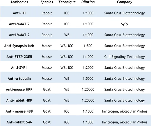
Table 1. Primary and secondary antibodies used for immunocytochemical and western blot techniques, the respective species, dilution, and company ... 22
xix
Acronyms list
6-OHDA - 6-hydroxydopamine
α7nAChR - α7 nicotinic acetylcholine receptor
AMPAR - alpha-amino-3-hydroxy-5-methyl-4-isoxazole propionic acid glutamate receptors BDNF - Brain-derived neurotrophic factor
BSA - Paraformaldehyde CNS - Central Nervous System
CREB - cAMP response element binding protein Cys - Cystein
D1DR - Dopamine receptor D1 DA - Dopaminergic neurons
DARPP-32 - Dopamine- and cAMP-regulated phosphoprotein DAT - Dopamine Transporter
DOPAL - 3, 4‐dihydroxyphenylacetaldehyde Elk1 - ETS Like-1 protein
ER - Endoplasmic Reticulum
ERK1/2 - Extracellular signal-Regulated Kinase 1/2 GluA1/GluA2 - subunit of the AMPAR
GluN2B - subunit of the NMDAR FBS - Foetal Bovine Serum
IP3R - 1,4,5-trisphosphate receptor IPD - Idiopathic Parkinson's disease KIM - Kinase Interaction Motif KO - Knockout
LTP – Long-term Potentiation LTD – Long-term Depression
xx
MAO-B - Monoamine Oxidase B
MAPK - Mitogen-Activated Protein Kinase MEM - Minimum Essential Medium
mGluR5 – Metabotropic Glutamate Receptor subtype 5 MPP+ - 1-methyl-4-phenylpyridinium
MPTP - 1-methyl-4-phenyl-1,2,3,6-tetrahydropyridine mRNA - Messenger RNA
NMDAR - N-methyl-D aspartate receptor OCT-3 - Organic Cation Transporter 3 PBS - Phosphate Buffered Saline PKA - Protein Kinase A
PD - Parkinson's disease PDL - Poly-D-lysine
p-ERK1/2 – phosphorylated Extracellular signal-Regulated Kinase 1/2 PFA - Paraformaldehyde
PINK1 - PTEN-induced kinase 1 PLCγ – Phospholipase C γ PP - Polyproline
PP1 - Protein phosphatase 1 PP2B - Protein Phosphatase 2B PSA - Ammonium Persulphate PSD - Postsynaptic Density
PTP - Protein Tyrosine Phosphatase PVDF - Polyvinylidene Difluoride Pyk2 - Proline-rich tyrosine kinase 2 SDS - Sodium Dodecyl Sulfate Ser - Serine
xxi
SVs - Synaptic Vesicles SYN - Synapsin
SYP - Synaptophysin TBS - Tris Buffer Saline
TC-2153 - 8-(Trifluoromethyl)-1,2,3,4,5-benzopentathiepin-6-amine hydrochloride TEMED - Tetramethylethylenediamine
TH – Tyrosine Hydroxylase TM - Transmembrane domains
TrkB - Tropomyosin receptor kinase B UPS - Ubiquitin/Proteasome System VM – Ventral Midbrain
VMAT – Vesicular Monoamine Transporter WT – Wild type
1
1 Introduction
1.1 Striatal-enriched protein tyrosine phosphatase
1.1.1 Expression, domains and structure
Striatal-enriched protein tyrosine phosphatase (STEP), encoded by the PTPN5 gene, exists in two major isoforms originated by alternative splicing, STEP61 and STEP46 (Boulanger et
al., 1995; Bult et al., 1996). These two STEP isoforms are differentially distributed in brain regions. Both isoforms main are present in the striatum, central nucleus of the amygdala and in the optic nerve, while STEP61 is expressed in the hippocampus, neocortex, spinal cord and
lateral amygdala (Boulanger et al., 1995; Lorber et al., 2004). STEP46 is a cytosolic variant,
whereas STEP61 is directed to endomembranes, like postsynaptic density (PSD) and endoplasmic
reticulum (ER), by the 172 amino acids sequence tagged in their amino terminal portion (Fig. 1) (Bult et al., 1996). This spatial distribution is imperative for its function, particularly for glutamate receptors trafficking (Snyder et al., 2005). Besides the mentioned isoforms, there are two less expressed STEP variants, STEP38 and STEP20 (Fig. 1) (Sharma et al., 1995).
The pattern of STEP isoforms expression changes during development (Raghunathan et al., 1996). Although the levels of membrane isoform are relatively high at birth and in adulthood, the cytosolic form does not appear until the sixth postnatal day. The profile of STEP46 expression suggests a role in synaptogenesis, as it increases from day 14 to 4 weeks of
age and remains stable throughout adulthood (Raghunathan et al., 1996). According to Kim et al., 2008, beyond the expression pattern, STEP colocalization with some proteins also change during development (Kim et al., 2008). At the tissue level, STEP is expressed mostly by neurons, but glial cells also exhibit some expression, however its expression has been reported after ischemic injury (Hasegawa et al., 2000).
The active isoforms, STEP61 and STEP46, share a protein tyrosine phosphatase (PTP)
catalytic domain at the C-terminal region (Fig. 1). The PTP domain contains a conserved sequence with a cysteine residue fundamental for its catalytic activity (Bult et al., 1996). STEP, such as PTP-SL and HePTP, contains a kinase interaction motif (KIM) (Hendriks et al., 2009; Mustelin et al., 2005). The two other STEP isoforms, STEP38 and STEP20, are catalytically inactive
because they do not possess the PTP domain. Their functions remain unknown (Bult et al., 1996; Sharma et al., 1995). The KIM domain is required for the interactions between STEP and his substrates, and it is only present in the catalytic active isoforms (Fig. 1).
2
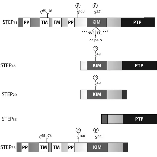
Figure 1. Representative scheme of STEP isoforms. The four main STEP variants are, STEP61, STEP46,
STEP38 and STEP20. STEP61 and STEP46 are the mostly expressed isoforms in the central nervous system (CNS). The scheme represents the KIM domain for substrate association and the consensual PTP sequence, [I/V] HCxAGxxR [S/T] G, which is necessary for the phosphatase activity. The other variants represented, STEP38 and STEP20, are inactive as they do not contain the PTP sequence. Their functions are unknown. STEP33 is generated by the cleavage by calpain in the domain of substrate association, KIM, thus interrupting the ability of STEP33 to interact with its substrates. STEP61 also contains 172 additional amino acids in its amino-terminal region, which contains two transmembrane domains (TM), two polyproline (PP) rich regions and an adjacent PEST sequence. The TM regions direct the STEP61 to the ER as well as to the PSD. For the KIM domain, the PP regions give specificity to the substrate. Finally, the two Cys65 and Cys76 cysteine residues found in the TM region mediate STEP dimerization and diminishing its phosphatase activity (Adapted from Goebel-Goody et al., 2012).
3
The KIM ability to interact with substrates, is given by a serine residue, and is lost when this residue is phosphorylated (Fig. 1) (Paul et al., 2000). The amino acid sequence at the STEP61
amino terminal contains two essential hydrophobic domains capable to direct it to the neuronal membranes. Moreover, the N-terminal region of STEP61 contains also two domains rich in
polyproline (PP) and PEST motives rich in proline (P), glutamic acid (E), serine (S) and threonine (T) (Bult et al., 1996). The PP domains are required to the association between the phosphatase and two of STEP substrates, the first one for Fyn kinase (Nguyen et al., 2002), while the second enables the binding to the proline-rich tyrosine kinase 2 (Pyk2) (Xu et al., 2012). The PEST sequences known to mediate a fast degradation (Rechsteiner & Rogers, 1996), contribute to STEP recognition and undergo proteolytic cleavage or ubiquitination (Spencer et al., 2004). STEP is found in excitatory and inhibitory neurons, in which it regulates the phosphorylation of their substrates, thus performing a key role at the synaptic level (Goebel-Goody et al., 2012).
1.2 STEP synaptic functions
At the beginning of the 20th century, scientists named the communication between nerve cells as synaptic transmission. The neuron soma gives rise to dendrites; structures specialized in receiving information from other neurons. Most synapses have no physical continuity between the pre- and postsynaptic terminals (Fig. 2). Instead, they communicate through the secretion of molecules which cross an extracellular gap named synaptic cleft and binds to receptors at the postsynaptic side. Pre- and postsynaptic terminals compose chemical synapses, in which synaptic vesicles (SVs) containing, i.e. neurotransmitters, release their contents to the synaptic cleft (Fig. 2). Neurotransmitter release is regulated by numerous proteins which are linked either to the vesicles or to the PSD, such as, synapsin, synaptophysin, synaptotagmin, among others. Each cell receives thousands of synaptic contacts, integrates, processes and transmits the information to the next cell (Fig. 2) (Purves et al., 2004).
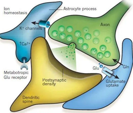
Beyond the known synaptic transmission occurring between neurons, there is also an exchange of information between neurons and astrocytes. Astrocytes respond to neuronal activity and regulate neuronal transmission. The term "tripartite synapses" arise in the 90s, when this bidirectional communication system between neurons and astrocytes was reported (Fig. 2). The astrocytes are indispensable for the brain development and functioning, regulating neuronal metabolism, synaptogenesis, homeostasis, and microcirculation. However, through gliotransmitter release they also play a crucial role in synapse physiology, modulating signal integration and processing, as well as plasticity (Perea et al., 2009).
4
Figure 2. Illustration of tripartite synapses. Communication between pre- and postsynaptic terminal and the regulation performed by astrocytes (Adapted from (Eroglu & Barres, 2010)).
The role played by STEP at the synaptic level was extensively studied. Previous research has shown that STEP opposes to the development of synaptic strengthening through dephosphorylation of neuronal signalling molecules at the postsynaptic level, such as the GluN2B subunit of the N-methyl-Daspartate receptor (NMDAR) and ERK1/2, and also mediates the internalization of GluA1/GluA2 containing alpha-amino-3-hydroxy-5-methyl-4-isoxazole propionic acid glutamate receptors (AMPAR). Additionally, STEP also dephosphorylates Fyn, p38 kinase and proline rich tyrosine kinase 2 (Pyk 2), controlling the duration of their signals. The identification of STEP substrates is important to understand how this phosphatase regulates neuronal mechanisms (Baum et al., 2010).
1.2.1 STEP substrates
1.2.1.1 Mitogen activated protein kinases (MAPK) – ERK1/2 and p38
ERK1/2 and p38 (Fig. 3), both members of the MAPK family, are STEP substrates (Paul et al., 2003; Semenova et al., 2007). ERK1/2 is involved in synaptic plasticity and memory formation through the stabilization of dendritic spines, regulation of local protein synthesis and nuclear transcription (Sweatt, 2004). STEP dephosphorylates ERK1/2 in its tyrosine residues (Tyr) inactivating it (Paul et al., 2000). It has been reported that ERK1/2 is involved in the development of synaptic strength and memory consolidation in the lateral amygdala. These link between STEP and ERK1/2 was provided by studies with STEP knockout mouse (STEP KO), which
5
show significantly elevated phospho-ERK1/2 levels and consequently increased phosphorylation of its substrates, CREB and Elk1 transcription factors (Fig. 3) (Venkitaramani et al., 2009; Venkitaramani et al., 2011). In addition, behavioral tests of STEP KO mice show facilitated hippocampal and amygdala-dependent learning (Venkitaramani et al., 2011; Olausson et al., 2012). Together, this may indicate that STEP generally regulates the duration of ERK1/2 signaling and raises the hypothesis that elevated levels of STEP may disrupt synaptic plasticity and memory formation (Paul et al., 2003).
p38 is also a member of the MAPK family and a STEP substrate. Contrary to ERK1/2, p38 is involved in NMDAR-mediated excitotoxicity and regulates cell death pathways (Ivanov et al., 2006; Semenova et al., 2007). STEP usually dephosphorylates p38, inactivating it.
Activation of the extra-synaptic NMDAR(GluN2B) promotes the activation of calpain, leading to the cleavage of STEP61 within the KIM domain. This cleavage results in a
non-functional STEP variant (STEP33), which is unable to bind to their substrates. Consequently, this
leads to p38 activation and stimulates cell death pathways (Fig. 3). Use of a peptide containing the site cleaved by calpain prevented STEP61 cleavage and promoted significant
neuroprotection from glutamate-mediated excitotoxicity (Xu et al., 2009).
ERK1/2 and p38 regulates STEP levels by mediating phosphorylation in sites near the KIM domain. The dephosphorylation of these sites leads to ubiquitination and degradation of STEP active form (Mukherjee et al., 2011). When synaptic- or extrasynaptic-NMDARs are activated is triggered respectively STEP61 ubiquitination (via ERK1/2 activation) or
calpain-mediated cleavage (via p38 signalling). This distinct response may occur due to the location of ERK and p38 in the terminals (Xu et al., 2009).
1.2.1.2 Glutamate receptors
STEP regulates the phosphorylation of the GluN2B subunit of NMDARs in two ways, either directly by dephosphorylation, or indirectly by Fyn-mediated phosphorylation (Nakazawa et al., 2001; Pelkey et al., 2002). When dephosphorylated directly by STEP, the Tyr1472 residue
of GluN2B binds to clathrin-adapting proteins that will promote the internalization of the GluN1/GluN2B receptor (Fig. 3) (Lavezzari et al., 2003; Snyder et al., 2005). Accordingly, it was reported that STEP KO mice present increased expression of GluN1/GluN2B receptors (Venkitaramani et al., 2011). The effect of STEP on NMDA receptor inhibits NMDA-mediated-LTP. Moreover, administration of a NMDA receptor antagonist and a Src kinase inhibitor prevents the effects mediated by STEP suggesting that STEP works as a "tonic brake" counteracting the NMDARs Src-dependent upregulation (Pelkey et al., 2002).
AMPAR are ligand-dependent ion channels composed of GluA1 to GluA4 subunits, and regulate synaptic strengthening and memory consolidation. The trafficking of AMPAR occurs in long-term depression (LTD) and is regulated by tyrosine phosphatases, including STEP.
GluA2-6
AMPAR subunit dephosphorylation- and internalization-mediated by STEP locally translated is triggered by metabotropic glutamate receptor (mGluR5) activation (Fig. 3) (Zhang et al., 2008). In addition, some studies suggest that STEP KO mice present increased surface expression of AMPARs containing GluA1/GluA2 (Venkitaramani et al., 2011).
Together these data indicate that prolonged neuronal stimulation results in a STEP increased activity, leading to a removal of NMDAR and AMPAR from synaptic membranes. Prolonged neuronal inhibition has the opposite effect, leading to the hypothesis that fine-tuning of STEP activity is necessary to maintain adequate levels of glutamate receptors at synapses.
Figure 3. Representative image of STEP activity and regulation mechanisms at the postsynaptic level. (From the left to the right- activation of extrasynaptic NMDA triggers proteolytic cleavage of STEP by calpain allowing p38 activation and triggering death pathways. TrkB-PLC activation by BDNF triggers UPS-mediated STEP degradation. PTPα activates Fyn and boosts NMDA receptor signalling when STEP is inactivated. Fyn is a STEP substrate, when STEP is absent Fyn increases NMDAR surface levels. Pyk2 is also a STEP substrate, when phosphorylated leads to Fyn activation and NMDAR phosphorylation. D1DR activation activates PKA, which phosphorylates STEP61 inhibiting it. PKA also leads to DARPP-32 activation, inactivating PP1 and promoting STEP interaction with its substrates. Conversely, NMDAR or α7nAChR- activation enable calcineurin/PP2B to inactivate DARPP-32 which mediates PP1 inhibition and increases STEP61 activity. STEP61 dephosphorylates GluN2B directly promoting his interaction with clathrin adaptor proteins and leads to endocytosis of these receptors. STEP61 is also required for the internalization of GluA1/GluA2-containing AMPARs. The mGluR5 leads to STEP local translation, enabling fast local response. The dimerization occurs under an oxidative stress condition, enabling the dimers formation and reducing the active STEP available).
7
1.2.1.3 Fyn
Fyn is a member of the Src kinases family and is associated with the cytoplasmic side of the plasma membrane. Fyn is directed to postsynaptic density where it regulates neuronal signalling (Ali & Salter, 2001). Fyn is inactivated by a C-terminal Src kinase. Fyn is involved in NMDARs trafficking (Snyder et al., 2005), NMDAR membrane insertion (Nakazawa et al., 2001) and regulation of synaptic strengthening. STEP binds Fyn through the first polyproline domain and the KIM domain and dephosphorylates Fyn on Tyr420 residue (Fig. 3) (Nguyen et al., 2002).
STEP KO mice show increased Fyn phosphorylation and increased phosphorylation of the GluN2B subunit on Tyr1472. In addition, STEP KO mice exhibit increased NMDAR levels, and improved
hippocampal-dependent memory (Venkitaramani et al., 2011; Zhang et al., 2010). Together, these results suggest that the regulation of Fyn by STEP contributes to suppression of synaptic plasticity and memory consolidation.
1.2.1.4 PTPα
PTPα is a widely expressed receptor-type PTP. In the CNS PTPα is involved in the regulation of synaptic plasticity and LTP. PTPα KO mice present decreased Fyn activity as well as deficits in memory consolidation and LTP (Petrone et al., 2003). This is due to PTPα ability to activate Fyn and boost the NMDA receptor signaling. STEP KO rats and WT animals treated with pharmacological inhibitors of STEP present increased phosphorylation of PTPα Tyr789, while overexpression of STEP61 decreased PTPα phosphorylation. Inactivation of STEP61 lead to
increased phosphorylation of PTPα and subsequent translocation in lipid raft fractions, leading to activation of Fyn (Fig. 3) (Xu et al., 2015). Together these data support PTPα as a STEP61
substrate.
1.2.1.5 Pyk2
Pyk2 is activated by phosphorylation of the Tyr402 residue. When activated Pyk2
phosphorylates and activates Fyn, which in turn phosphorylates GluN2B subunit of NMDARs (Fig. 3). So, Pyk 2 activation triggers NMDARs phosphorylation and an increase in the surface levels of these receptors (Besshoh et al., 2005). Besides that, it leads to an increase of ERK1/2 phosphorylation (Nicodemo et al., 2010). Pyk2 inhibitors block LTP (Huang et al., 2001), highlighting its importance to synaptic plasticity. Moreover, it was shown that Tyr402 residue of
Pyk2 is highly phosphorylated in STEP KO mice. Accordingly, Pyk2 was suggested as a substrate of STEP (Venkitaramani et al., 2011).
1.2.2 STEP regulation
Several mechanisms have been reported as relevant in the regulation of STEP levels and activity, namely phosphorylation, ubiquitination, local translation, proteolytic cleavage and dimerization. Some of these mechanisms are disturbed in neurodegenerative and neuropsychiatric diseases and may lead to anomalous STEP activity (Goebel-Goody et al., 2012).
8
1.2.2.1 Phosphorylation
As already discussed, STEP interaction with their substrates is regulated by phosphorylation in a KIM domain-serine residue. Phosphorylation of STEP induced by PKA-dopamine-D1-receptor-dependent activation reduces the affinity of STEP to its substrates (Fig. 3) (Paul et al., 2000). Conversely, STEP affinity to his substrates increases when STEP dephosphorylation occurs in PKA-phosphorylation site by protein phosphatase 2B activation (PP2B) induced by NMDAR- or α7 nicotinic acetylcholine receptor (α7nAChR) activation (Fig. 3) (Paul et al., 2003; Snyder et al., 2005). DARPP-32 is a modulator of PP1 activity. When phosphorylated by PKA, DARPP-32 inhibits PP1 impeding STEP dephosphorylation maintaining this phosphatase inactive (Fig. 3) (Valjent et al., 2005). Hence, these two mechanisms appear to modulate STEP phosphorylation and the binding to its substrates.
1.2.2.2 Ubiquitination
The ubiquitin-proteasome system is a fundamental tool for normal cell function. Disruption of this system has been reported in neurodegenerative and neuropsychiatric disorders such as, AD, HD, Schizophrenia, and Parkinson's disease (PD) (Cheon et al., 2018; Zheng et al., 2016). STEP61 ubiquitination and degradation triggered by NMDAR synaptic
activation leads to a reduction of its levels and promotes synaptic plasticity through the increase of the phosphorylated form of its substrates. As reported by Xu et al., 2009, after the synaptic activation of NMDAR, p-ERK1/2 levels and STEP61 ubiquitination and degradation
increases, which leads to ERK1/2 nuclear translocation and intensify gene transcription (Fig. 3) (Xu et al., 2009). The ubiquitin ligase Parkin in responsible for STEP61 ubiquitination and
degradation (Kurup et al., 2015b).
1.2.2.3 Local translation
Local translation (Fig. 3) enables rapid effects at distinct synaptic locations in order to induce or sustain synaptic plasticity. Therefore, processes like long-term potentiation (LTP) and long-term depression (LTD) require local dendritic translation which can be triggered by mGluR activation (Bramham & Wells, 2007; Costa-Mattioli et al., 2009). Some studies indicate that mRNAs, which are key for these processes, are transferred through the dendrites, where they remain until the appropriate stimulus allows its translation (Bramham & Wells, 2007). Findings suggest that STEP is a member of the family of local translated proteins, furthermore STEP mRNA and protein are present in the puncta over dendrites and close to the PSD. In addition, STEP translation is regulated within synaptoneurosomes, suggesting that STEP is dendritically translated (Zhang et al., 2008). STEP mRNA associated to RNA binding-proteins remains suppressed in dendrites until to be released and locally translated.
9
1.2.2.4 Proteolytic cleavage
As previously mentioned, STEP33 is a truncated and non-active product resultant from
the proteolytic cleavage of STEP61-KIM domain by calpain (Fig. 3). Proteolytic cleavage is a
consequence of p38 activation derived from activation of extra-synaptic NMDAR which triggers the cell death signaling cascade (Xu et al., 2009). As mentioned earlier, NMDAR extra-synaptic activation lead to STEP33 production.
1.2.2.5 Dimerization
STEP oligomerization occurs under oxidative stress and reduces its activity. In part, STEP61 dimerization (Fig. 3) is mediated by the formation of intermolecular disulphide bonds
between two cysteine residues, Cys65 and Cys76, present in the hydrophobic region of the amino-terminal (Deb et al., 2011). Studies have shown that STEP61 dimerization occurs under
basal conditions suggesting that it is a STEP61 inherent property. When subjected to H2O2
-induced oxidative stress, both STEP61 and STEP46 suffer a dose-dependent oligomerization (Deb
et al., 2011). It was also shown that aging, which is characterized by the depletion of glutathione, is associated with increased dimerization of STEP. The increase in oxidative stress occurring in aging may increase brain susceptibility to age-related neurological disorders and to their fast progression. The presented data reveals a link between this brain specific PTP and the aging process (Rajagopal et al., 2016).
1.2.2.6 Pharmacologic regulation of STEP
The catalytic cysteine in PTPs is prone to several reactions which can inhibit its activity. Some studies reported that 8-(Trifluoromethyl)-1,2,3,4,5-benzopentathiepin-6-amine hydrochloride (TC-2153) (Fig. 4) irreversible inhibits STEP. In absence of TC-2153, a disulphide bridge between Cys465 and Cys472 is present in the catalytic cysteine of STEP. Incubation of WT STEP with TC-2153 resulted in the presence of a new trisulphide on the Cys465/Cys472 bridge, which was not observed when the cysteine of the catalytic site (Cys472) was mutated to serine.
These results indicate that the cysteine in the active site is probably modified by TC-2153 and suggest that the sulphur from the benzopentathiepine nucleus is maintained, giving rise to the trisulphide, which was identified by mass spectrometry (Xu et al., 2014).
Figure 4. Structure of the STEP pharmacologic inhibitor known as TC-2153 (Adapted from Xu et al., 2014).
10
1.2.2.7 Regulation by brain-derived neurotrophic factor (BDNF)
Brain-derived neurotrophic factor (BDNF) is one of the neurotrophic factors that supports differentiation, maturation and survival of neurons in the nervous system and has also neuroprotective effects under adverse conditions. BDNF levels are decreased in some neurodegenerative diseases, such as PD, multiple sclerosis and Huntington's disease (Bathina & Das, 2015). In primary cortical, striatal and hippocampal neurons BDNF induces STEP61
degradation via the PLCγ-UPS pathway (Fig. 3), which leads to an increase of phosphorylation levels of GluN2B and ERK1/2 (Saavedra et al., 2016). It was also reported that reduction of BDNF leads to increased expression of STEP61 in mice. Both STEP inhibitor and a TrkB agonist
reverses biochemical and motor changes observed in BDNF+/- type mice. These findings suggest
that BDNF and STEP61 reciprocally regulate their expression patterns in several neuropsychiatric
and neurodegenerative diseases (Xu et al., 2016).
The information gathered so far focuses on the activity and regulation of STEP present postsynaptically. From this point forward, we center on the presynaptic role of STEP.
1.3 Presence of STEP in presynaptic terminals
1.3.1 Glutamatergic synapses
Most of the work on STEP focuses on its function in the postsynaptic region. Recently, Bosco and collaborators described the presence of STEP in presynaptic terminal and reported its role as a regulator of calcium levels in glutamatergic terminals (Bosco et al., 2018). STEP KO mice showed increased activity of Fyn kinase. A substrate of this kinase is the inositol 1,4,5-trisphosphate receptor (IP3R), present in the RE membrane. When phosphorylated by Fyn, IP3R triggers an increase in calcium levels in the terminal (Fig. 5). The function of calcium homeostasis in the release of neurotransmitters is well established, as well as in the activation of proteins whose activity depends on the presence of calcium, such as Ca2+
/calmodulin-dependent protein kinase II and calcineurin. Synapsin I (Syn I) has several phosphorylation sites and can also be phosphorylated by CaMKII. This phosphorylation leads to Syn I disassociation from actin, leaving the synaptic vesicles ready for synaptic synchronous transmission (Fig. 5). As noted above, the increase in calcium levels leads also to the activation of the phosphatase calcineurin, which has Syn I as substrate, leading to its dephosphorylation and consequent inactivation. On the other hand, it was reported that STEP KO present increased activity of ERK kinase, which phosphorylates and activate Syn I at the same sites of calcineurin. It was proposed that STEP is a regulatory element and helps to maintain the balance in the presynaptic terminal, although more data is needed to confirm this hypothesis (Bosco et al., 2018).
11
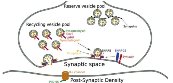
Figure 5. Role of STEP in the regulation of neurotransmitter release. The loss of STEP activity at presynaptic terminals leads to hyperphosphorylation of Fyn, which increases the activation of IP3R and the consequent release of Ca2+from the ER. Cytosolic Ca2+activates Ca2+/calmodulin-dependent protein kinase II enabling Syn I phosphorylation, which leads to an increase of the availability of SVs and the increase of the readily releasable pool size. Glutamate release and synaptic plasticity are consequently enhanced in STEP-deficient presynaptic terminals (Adapted from Bosco et al., 2018).Synapsin (Syn) is the most abundant presynaptic phosphoprotein, it interacts reversibly with synaptic vesicles and regulates the synaptic vesicles clustering (Fig. 6) and plasticity (Bykhovskaia, 2011). The Syn family contains several proteins (Cesca et al., 2010) and it has been reported that its important role in the regulation of neuronal development and synapse formation (Valtorta et al., 1995; Ferreira et al., 1998). The other protein analyzed, synaptophysin (SYP), is the most abundant protein in SVs (Fig. 6) (Takamori et al., 2006) and because of that it is widely used as a marker for presynaptic terminals (Chen et al., 2005). Therefore, SYP was associated to the regulation of exocytosis, synapse formation and endocytosis of SVs (Eshkind & Leube, 1995; Tarsa & Goda, 2002).
Figure 6. Schematic representation of the synapse with identification of presynaptic proteins (Adapted from Osimo et al., 2019).
12
1.3.2 Dopaminergic synapses
Although there is no information on the role of STEP in dopaminergic synapses, it was reported that the levels of tyrosine hydroxylase (TH) the striatum of STEP KO mice are increased when compared to WT mice, THSer31 phosphorylation and dopamine levels are also increased (Kurup et al., 2015a), suggesting that STEP may act as inhibitor of dopaminergic transmission.
1.3.2.1 Idiopathic Parkinson's disease and STEP
Idiopathic Parkinson's disease (IPD) is a progressive neurodegenerative disorder manifested over a wide range of motor and non-motor symptoms (Antony et al., 2013). It was first characterized in 1817 by the English physician James Parkinson (Parkinson, 1817). IPD is the second most common neurodegenerative disorder after Alzheimer's disease affecting populations worldwide (Meenalochani et al., 2016). Motor symptoms that characterize the clinical syndrome of IPD are bradykinesia, rest tremor, rigidity, and postural instability (Parkinson, 1817). The non-motor symptoms may precede the onset of movement disorders for many years and the most frequent are depression, olfactory deficit, constipation and idiopathic REM sleep disorder (Park & Stacy, 2009). The continuous deterioration of PD-associated motor functions is caused by the progressive loss of dopaminergic neurons (DA) in the substantia nigra
pars compacta (SNpc), that innervate the basal ganglia (Hornykiewicz, 2002; Greffard et al.,
2006). Although the central cause of this neuronal cell death remaining uncertain, it is known that aging, genetic alterations, and environmental factors appear to be associated (Antony et al., 2013).
Population-based prevalence and incidence studies show a strong correlation of IPD occurrence with age. In familial forms of PD, an earlier onset is possible, but, with increasing age, the risk of disease onset rises as well (Reeve et al., 2014). Indeed, to date, aging represents the most significant risk factor for developing PD (Hindle, 2010). Genetic factors seem to be the main cause of about 5-10% of PD patients. In recent decades, genes linked to common monogenic forms of PD have been identified, such as PINK1, Parkin and DJ-1 (Lesage et al., 2009). These alterations are involved in mechanisms of oxidative stress and are associated with clinical characteristics of early-onset PD subtypes (Lesage et al., 2009). A link has been reported between exposure to environmental toxins and increased risk of developing PD. Subsequently, the evidence emerged when a secondary product of the narcotic meperidine drug synthesis, namely 1-methyl-4-phenyl-1,2,3,6-tetrahydropyridine (MPTP), gave rise to irreversible parkinsonism, with all the clinics characteristics of the disease (Langston & Ballard, 1983) and is now widely used to develop PD mice models.
MPTP is highly lipophilic and crosses the blood-brain barrier (Markey et al., 1984), after that it is absorbed by astrocytes and metabolised by monoamine oxidase B (MAO-B) into MPP+,
its active metabolite, (Fig. 7) (Dauer & Przedborski, 2003). When released from astrocytes by the organic cation transporter 3 (OCT-3) into the extracellular space (Cui et al., 2009), the
13
MPP+ is transported into neurons by the dopamine transporter (DAT) (Fig. 7). It may be
accumulated in the mitochondria (Ramsay & Singer, 1986) or can be stored in vesicles through the monoamine vesicular transport (VMAT2) (Chen et al., 2008). In the mitochondria, as mentioned above, it blocks complex I and start other intracellular reactions (Fig. 7). The effect of MPTP as inhibitor of the mitochondrial electron transport chain complex I (Mizuno et al., 1988) was crucial to identify the key role of mitochondria in the pathogenesis of PD.
Figure 7. Schematic representation of the MPTP absorption and intracellular effects. After crossing the blood-brain barrier, the MPTP is absorbed by the glial cells. It is then transported out of the glial cells by OCT-3 and enters the dopaminergic neurons via the dopamine transporter (DAT). In dopaminergic neurons it will be responsible for several oxidative processes, among them inhibition of mitochondrial complex I (Adapted from Dauer & Przedborski, 2003).
When stored in the vesicles, MPP+ can extrude DA from the vesicles fostering its
metabolization into toxic compounds, such as DOPAL (Panneton et al., 2010), and promoting the formation of superoxide radicals (5-cysteine AD) and hydroxyl radical attack (6-OHDA).
As mentioned above, it is not yet known what causes the dopaminergic neuronal retrograde death responsible for the motor symptoms that appear in PD. It has been reported that STEP levels are high in the disease model and in PD patients with the parkin mutation. Parkin is a ligase involved in STEP ubiquitination, meaning that a parkin dysregulation may lead to an increase in STEP levels, which may contribute to the dopaminergic neuronal death occurring in PD.
15
2 Aims
The main goal of this work is to understand if presynaptic STEP plays a role in dopaminergic synaptic transmission and how its inhibition can affect the formation of new synapses. For this purpose, the following objectives were defined:
• To determine if STEP is located at dopaminergic terminals;
• To verify how STEP modulation can affect dopaminergic neurons morphology;
•
To demonstrate if the presynaptic STEP can affect dopaminergic synapse formation;17
3 Materials and methods
3.1 Cells cultures
3.1.1 Neuron and neuron-glial cells midbrain co-cultures
Neuron and neuron-glial cells cultures (Fig. 8A) were prepared from Wistar rat embryos. A pregnant female on the 15th or 16th day of gestation was anesthetized with Ketamine:Xylazine, the embryos were removed, and the brains collected. Meninges were gently removed, and the ventral midbrains dissected. The tissue obtained was placed in cold phosphate buffered saline (PBS: NaCl 140 mM, KCl 2.7 mM, KH2PO4 1.5 mM and Na2HPO4 8.1 mM, pH 7.4). The tissue was then mechanically dissociated with a 5 mL pipette and then with
micropipette tips in order to achieve a homogeneous suspension. The cellular suspension was centrifuged for 3 minutes at 405g (3K18C Bioblock Scientific; Sigma Laboratory Centrifuges). The pellet obtained was resuspended in Neurobasal™ Medium (Gibco®, Paisley, Scotland, UK) supplemented with B27TM 10% (Gibco®, Paisley, Scotland, UK), L-glutamic acid 25 µM/mL (G8415, Sigma-Aldrich, St. Louis, USA), L-glutamine 0.5 mM/mL (G3126, Sigma-Aldrich, St. Louis, USA) and Gentamicin 120 µg/mL (G1272, Sigma-Aldrich, St. Louis, USA), and heat-inactivated fetal bovine serum (Biochrom®) 10% (HiFBS) (only in the case of neuron-glial cells
cocultures). Viable cells were counted using a Neubauer chamber, from a dilution of 1:1 of the final cell suspension with Trypan blue (0.4% in 0.9% NaCl). Finally, the cells were plated in 12-multiwell plates (Orange Scientific) coated with poly-D-lysine (PDL) (Sigma-Aldrich, St. Louis,
USA) at a density of 0.8 x 10⁶ cells/well (Fig. 8A) or in micro-islands of neurons (Fig. 8B) plated
in 12mm-coverslips coated with PDL. In each coverslip 3 micro-island were plated by adding a drop of the cell suspension (3µL with 0.1 x 10⁶ cells/micro-island). After cell adhesion, nearly
90 minutes after plating, 0.6 mL of culture medium were added to each wells. Cultures were maintained at 37º C under an atmosphere of 5% CO2 and 95% air over 4-5 days. After cells
achieved confluence, fluorodeoxyuridine was added to the culture (FDU: uridine 68μM and
5-Fluoro-5′-deoxyuridine 27 μM; Sigma-Aldrich, St. Louis, USA) to inhibit cell proliferation.
3.1.2 Ventral midbrain (VM) glial cells cultures
Glial cells cultures (Fig. 8B) were performed using ventral midbrain (VM) from postnatal day 2-5 Wistar rats. After beheading, the brain was removed and placed in cold PBS. The meninges were carefully pulled out and the region corresponding to the VM was placed in PBS. The tissue was mechanically dissociated in M10C-G medium consisting of MEM (M0268,
Sigma-Aldrich, St. Louis, USA) and supplemented with 2.2 g / L sodium bicarbonate (NaHCO3), 0.75% glucose 45%, 0.12% antibiotic (Penicillin and Streptomycin, Sigma), 0.02% insulin (I5500, Sigma-Aldrich, St. Louis, USA) and 10% FBS (Biochrom®), and pelleted by centrifugation. As previously
18
were cultured at a density of 0.132x10⁶ cells/well in 24-multiwell plates (Orange Scientific),
previously coated with PDL. Culture was maintained at 37°C in a 5% CO2, 95% air atmosphere
for 6-7 days. After reaching confluence, they were used to perform the bunker cultures, described later.
3.1.3 Neuron-micro-islands and VM-glial cells bunker cocultures
As reported over the years, glial cells are crucial for neurons development, maturation and survival. Due to that, and as already been frequently performed by our group, bunker cultures were used to perform some of the experiments. Bunker cultures (Fig. 8B) consist in putting different cell cultures in contact only through the medium. VM-glial cells were plated in a multi well plate, as previously described, and on top of these cells were added neuron micro-islands plated on coverslips. To avoid direct physical contact between the two cultures small paraffin spheres were placed between the coverslip and the bottom of the multiwell (Fig. 8).
3.1.4 Treatments
Neuron-enriched cultures, neuron-glia cocultures (Fig. 8A), and bunker cultures of neurons-micro-islands and VM-glial cells (Fig. 8B) were treated with TC-2153 (1 µM). One hour after adding TC-2153 the MPP+ toxin (10 µM) was added and maintained for further 24h. At the
end of the treatment samples were processed for western blot (Fig. 8A) or for immunocytochemistry (Fig. 8B).
3.1.5 Protein extraction
At the first stage of the study, the primary neuron cultures and neuron-glial cells cocultures have been washed twice with PBS and later lysed on ice using a cold lysis buffer containing Triton X-100 1%, Tris-HCl 50 mM, pH 7.5, EGTA 10 mM, NaCl 150 mM, protease inhibitors (phenylmethylsulphonyl fluoride 2 mM, aprotinin 10 μg/μl, and leupeptin 1μg/μl), and the phosphatase inhibitor sodium orthovanadate. The cellular extracts were centrifuged at
405 g for 30 minutes at 4ºC (Mikro 200R, Hettich Zentrifugen, Tuttlingen, Germany) and subsequently, the supernatants were collected. The Bradford protein assay was used to determine the total protein concentration of the samples by using bovine albumin (Bovine
19
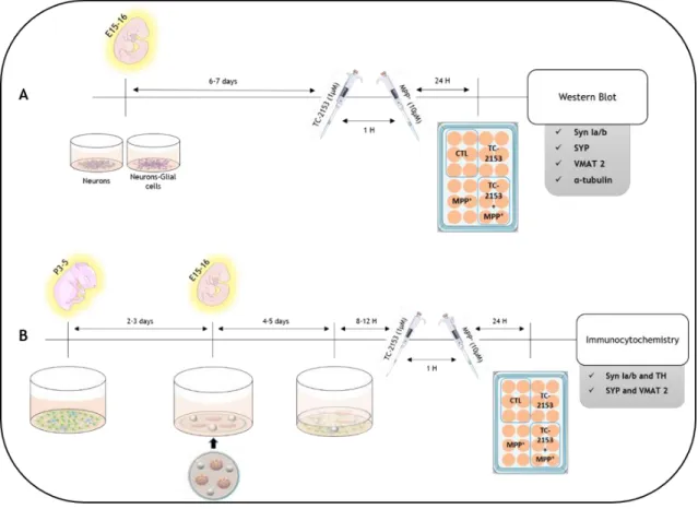
Figure 8. Schematic representation of the experimental procedures. (A) Cocultures of neuronal-enriched cells and neuronal-glial cells, obtained from E15-16 embryos. They were allowed to reach confluence and then stimuli were carried out, first using the inhibitor, TC-2153 (1mM), and one hour later with the dopaminergic toxin, MPP+ (10mM). The total protein was extracted from these cultures and quantified, in order to perform the western blot. (B) For Bunker-type cultures, glial cells from postnatal Wistars with P2-5 were plated and two-three days later neurons were isolated and cultured in micro-islands. About 12 hours after putting the 2 cultures in contact, we proceeded to the stimuli, as described above. Paraffin spheres placed in the coverslips were used to keep the two cultures physically separated. After the stimulus, the cells were fixed in order to perform immunocytochemical tests.3.2 Western blot
After quantifying total protein levels, samples were denatured in 0.5M Tris-HCl, pH 6.8, 10% (w/v) Sodium Dodecyl Sulfate (SDS), 10% glycerol, 140mM 2-mercaptoethanol, and 0.1% (w/v) bromophenol blue and heated at 100°C for 5 minutes. A volume of sample corresponding to 30 μg was loaded in each well. The gel consisted of a stacking gel (4% acrylamide, 0.5 mM Tris-HCl (pH 6.8), 10% SDS, 0.05% ammonium persulphate (PSA) and 0.05% tetramethylethylenediamine (TEMED)) and a resolving gel (acrylamide 12% in 1.5 M Tris-HCl (pH 8.8), containing 10% SDS, 0.05% PSA and 0.05% TEMED).
Electrophoresis of samples and colored molecular weight marker were performed under 160 V for1 hour, at room temperature, using a buffer with 960 mM Glycine, 125mM Tris and 0.5% SDS. After the electrophoresis, the stacking gel was removed and the resolving gel was
20
immersed in Transfer Buffer (200mL of Trans-Blot® Turbo™ 5x Transfer Buffer, 200mL Methanol and 600mL H2OmQ). In order to enable the proteins access to antibody detection, they were
transferred from the gel to a polyvinylidene difluoride (PVDF - GE Healthcare, Amersham, UK) membrane. Prior to transfer, the PVDF membranes was activated by immersion for 5 seconds in 100% methanol, followed by 5 minutes in water and 10 minutes in Transfer Buffer. Then, a semi-dry transfer was performed. The gel in contact with the membrane was placed between two sponges previously impregnated in a Transfer Buffer. The electrotransfer was carried out at 25 V for about 45 minutes.
After electrotransfer, the membranes were blocked by an incubation in Tris buffer saline with 0.1% of Tween 20 (TBS-T) and 5% skimmed milk, at room temperature for 1 hour. Following blocking, membranes were incubated with the primary antibodies (diluted in TBS-T according to Table 1) overnight at 4°C. The membranes were washed thrice in TBS-T for 10 minutes each followed by an overnight incubation with the corresponding secondary antibody (diluted in TBS-T according to Table 1). After washing, membranes were incubated with peroxidase substrate (LuminataTM Growing Western HRP Substrate, Millipore Corporation, Billerica, MA). The luminescence of the peroxidase reaction product was detected with a ChemiDoc XRS+ system (Bio-Rad, Munich, Germany). To normalize for sample loading the
house-keeping protein α-tubulin was quantified in each membrane. A ratio of the optic density (OD) of each band and the OD of α-tubulin for the same well was calculated. The results are expressed as percentage of the values obtained under control conditions.
3.3
Immunocytochemistry
At the end of each experimental procedure the cells were washed with PBS and fixed with 4% paraformaldehyde (PFA) for 10 minutes. After washing with PBS to remove excess PFA cells were permeabilized with Triton X-100 1.0% in PBS for 10 minutes. After permeabilization, nonspecific bonds were blocked by incubation with a PBS solution containing 0.1%-Tween (PBS-T) 20% FBS and 0.3% BSA, for two hours at room temperature. After blocking, we proceeded to a PBS-T wash and then primary antibodies diluted in PBS-T+1% FBS (Table 1) were incubated for 72 hours at 4°C. Unlinked antibodies were removed by washing 6 times, 15 minutes each, with PBS-T and were then incubated with the corresponding secondary antibodies conjugated to Alexa® 488 or Alexa® 594 fluorophores (according to Table 1). After incubation with the secondary antibodies, the cells were washed six times with PBS-T, and counterstained with Hoechst 2μM (Invitrogen, CA, USA) for 10 minutes and finally mounted in fluorescent mounting medium (DAKO, Glostrup, Denmark). The images were acquired with a Zeiss Axio Imaging Microscope (Axiobserver Z1, Zeiss). Immunocytochemical assays were carried out on at least three different cellular preparations with two or three coverslips for each experimental condition.
21
3.3.1 Neurites analysis and Sholl analysis
After acquiring the images several analyses were performed, including cell counting and analysis of neurites in dopaminergic neurons. TH+ labelling was used to draw the neurites
(Fig. 9).
Figure 9. Flow diagram of the analysis performed from immunocytochemical analysis of Syn++TH+,
and SYP++VMAT 2+.
3.3.1.1
Cell counting
Images for the entire micro-island were acquired with a 40X magnification. TH positive cells were counted and the mean values of all micro-islands in each coverslip were determined.
3.3.1.2
Neurite analysis
Resorting to a plugin for the FIJI software, Simple Neurite Tracer, the morphological analyses were conducted. After drawing the neurons maximum and the mean length of neurites as well as the mean of total neurites per neuron was determined. In each replicate experimental replicate 10 neurons per micro-island were drawn.
3.3.1.3
Sholl Analysis
The Sholl analysis (Sholl, 1953) is a widely used method in neurobiology to evaluate the complexity of the dendritic tree. A Sholl profile is obtained by plotting the number of dendritic intersections against the radial distance from the center of the cell body. The number of intersections is plotted against the radial distance from where a summary estimation (area under the Sholl profile) can be derived as a single measure of dendritic complexity (Fig. 10).
22
After the neurite’s drawings, an image of all pathways is generated and, from this file, the neurons arborization was assessed by Sholl's method (Binley, 2014).
Figure 10. Representation of Sholl analysis. Concentric circles are drawn starting at the neuron's center to evaluate its arborization.
Table 1. Primary and secondary antibodies used for immunocytochemical and western blot techniques, the respective species, dilution, and company.
Antibodies Species Technique Dilution Company
Anti-TH Rabbit ICC 1:1000 Santa Cruz Biotechnology
Anti-VMAT 2 Rabbit ICC 1:1000 SySy
Anti-VMAT 2 Rabbit WB 1:1000 Santa Cruz Biotechnology
Anti-Synapsin Ia/b Mouse WB, ICC 1:500 Santa Cruz Biotechnology
Anti-STEP 23E5 Mouse WB, ICC 1:1000 Cell Signaling Technology
Anti-SYP I Mouse WB, ICC 1:2000 Santa Cruz Biotechnology
Anti-α tubulin Mouse WB 1:5000 Santa Cruz Biotechnology
Anti-mouse HRP Goat WB 1:20000 Santa Cruz Biotechnology
Anti-rabbit HRP Goat WB 1:20000 Santa Cruz Biotechnology
Anti- mouse 488 Goat ICC 1:1000 Invitrogen, Molecular Probes
23
3.3.2 Dopaminergic terminals puncta analysis
To analyze specifically dopaminergic terminal co-labeling of a presynaptic synaptic marker and a dopaminergic marker were used: Syn Ia/b and TH or SYP and anti-VMAT 2. To allow identification of isolated terminals images were acquired in the periphery of micro-islands with a 63x magnification. The integrated densities of the puncta present at the dopaminergic marker-positive terminals were analyzed. The mean values of 3-5 terminals/ micro-island was determined.
3.4 Statistical analysis
Data is expressed as percentages of the values obtained in control conditions and presented as means ± SEM for the indicated number of experiments, performed with independent cell cultures. The statistical analysis was performed using Unpaired t test followed by One-tailed, as indicated in figure legends. Values of p<0.05 were considered statistically significant. All statistical procedures were performed using GraphPad Prism 5 software (GraphPad Software Inc.).
25
4 Results
4.1 STEP at the dopaminergic synapses
As reported previously STEP protein is present presynaptically in glutamatergic hippocampal neurons. To determine whether this protein is also present presynaptically in dopaminergic neurons we performed an immunocytochemical assay for a presynaptic dopaminergic marker (VMAT 2) and for STEP. As shown in Figure 11, STEP (green) and VMAT 2 (red) labelling occurred in the same region (yellow, highlighted by the arrow). This data confirmed the presence of STEP in presynaptic terminals of dopaminergic neurons and permited to continue with our aims of analysing the role of STEP in the modulation of dopaminergic synapses.
Figure 11. Presence of STEP protein in the presynaptic region of dopaminergic neurons. Representative image (63x magnification) of an immunocytochemistry performed against VMAT 2 (red) and STEP (green). Nuclei were stained with Hoechst 33342 (blue). The arrows in the right panel (merge) indicates spots in which VMAT 2 and STEP labelling overlap.
26
4.2 STEP as a modulator of synapses
To determine whether the activity of STEP regulates synaptic terminals we started by analysing two general presynaptic markers by western blot, synaptophysin (SYP) and synapsin I, both widely used as neuronal specific markers (Calhoun et al., 1996; Gitler et al., 2004). In addition to these two markers the VMAT 2 was used to evaluate the lesion extent because it is a specific marker for dopaminergic neurons. To modulate STEP activity, we used the specific STEP inhibitor TC-2153 (1µM). As previously reported, STEP levels are increased in MPTP treated mice, so to understand the STEP modulation effect on an injured condition we also used MPP+(10µM), a dopaminergic toxin, and an in vitro model of PD. Since a large amount of data
indicates that astrocytes play an important role in the modulation of synapses the study was carried out in two types of cell cultures, neuron-enriched and neuron-glia cultures.
4.2.1 Neuron-enriched culture
In control conditions inhibition of STEP in neuron-enriched cultures lead to an increase in the levels of SYP (25.86±13.58%) and SYN Ia/b, although not statistically significant in the latter case (Fig. 12 A, B). Whereas for the dopaminergic marker VMAT 2 no changes were observed in the presence of the STEP inhibitor (Fig. 12C). Exposure to the dopaminergic toxin MPP+, per se, did not alter the levels of SYP, SYN or VMAT 2, and STEP inhibition also did not