M.Sc in Structural and Functional Biochemistry
Protein-carbohydrate recognition in the
biodegradation of the plant cell wall:
Functional and structural studies using
carbohydrate microarrays and X-ray
crystallography
Thesis submitted for the degree of Doctor of Philosophy in
Biochemistry
Supervisor: Angelina Sá Palma, PhD
Assistant Researcher UCIBIO
Faculdade de Ciências e Tecnologia, NOVA
Co-Supervisor: Ana Luísa Carvalho, PhD
Assistant Researcher UCIBIO
Faculdade de Ciências e Tecnologia, NOVA
Co-Supervisor: Ten Feizi, MD
Full Professor and Director of Glycosciences Laboratory
Imperial College London
Examination Committee:
President: Professor Maria João Lobo de Reis Madeira Crispim Romão Main Examiners: Doctor Serge Perez
Doctor Isabel Maria Travassos de Almeida de Jesus Bento Examiners: Professor Carlos Mendes Godinho de Andrade Fontes
Doctor Maria Angelina de Sá Palma
Doctor Filipa Margarida Barradas Morais Marcelo
March 2020
Faculdade de Ciências e Tecnologia
Diana de Oliveira Ribeiro
M.Sc in Structural and Functional Biochemistry
Protein-carbohydrate Recognition in the Biodegradation
of the Plant Cell Wall:
Functional and Structural Studies Using Carbohydrate
Microarrays and X-ray Crystallography
Thesis submitted for the degree of Doctor of Philosophy in Biochemistry
I
Protein-carbohydrate Recognition in the Biodegradation of the Plant Cell Wall:
Functional and Structural Studies Using Carbohydrate Microarrays and X-ray Crystallography
“Copyright” em nome de Diana de Oliveira Ribeiro, FCT-NOVA
A Faculdade de Ciências e Tecnologia e a Universidade Nova de Lisboa têm o direito, perpétuo e sem limites geográficos, de arquivar e publicar esta dissertação através de exemplares impressos reproduzidos em papel ou de forma digital, ou por qualquer outro meio conhecido ou que venha a ser inventado, e de a divulgar através de repositórios científicos e de admitir a sua cópia e distribuição com objectivos educacionais ou de investigação, não comerciais, desde que seja dado crédito ao autor e editor.
III
BEP/0869/2014, PTDC/BIA-MIC/5947/2014, RECI/BBB-BEP/0124/2012, funded by Fundação para a Ciência e a Tecnologia - Ministério da Ciência, Tecnologia e Ensino Superior (FCT-MCTES), in the research unit UCIBIO, funded by FCT-MCTES, and complemented with a stay at the Glycosciences Laboratory, Imperial College London, co-funded by Welcome Trust.
From the work developed have resulted the following publications:
1. Ribeiro, D.O., Pinheiro, B.A, Carvalho, A.L., Palma, A.S., Targeting protein-carbohydrate
interactions in plant cell-wall biodegradation: the power of carbohydrate microarrays in Carbohydrate Chemistry: Chemical and biological approaches, eds. A. P. Rauter, T. Lindhorst and Y. Queneau, Royal Society of Chemistry, 2018, vol. 43, pp. 159–176 (DOI: 10.1039/9781788010641-00159).
2. Ribeiro, D.O., Viegas, A., Pires, V.M.R., Medeiros-Silva, J., Bule, P., Chai, W., Marcelo, F.,
Fontes, C.M.G.A., Cabrita, E.J., Palma, A.S., Carvalho, A.L., Molecular basis for the preferential recognition of β1,3-1,4-glucans by the family 11 Carbohydrate-Binding Module from Clostridium thermocellum, FEBS Journal, 2019, in press (DOI: 10.1111/febs.15162).
V
This Thesis is dedicated to my partner in life, Nuno Mariani. For always being at my side, since day one, no matter what, pushing me further. For all the love, unconditional strength and support, and all the cooked meals! I wouldn’t have done it without you. And the best is yet to come! To my supervisors, Doctor Angelina Sá Palma and Doctor Ana Luísa Carvalho, a profound thank you for the opportunity, the guidance, encouragement and all the support even through the hardest moments, it has been 5 years of professional but also personal enrichment.
A big and warm thank you to Prof. Ten Feizi for receiving me with open arms in her group and sharing her expertise and knowledge, and always giving her tirelessly support.
A special thank you to Doctor Wengang Chai, for eagerly supervising me and for all his continued guidance, during my stay and even after from a distance.
I would like to express my gratitude to Prof. Maria João Romão, for opening the doors of her lab to me all those years ago, which eventually lead me to this path.
To our collaborators, Prof. Carlos Fontes, Doctor Joana Brás, Doctor Virgínia Pires, Doctor Natércia Brás, Prof. Manuel Coimbra, Doctor Eurico Cabrita and Doctor Filipa Marcelo.
To Doctor Benedita Pereira, Doctor Márcia Correia and Doctor Yibing Zhang for always being available to help and share their knowledge and guidance.
To Viviana Correia and Raquel Costa for all the support, companionship and help keeping my mental sanity during these years. Also, for Raquel’s valuable contributions to this work, it was a pleasure teaching you, learning with and from you.
A thank you to my colleagues, past and present, Lisete, Francisco, Raquel S., Marino, Filipa, Filipe, Catarina and Cecília.
To my friends Ana, Rita, Joana, Liliana, Cláudia, and all others that have accompanied me and given their support during this journey.
To my mother, the biggest thank you, for always believing in me, unconditionally supporting me every step of the way. I am where I am because of you.
To Carlos, for contributing more that he probably realises.
To my father, for all his support, and my sisters and brother Patrícia, Susana and Hugo for all that I’ve learned from you.
To my dearest in-laws, Idalécio and São, for all their love and kindness. Finally, to my grandfather Artur, that will always be present in my thoughts.
VII
“The beauty of a living thing is not the atoms that go into it, but the way those atoms are put together”
IX
The plant cell wall is, in its majority, constituted by complex and structurally diverse polysaccharides that are valuable resources for industrial and biotechnological applications. Anaerobic microbial organisms are highly efficient for plant cell wall polysaccharide biodegradation and have evolved a multi-enzyme complex system, the Cellulosome, where catalytic enzymes have non-catalytic Carbohydrate Binding Modules (CBMs) appended that highly potentiate the enzymes’ catalytic efficiency. Deciphering at molecular level the mechanisms underlying plant cell wall carbohydrate recognition and deconstruction by different cellulolytic bacteria is crucial to elucidate these complex biological systems, as well as to further promote novel potential applications. The work developed in this Thesis focused on the unique approach of combining carbohydrate microarrays with X-ray crystallography, to uncover carbohydrate ligands for CBMs and to structurally characterize novel CBM-carbohydrate interactions of two anaerobic bacteria that reside in different ecological niches: Clostridium
thermocellum, found in soils, and Ruminococcus flavefaciens FD-1, present in the rumen of
herbivorous. To this end, microarrays featuring carbohydrate probes with polysaccharide and oligosaccharide sequences representative of the structural diversity found on plant cell walls, but also in fungal and bacterial cell walls, were developed and then used to screen the carbohydrate-binding and ligand-specificity of 150 CBMs of C. thermocellum and R. flavefaciens CBMomes. The groups of polysaccharides that are differentially recognised were revealed for 59 CBMs and novel CBM-ligand specificities were identified for 23 modules from C. thermocellum and 21 from R. flavefaciens. Overall, the two bacteria differentially expressed CBM families with different carbohydrate-binding specificities, which may reflect adaptation to substrate availability in their specific ecological niche or the complexity of their Cellulosome. Using the information derived from the high-throughput microarray analysis, and according to their biotechnological relevance or novelty, CBMs and the respective ligands were selected for further structural studies. The novel CBM structures solved, complemented with biochemical and biophysical data, enabled the characterization of the molecular determinants for the recognition of mixed-linked β1,3-1,4-glucans by C. thermocellum family 11 CBM, chitin and peptidoglycan-derived sequences by a novel LysM domain from C. thermocellum family 50 CBMs, and pectic arabinans by
R. flavefaciens family 13 CBM. The results reported here allow to assign a functional role for these
CBMs and CBM families and contribute to the classification of the novel CBMs identified in the genome of the two bacteria, particularly those from R. flavefaciens FD1. Furthermore, the information derived from this integrative study, can promote a better understanding of cellulolytic capabilities of these bacteria, as well as to potentiate biotechnological applications of CBMs.
Keywords: Carbohydrate-Binding Modules (CBMs) • Protein-carbohydrate recognition •
Clostridium thermocellum • Ruminococcus flavefaciens FD-1 • Carbohydrate microarrays • X-ray
XI
A parede celular das plantas é maioritariamente constituída por polissacáridos complexos e estruturalmente diversos, também designados de hidratos de carbono, que são importantes recursos para aplicações industriais e biotecnológicas. Alguns microorganismos que residem em ambiente anaeróbio são altamente especializados e eficientes na degradação de hidratos de carbono da parede celular vegetal, tendo desenvolvido para esta função um complexo multi-enzimático de tamanho megadalton à superfície celular, designado de Celulossoma. Neste complexo, os módulos catalíticos encontram-se adjacentes a módulos não-catalíticos de ligação a hidratos de carbono (CBMs), que potenciam a eficiência catalítica das enzimas. De forma a elucidar estes sistemas biológicos complexos e promover novas aplicações, é crucial decifrar ao nível molecular os mecanismos subjacentes ao reconhecimento e desconstrução de hidratos de carbono por diferentes bactérias celulolíticas. Neste sentido, esta Tese combina a tecnologia dos
microarrays de hidratos de carbono e a cristalografia de raios-X para identificar ligandos e
caracterizar estruturalmente novas interacções CBM-hidratos de carbono de duas bactérias anaeróbias que residem em diferentes nichos ecológicos: Clostridium thermocellum, encontrada maioritariamente no solo, e Ruminococcus flavefaciens FD-1, presente no rúmen de herbívoros. No trabalho desenvolvido, foram aplicados microarrays constituídos de hidratos de carbono (polissacáridos e oligossacáridos) com sequências representativas da diversidade estrutural da parede celular das plantas, e outras de fungos ou bactérias, para investigar o reconhecimento e a especificidade de 150 CBMs identificados no genoma destas bactérias. Os resultados identificaram diferentes grupos de polissacáridos reconhecidos por 59 CBMs e novas especificidades para 23 CBMs de C. thermocellum e 21 CBMs de R. flavefaciens. Os CBMs das duas bactérias exibiram especificidades diferentes para ligação a hidratos de carbono, o que poderá reflectir a adaptação ao seu nicho ecológico e a complexidade dos seus celulossomas. Utilizando a informação derivada dos microarrays, e de acordo com a relevância biotecnológica ou novidade, CBMs e respectivos ligandos foram seleccionados para caracterização estrutural. As novas estruturas de CBMs resolvidas permitiram a caracterização dos determinantes moleculares para o reconhecimento de β1,3-1,4-glucanos pelo CBM da família 11 de
C. thermocellum, de quitina e peptidoglicano por um novo domínio LysM da família de CBMs 50
de C. thermocellum, e de arabinanos pécticos por CBMs da família 13 de R. flavefaciens. Os resultados permitiram atribuir um papel funcional a estes CBMs e famílias de CBMs nas duas bactérias, contribuindo para a classificação dos novos CBMs identificados, particularmente de
R. flavefaciens. Este estudo integrativo permitiu uma melhor compreensão das capacidades
celulolíticas destas bactérias, e servirá para potenciar aplicações biotecnológicas destes CBMs.
Palavras-chave: Módulos de ligação a hidratos de carbono (CBMs) • Reconhecimento
proteína-hidrato de carbono • Clostridium thermocellum • Ruminococcus flavefaciens FD-1 •
XIII
Acknowledgments……….……….……… V Abstract……….……….………. IX
Resumo……….……….……… XI
Table of contents……….……….……….. XIII List of figures……….……….………. XIX List of tables……….……….……… XXIII Abbreviations and symbols……….……… XXV Thesis outline……….……….………. XXIX
Chapter 1……….……….……….………. 1
1 General Introduction……….……….………..3
1.1 Structural diversity of plant cell wall polysaccharides………... 3
1.2 Cellulolytic microorganisms’ express proteomes highly efficient in plant cell wall biodegradation……….……….……….. 4
1.2.1 Carbohydrate-binding modules: the non-catalytic domains associated with Carbohydrate Active enZymes……….……….………... 7
1.2.1.1 Classification of CBMs……….……….. 7
1.2.1.2 Functional Roles of CBMs……….……… 8
1.2.2 Biotechnological applications of carbohydrate-binding proteins………... 10
1.2.3 Clostridium thermocellum and Ruminococcus flavefaciens FD-1……….11
1.3 Methods for characterizing protein-carbohydrate interactions……….………. 12
1.3.1 Carbohydrate microarrays……….……….……….………12
1.3.1.1 Carbohydrate microarray platforms……….……….………. 13
1.3.1.2 Microarrays focused on plant carbohydrates for recognition studies…………...17
1.3.1.3 Combining microarray analysis with mass spectrometry……….………….. 18
1.3.2 Protein crystallography……….……….……….………. 19
1.3.2.1 Protein crystallization……….……….……….……… 20
1.3.2.2 X-ray diffraction……….……….……….……….……… 23
1.3.2.3 3D structure determination……….……….……….……….. 25
1.3.2.4 Model building and validation……….……….……….……….. 26
1.4 Structural characterization of protein-carbohydrate interactions……….…………. 27
1.5 Thesis main objectives……….……….……….……….……… 31
Chapter 2……….……….……….……….……….……….……… 33
2 Development of glucan and hemicellulose oligosaccharide microarrays applied to plant cell wall carbohydrate recognition……….……….……….……….. 35
2.1 Introduction……….……….……….……….……….……….. 35
2.2 Results……….……….……….……….……….……….. 37
2.2.1 Construction of glucan and hemicellulose oligosaccharide microarrays…………. 37
XIV
2.2.2.2 Recognition of linear β-xylans, branched arabinoxylans and α-arabinans……..42
2.2.2.3 Differential recognition of linear β1,4-mannans and branched galactomannans……….……….……….……….………... 44
2.2.2.4 Recognition studies of xyloglucan oligosaccharides using complex oligosaccharide mixtures and a deconvolution strategy……….……….……….. 46
2.3 Discussion……….……….……….……….……….……… 50
2.4 Conclusions……….……….……….……….……….………. 54
2.5 Experimental procedures……….……….……….……….……… 55
2.5.1 Monoclonal antibodies, CBMs and lectins used for probe structural validation and microarray quality control……….……….……….……….……… 55
2.5.2 Sources of carbohydrates……….……….……….……….……... 55
2.5.3 Preparation of oligosaccharide fractions……….……….……… 55
2.5.4 Preparation of AO-NGLs by oxime-ligation……….……….……… 56
2.5.5 Preparation of DAN-conjugated xyloglucan oligosaccharides by reductive amination……….……….……….……….……….……….. 56
2.5.6 Preparation of DHPA-NGLs by reductive amination……….……….……. 57
2.5.7 MALDI Mass Spectrometry……….……….……….……….. 57
2.5.8 Electrospray Mass Spectrometry……….……….……….……… 57
2.5.9 Carbohydrate microarrays construction and analysis……….………58
2.5.10 Microarray data analysis and presentation……….……….……… 59
2.6 Work contributions……….……….……….……….………... 59
Chapter 3……….……….……….……….……….………. 61
3 Cellulolytic bacteria express CBMomes that dictate their ecological niche polysaccharide utilization……….……….……….……….……… 63
3.1 Introduction……….……….……….……….……….……….. 63
3.2 Results and Discussion……….……….……….……….………... 64
3.2.1 Multi-step strategy to assign carbohydrate-binding specificities of CBMs in a high-throughput manner……….……….……….……….………..64
3.2.2 Bacterial CBMomes from different ecological niches……….……….…... 66
3.2.3 Carbohydrate microarray platforms for ligand discovery……….……….. 67
3.2.4 Screening C. thermocellum and R. flavefaciens FD-1 CBMomes for carbohydrate-binding specificity……….……….……….……….………… 69
3.2.4.1 Recognition of α-glucans and β-glucans with linear or branched chains……… 75
3.2.4.2 Recognition of linear β1,4 mannans and branched galactomannans………….. 77
3.2.4.3 Recognition of α-arabinose- and galactose-containing sequences in different polysaccharides……….……….……….……….……….…………...78
3.2.4.4 Assignment of C. thermocellum family 50 CBMs ligand specificity towards β1,4-GlcNAc oligosaccharides……….……….……….……….………...79
3.2.4.5 Assignment of ligand specificity and chain-length requirement for families 6 and 22 CBMs towards β-xylans……….……….……….……….………. 80
XV
3.2.6 CBMs spectrum of carbohydrate recognition reflects the bacteria’s ecological
niche……….……….……….……….……….……….……….82
3.3 Conclusions……….……….……….……….……….………. 83
3.4 Experimental procedures 84 3.4.1 Monoclonal antibodies, CBMs and lectins used for microarray quality control….. 84
3.4.2 High-throughput cloning, expression and purification of C. thermocellum and R. flavefaciens FD-1 CBMs……….……….……….……….……… 84
3.4.3 Sources of carbohydrates……….……….……….……… 85
3.4.4 Carbohydrate microarray analysis……….……….……….………….. 85
3.4.5 Microarray data analysis and presentation……….……….……….86
3.4.6 Affinity gel electrophoresis with soluble polysaccharides……….………. 87
3.5 Work contributions……….……….……….……….………87
Chapter 4……….……….……….……….……….………. 89
4 Molecular basis for the preferential recognition of β1,3-1,4-glucans by the family 11 CBM from Clostridium thermocellum……….……….……….. 91
4.1 Introduction……….……….……….……….……….……….. 91
4.2 Results and Discussion……….……….……….……….………... 93
4.2.1 Specificity assignment using carbohydrate microarrays……….………... 93
4.2.2 Crystal structure of CtCBM11 bound to β1,3-1,4-gluco-oligosaccharides……….. 95
4.2.3 CtCBM11 binding mode……….……….……….………... 98
4.2.4 The CH-π stacking and hydrogen bonding network as determinants of the ligand-specificity……….……….……….……….……….……… 101
4.2.5 CtCBM11 ligand specificity in the context of CAZy CBMs……….…………. 104
4.3 Conclusions……….……….……….……….……….………105
4.4 Experimental procedure……….……….……….……….……… 105
4.4.1 Gene cloning, mutagenesis and protein purification……….………105
4.4.2 Sources of carbohydrates……….……….……….……….. 106
4.4.3 Mass spectrometry analysis of barley hexasaccharide……….………...106
4.4.4 Carbohydrate microarray analysis……….……….……….………… 106
4.4.5 Crystallization and X-ray Diffraction Data Collection……….………... 107
4.4.6 Phasing, model building, and refinement……….……….………. 107
4.4.7 Isothermal titration calorimetry……….……….……….……….. 108
4.5 Work contributions……….……….……….……….………. 109
Chapter 5……….……….……….……….……….……….…………. 111
5 Unravelling family 50 CBMs of Clostridium thermocellum: Structural and functional characterization of a new LysM domain……….……….……….. 113
5.1 Introduction……….……….……….……….……….……… 113
XVI
5.2.3 Binding affinity of CtCBM50 to β1,4-GlcNAc oligosaccharides and influence
of chain-length……….……….……….……….……….………... 120
5.2.4 Molecular determinants of CtCBM50 ligand recognition and chain-length dependency……….……….……….……….……….………122
5.2.5 CtCBM50 interaction with peptidoglycan sequences……….……….. 124
5.2.6 Clostridium thermocellum family 50 CBMs in the context of LysM domains…… 128
5.3 Conclusions……….……….……….……….……….………130
5.4 Experimental procedure……….……….……….……….……… 131
5.4.1 Gene cloning, mutagenesis and protein purification……….………131
5.4.2 Sources of carbohydrates……….……….……….……….……. 131
5.4.3 Carbohydrate microarray analysis……….……….……….………… 132
5.4.4 Crystallization and X-ray Diffraction Data Collection……….………... 132
5.4.5 Phasing, Model Building, and Refinement……….……….………... 132
5.4.6 Isothermal titration calorimetry……….……….……….……….. 133
5.4.7 Molecular modelling……….……….……….……….…………...133
5.4.8 Minimization, molecular dynamics simulations and binding energies………134
5.4.9 Binding to insoluble polysaccharides by co-precipitation assays………... 134
5.5 Work contributions……….……….……….……….………. 135
Chapter 6……….……….……….……….……….……….…………. 137
6 Assigning the carbohydrate specificity of Ruminococcus flavefaciens family 13 CBMs: Recognition of pectic arabinans by a novel CBM13……….……….……...139
6.1 Introduction……….……….……….……….……….……… 139
6.2 Results and Discussion……….……….……….……….………. 142
6.2.1 Ligand specificity of R. flavefaciens family 13 CBMs……….……….. 142
6.2.2 Crystal structure of RfCBM13-1 revealing the putative binding sites………. 144
6.2.3 Characterization of RfCBM13-1-ligand interaction……….……….. 145
6.2.4 R. flavefaciens family 13 CBMs in the context of plant cell wall recognition…….150
6.3 Conclusions……….……….……….……….……….………153
6.4 Experimental procedure……….……….……….……….……… 154
6.4.1 Gene cloning, mutagenesis and protein purification……….………154
6.4.2 Sources of carbohydrates……….……….……….……….. 155
6.4.3 Carbohydrate microarray analysis……….……….……….………… 155
6.4.4 Crystallization and X-ray Diffraction Data Collection……….……….….. 155
6.4.5 Phasing, Model Building, and Refinement……….……….………... 155
6.4.6 Isothermal titration calorimetry……….……….……….……….. 156
XVII
7 Conclusions and future perspectives……….……….……….………. 159
7.1 General conclusions……….……….……….……….……….. 159
7.2 Future perspectives……….……….……….……….……….….. 161
References……….……….……….……….……….……….……….. 163
Appendix……….……….……….……….……….……….………….. 177
Chapter 2 - Supplementary Information……….……….……….………. 179
Supplementary Figures……….……….……….……….……….……… 179
Supplementary Tables……….……….……….………181
Chapter 3 - Supplementary Information……….……….……….………. 199
Supplementary Figures……….……….……….……….. 199
Supplementary Tables……….……….……….………... 202
Chapter 4 - Supplementary Information……….……….……….………. 233
Supplementary Figures……….……….……….……….. 233
Supplementary Tables……….……….……….……….. 234
Chapter 5 - Supplementary Information……….……….……….………. 237
Supplementary Figures……….……….……….……….. 237
Supplementary Tables……….……….……….……….. 239
Chapter 6 - Supplementary Information……….……….……….………. 245
Supplementary Figure……….……….……….……… 245
XIX
Chapter 1
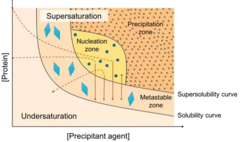
Figure 1.1. Illustrative representation of the diversity of major polysaccharides in the plant cell wall. ..……… 3 Figure 1.2. Examples of structures of polysaccharides found in the plant cell wall. ..……….5 Figure 1.3. The anaerobic bacterial cellulosome. .………... 6 Figure 1.4. Carbohydrate recognition by CBMs. .………. 9 Figure 1.5. Schematic overview of the main steps comprising the analysis using neoglycolipid (NGL)-based carbohydrate microarrays. ..………...14 Figure 1.6. Graphic representation of examples of immobilization strategies used to generate carbohydrate microarrays. .……… 16 Figure 1.7. Schematic overview of the main steps comprised in the determination of protein and protein-ligand structures by X-ray crystallography. ..………..20 Figure 1.8. Phase diagram for protein crystallization. .……….. 21 Figure 1.9. Assembly of unit cells in a three-dimensional crystal lattice. .……….. 23 Figure 1.10. Bragg's Law. .………. 24 Figure 1.11. Examples of carbohydrate-aromatic CH-π interactions. .………... 28
Chapter 2
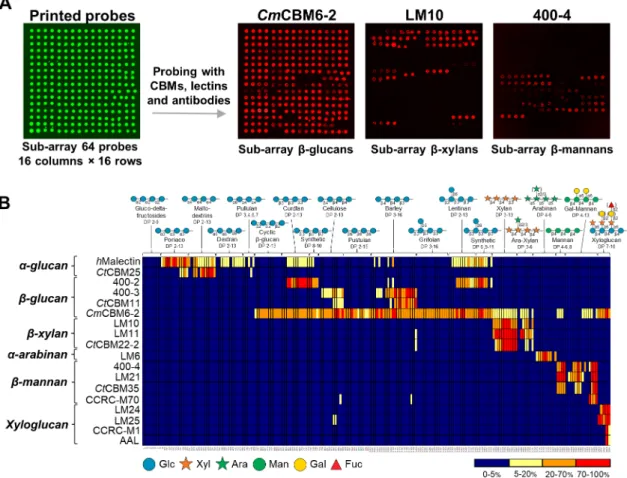
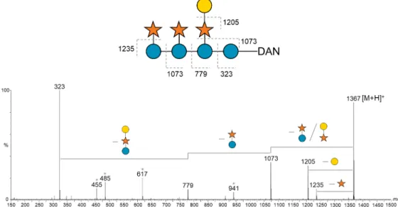
Figure 2.1. Binding patterns revealed by probing the glucan and hemicellulose oligosaccharide microarrays with sequence-specific carbohydrate-binding proteins. ……….. 39 Figure 2.2. Microarray analysis of glucan-binding proteins. ………. 41 Figure 2.3. Microarray analysis of xylan- and arabinan-binding proteins. ………..43 Figure 2.4. Microarray analysis of mannan-binding proteins. ……….. 45 Figure 2.5. Deconvolution of the xyloglucan oligosaccharides from tamarind.……….. 47 Figure 2.6. Positive-ion ESI-MS/MS product-ion spectra used for sequencing of xyloglucan-DAN DP-8. ……….……….……….……….. 48 Figure 2.7. Preparation of the xyloglucan-DAN-NGL probes from tamarind included in the new xyloglucan microarrays. ……….……….……… 49 Figure 2.8. Validation and analysis of the new xyloglucan microarrays. ……… 51
Chapter 3
Figure 3.1. Schematic representation of the multi-step strategy followed in this work. .……….. 65 Figure 3.2. Overview of the selected CBMomes of the two bacteria. ………. 66
XX
Figure 3.4. Analysis of C. thermocellum and R. flavefaciens FD-1 CBMs using polysaccharide microarrays – 1st screening for carbohydrate-binding activities. ………..70
Figure 3.5. Validation of the C. thermocellum CBMs microarray binding patterns using affinity gel electrophoresis. ……….……….………. 71 Figure 3.6. Validation of the R. flavefaciens CBMs microarray binding patterns using affinity gel electrophoresis. ……….……….………. 72 Figure 3.7. Analysis of C. thermocellum and R. flavefaciens FD-1 CBMs using oligosaccharide microarrays – 2nd screening for assigning carbohydrate-binding specificities. ………..73
Figure 3.8. Comparison of carbohydrate-binding specificities of families 6 and 22 CBMs from
C. thermocellum and R. flavefaciens FD-1 to xylan sequences. ………. 81
Chapter 4
Figure 4.1. Top view on the identified binding site of wild-type CtCBM11. ……… 92 Figure 4.2. Analysis of carbohydrate binding specificity using a microarray of sequence-defined gluco-oligosaccharides. ……….……….……… 94 Figure 4.3. Ribbon representation of the three-dimensional crystal structures of CtCBM11 complexes. ……….……….………. 96 Figure 4.4. Comparison of unbound and ligand-bound CtCBM11 structures. ……….. 98 Figure 4.5. CtCBM11-ligand interactions. ……….……….. 99 Figure 4.6. Representative isothermal calorimetry titrations of binding of CtCBM11 and its mutants to oligosaccharides. ……….……….……… 101 Figure 4.7. Alignment of CBM11 family members. ……….………. 104
Chapter 5
Figure 5.1. Modular architecture of proteins containing family 50 CBMs in the genome of
C. thermocellum. ……….……….………. 115
Figure 5.2. Oligosaccharide microarray analysis of C. thermocellum family 50 CBMs. ……… 116 Figure 5.3. Comparative analysis of C. thermocellum family 50 CBMs binding to β1,4-linked GlcNAc oligosaccharides. ……….……….……….. 117 Figure 5.4. Ribbon representation of the three-dimensional crystal structure of the
CtCBM50-GlcNAc3 complex. ……….……….……….118
Figure 5.5. Isothermal calorimetry titrations of binding of CtCBM50 and its mutant derivatives to β1,4-linked GlcNAc oligosaccharides. ……….……….. 121 Figure 5.6. Representation of the last simulation structure of the various GlcNAc oligosaccharides bound to the CtCBM50 complex of chains A and B. ……….……….. 123 Figure 5.7. Molecular dynamics simulations of CtCBM50 chain-length dependency. ..……… 125
XXI
GlcNAc residue in simulations with GlcNAc and MurNAc-GlcNAc pentasaccharides. ………..127 Figure 5.10. Alignment of CBM50 family members. ……….………... 128 Figure 5.11. Superposition of CtCBM50 with Thermus thermophilus LysM1. ……….129 Figure 5.12. Schematic representation illustrating the hypothesized cooperative binding by
CtCBM50 to short and long GlcNAc oligosaccharides. ………...130
Chapter 6
Figure 6.1. Schematic representation of the major pectic polysaccharide structural domains. 140 Figure 6.2. Modular architecture of R. flavefaciens proteins containing family 13 CBMs. …… 141 Figure 6.3. Carbohydrate microarray analysis of R. flavefaciens family 13 CBMs. ……… 142 Figure 6.4. Binding analysis of R. flavefaciens family 13 CBMs arabinan-derived oligosaccharides included in the carbohydrate microarrays. ……….………... 143 Figure 6.5. Ribbon representation of RfCBM13-1 three-dimensional structure. ………. 145 Figure 6.6. Isothermal calorimetry titrations of binding of RfCBM13-1 to α1,5-linked arabinan sequences. ……….……….………... 147 Figure 6.7. Isothermal calorimetry titrations of binding of RfCBM13-1 mutant derivatives to α1,5-linked arabinan sequences. ……….……….. 149 Figure 6.8. Alignment of CBM13 family members. ……….. 151 Figure 6.9. Superposition of RfCBM13-1 with Streptomyces avermitilis CBM13. ……….. 152
XXIII
Chapter 2
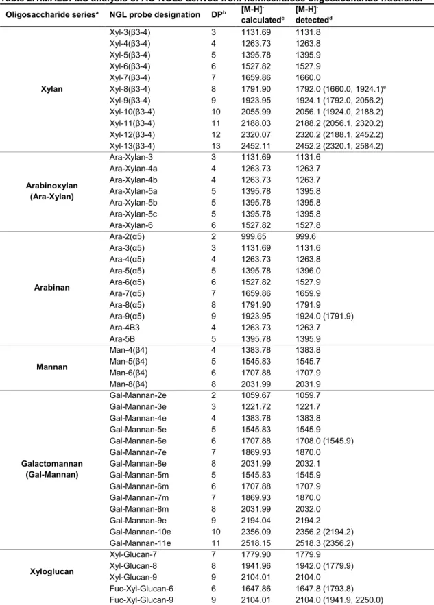
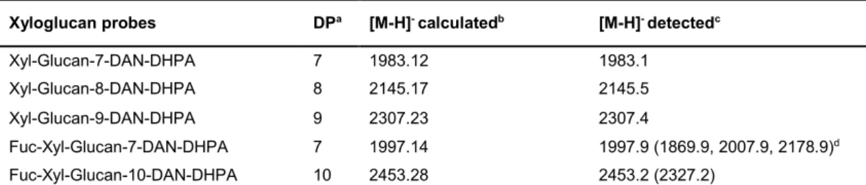
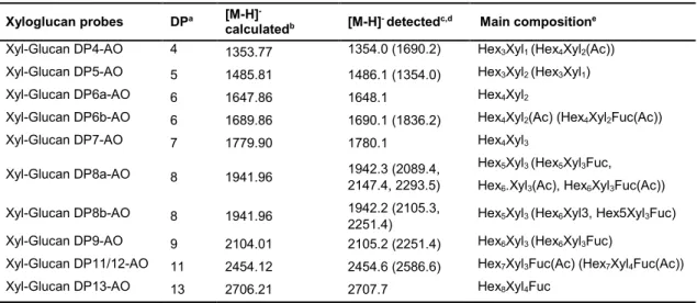
Table 2.1.MALDI-MS analysis of AO-NGLs derived from hemicellulose oligosaccharides. ……38 Table 2.2. MALDI-MS analysis of xyloglucan-DAN-DHPA NGLs investigated. ……… 50 Table 2.3. MALDI-MS analysis of the new xyloglucan-AO-NGL probes generated. ……… 52
Chapter 3
Table 3.1. Summary of the C. thermocellum CBMs ligand recognition and specificity obtained in the carbohydrate microarray screenings and affinity gel electrophoresis (AGE), cross-referencing with the available literature. ………74 Table 3.2. Summary of the R. flavefaciens FD-1 CBMs ligand recognition and specificity obtained in the carbohydrate microarray screenings and affinity gel electrophoresis (AGE). ………. 76
Chapter 4
Table 4.1. X-ray diffraction and structure refinement parameters and statistics for
CtCBM11-G4G4G3G and CtCBM11-G4G3G4G4G3G structures. ………. 97
Table 4.2. Thermodynamic parameters of the binding of CtCBM11 and its mutant derivatives to polysaccharides and oligosaccharides. ……….……… 102
Chapter 5
Table 5.1. X-ray diffraction and structure refinement parameters and statistics for
CtCBM50-GlcNAc3. ……….……….……….119
Table 5.2. Thermodynamic parameters of the binding of CtCBM50 and its mutant derivatives to polysaccharides and oligosaccharides. ……….……… 122 Table 5.3. Binding enthalpies and binding free-energies of the GlcNAc ligands to the CtCBM50 chains A and B. ……….……….……….. 124 Table 5.4. Binding enthalpies and binding free-energies of the peptidoglycan ligands to
CtCBM50. 127
Chapter 6
Table 6.1. X-ray diffraction and structure refinement parameters and statistics for
RfCBM13-1. ……….……….………. 146
Table 6.2. Thermodynamic parameters of the binding of RfCBM13-1 wild type and its mutant derivatives to polysaccharides and oligosaccharides. ……….………148
XXV
ACN Acetonitrile
AG-I Arabinogalactan type I
AG-II Arabinogalactan type II
AGE Affinity Gel Electrophoresis
AOPE (AO) Aminooxy-functionalized-1,2-dihexadecyl-sn-glycero-3-phosphoethanolamine
BSA Bovine Serum Albumin
CAZymes Carbohydrate Active enZymes
CBD Cellulose Binding Domain
CBM Carbohydrate Binding Module
CBMomes All CBM modules assigned to CAZy families up to 2015
CE Carbohydrate Esterase
CFG Consortium for Functional Glycomics
CID Collision-Induced Dissociation
CoMPP Comprehensive Microarray Polymer Profiling
Ct Clostridium thermocellum (C. thermocellum)
Cy3 Cyanine 3 fluorophore
DAN 1,5-diaminonaphthalene
DHPA N-(4-formylbenzamide)-1,2-dihexadecyl-sn-glycero-3-phosphoethanolamine
DHPE 1,2-dihexadecyl-sn-glycero-3-phosphoethanolamine
DOC Dockerin
DP Degree of Polymerization
E. coli Escherichia coli
ESI Electrospray Ionization
ESRF European Synchrotron Radiation Facility
GH Glycoside Hydrolases
HEC Hydroxyethyl cellulose
HEPES Hydroxyethyl piperazineethanesulfonic acid
HG Homogalacturonan
HPLC High Performance Liquid Chromatography
HPTLC High-Performance Thin Layer Chromatography
IMAC Ion Metal Affinity Chromatography
IPTG Isopropyl β-D-1-thiogalactopyranoside
ITC Isothermal Titration Calorimetry
LB Luria-Bertani
LIC Ligation Independent Cloning
Lic Lichenase
XXVI
MD Molecular Dynamics
MeOH Methanol
MIRAGE Minimum Information Required for A Glycomics Experiment
MOPS 3-morpholinopropane-1-sulfonic acid
MR Molecular Replacement
MS Mass Spectrometry
NGL NeoGlycoLipid
NHS N-hydroxysuccinimide
NMR Nuclear magnetic resonance
OD600nm Optical density at 600 nm
PDB Protein Data Bank
PEG Polyethylene glycol
PG Peptidoglycan
PL Polysaccharide Lyases
Rf Ruminococcus flavefaciens FD-1 (R. flavefaciens)
RG-I Rhamnogalacturonan I
RG-II Rhamnogalacturonan II
rmsd Root-mean-square deviation
rpm Rotation per minute
SDS-PAGE Sodium dodecyl sulphate - Polyacrylamide gel electrophoresis
SLS Swiss Light Source
SNFG Symbol Nomenclature for Glycans
TBA Tetrabutylammonium cyanoborohydride
UV Ultraviolet
WT Wild type
XXVII Arginine Arg R Asparagine Asn N Aspartate Asp D Cysteine Cys C Glutamate Glu E Glutamine Gln Q Glycine Gly G Histidine His H Isoleucine Ile I Leucine Leu L Lysine Lys K Methionine Met M Phenylalanine Phe F Proline Pro P Serine Ser S Threonine Thr T Tryptophan Trp W Tyrosine Tyr Y Valine Val V
Monosaccharide abbreviations and symbols
a3-deoxy-D-lyxo-2-heptulosaric acid Dha
3-deoxy-D-manno-2-octulosonic acid Kdo
Apiose Api
Arabinose Ara
Fucose Fuc
Galactose Gal
Galacturonic acid GalA
Glucose Glc
Glucosamine GlcN
N-acetyl-glucosamine GlcNAc
Glucuronic acid GlcA
Mannose Man
Muramic acid MurNAc
Rhamnose Rha
Xylose Xyl
3-C-carboxy-5-deoxy-L-xylose Aceric acid
aThemonosaccharide symbolic representation used was according to the updated Symbol Nomenclature for Glycans (SNFG)1 from the 3rd Edition of the Essentials of Glycobiology2.
XXIX
The work described in this Thesis focused on a unique approach combining carbohydrate microarrays with X-ray crystallography, to uncover the carbohydrate-binding specificity of CBMs from the cellulolytic bacteria C. thermocellum and R. flavefaciens FD-1 and to structurally characterize novel CBM-carbohydrate recognition mechanisms at a molecular level. To this end, sequential projects were followed, and the respective results and conclusions were divided into 5 chapters (Chapters 2-6) as outlined below.
Chapter 1 starts with a general introduction on the composition of plant cell wall polysaccharides and the systems that cellulolytic microorganisms employ for its biodegradation, including several biotechnological applications of CBMs. These sections are followed by an overview on the carbohydrate microarrays technology and on protein X-ray crystallography as the two main techniques applied in this Thesis, focusing on the characterization of protein-carbohydrate interactions. At the end of the chapter, the rationale and the main objectives of the Thesis work plan are presented.
Chapter 2 is the first results chapter and describes the construction and validation of an NGL-microarrays platform comprised of naturally-derived glucan- and hemicellulose-related oligosaccharides to address the need of increasing the diversity of plant-derived sequence-defined microarrays developed to this date. The application of the microarrays to assign carbohydrate ligands and the specificities of CBMs and plant carbohydrate-specific antibodies is demonstrated.
The application of these new microarrays to the high-throughput screening of C. thermocellum and R. flavefaciens CBMomes and to assign novel CBM-carbohydrate specificities is described in Chapter 3. Derived from the results obtained from the microarrays screening analysis, and considering their functional, biotechnological and industrial relevance, three representative CBMs, with the respective ligands, were selected for further biochemical and biophysical characterization, and are explored in the next three chapters.
Chapter 4 details the molecular determinants for the ligand recognition of family 11
C. thermocellum CBMto mixed-linked β1,3-1,4-glucans, describing the CBM 3D structures in complex with tetra- and hexa-saccharide ligands.
In the same trend, Chapter 5 is focused on the structural and functional characterization of a
C. thermocellum CBM from family 50, which is identified as a novel LysM domain binding to chitin
XXX
sequences is described.
Finally, in Chapter 7 the main conclusions of the Thesis work are drawn in an integrative manner, and future perspectives are presented.
C
HAPTER
1
G
ENERAL
I
NTRODUCTION
1
1Some sections of this Introduction were reproduced and updated from Ribeiro, D.O., Pinheiro,
B.A., Carvalho, A.L., Palma, A.S. (2018) Targeting protein-carbohydrate interactions in plant cell-wall biodegradation: the power of carbohydrate microarrays. In Carbohydrate Chemistry: Chemical and biological approaches (Rauter AP, Lindhorst T, & Queneau Y, eds), pp. 159–176. Royal Society of Chemistry (DOI: 10.1039/9781788010641-00159).
3
1 General Introduction
1.1 Structural diversity of plant cell wall polysaccharides
The plant cell wall is an intricate structure composed in its majority by complex polysaccharides and a smaller number of structural proteins. The composition in polysaccharides is highly variable, depending if the plant cell wall is expanding (primary cell wall) or if its role is to give additional structural support to the cell (secondary plant cell wall). It also differs between species, with distinct chemical compositions among the cell walls of grass and flowering plant species3,4. The
wider molecular and functional diversity of the polysaccharides is mainly observed in the primary cell wall with their configurations changing throughout the plant cell development, expansion and division5.
In plant cell walls, microfibrils of the major polysaccharide cellulose form a network embedded in a matrix of various complex polysaccharides, such as hemicelluloses, β-glucans and pectins (Figure 1.1). The hemicelluloses are interconnected with cellulose reinforcing the strength and resilience of the network, while the hydrated gels composed of pectin that intercalate this network, determine the porosity and thickness of the cell wall. The entire structure is maintained by non-covalent interactions, both spontaneous physico-chemical interactions and enzymatic crosslinking, that exist between these polysaccharides4,6.
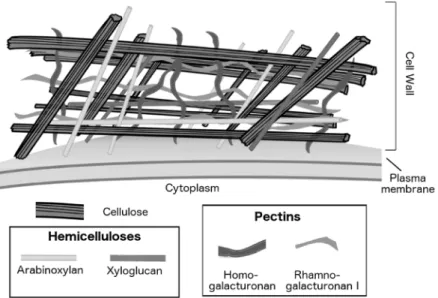
Figure 1.1. Illustrative representation of the diversity of major polysaccharides in the plant cell wall.
In combination with hemicelluloses, cellulose microfibrils form a network interspersed by pectin polysaccharides. The main hemicellulose polysaccharides found in plant cell walls are xyloglucan (dicot species) and arabinoxylan (grasses). The major pectin polysaccharides are rhamnogalacturonan I and homogalacturonan4.
The plant cell wall polysaccharides are structurally diverse (Figure 1.2). Cellulose is composed of aligned linear homopolymers of β1,4-D-linked glucosyl residues, organized in sheets packed in a
4
“parallel-up” fashion forming a structure that is, in its majority, crystalline7. Hemicelluloses
maintain a β1,4-D-linked backbone but are structurally more complex and composed of other
residues, in addition to glucose, such as mannose and xylose, in linear or branched sequences. Examples of hemicelluloses include xyloglucans, xylans, mannans, glucomannans and mixed-linked β1,3-1,4-D-glucans (β1,4-D-linked glucans with interspersed single β1,3-D-linkages).
All hemicelluloses have significant structural similarity as their backbone residues share the same equatorial configuration at C1 and C4 positions. While xyloglucans are widespread in land plants, the others are more species specific: xylans (dicots and commelinid monocots), mannans (charophytes) and β1,3-D-β1,4-D-glucans (grasses)8. Callose is a β1,3-D-glucan that is
widespread and occurs in specialized walls or wall-associated structures, specifically at stages of differentiation9. Pectins are a structurally diverse group of polysaccharides constituted by
galacturonic acid (GalpA) in their backbone sequences. The predominant pectins in the primary plant cell wall are: homogalacturonan (HG), a polysaccharide with an unsubstituted backbone of α1,4-D-linked GalpA residues, and rhamnogalacturonan-I (RG-I), a polysaccharide with a
backbone of the repeating disaccharide [-α1,2-L-Rhap-α1,4-D-GalpA-]n, substituted at the
rhamnose residue with different structural domains, such as galactans, arabinans or arabinogalactans (Figure 1.2). Other pectins such as xylogalacturonan (XGA), which has a homogalacturonan backbone substituted by xylose, and rhamnogalacturonan-II (RG-II), a highly ramified polysaccharide with a homogalacturonan backbone comprising 7 to 9 α1,4-D-linked
GalpA residues are present in smaller amounts. Rhamnogalacturonan-II is the most structurally complex cell wall known polysaccharide as it has 12 different monosaccharide residues interconnected by more than 20 glycosidic types of linkages10.
As consequence of this structural diversity, cellulolytic microorganisms that are highly specialised in degrading the plant cell wall polysaccharides have developed a consortium of Carbohydrate Active Enzymes (CAZymes) appended by Carbohydrate Binding Modules (CBMs) for which the diversity of activities and specificities matches the variety of polysaccharides.
1.2 Cellulolytic microorganisms’ express proteomes highly efficient in
plant cell wall biodegradation
The machinery for degrading plant cell wall polysaccharides differs from anaerobic to aerobic microorganisms, however the modular organization of the polysaccharide-degrading enzymes is maintained in both.
Due to energetic constraints and competition between the species found in anaerobic environments, most anaerobic cellulolytic microorganisms have arranged an efficient but rather elaborated system, where the produced and secreted CAZymes, such as endoglucanases, exoglucanases and β-glucosidases, are assembled in supramolecular complexes (molecular
5
Figure 1.2. Examples of structures of polysaccharides found in the plant cell wall. The Haworth
conformational structure of the main chain representative tetrasaccharide sequences is represented; –R, possible ramification position; the different possible sequences of the ramifications to the main backbone chain are depicted. In the mixed-linked β-glucans, the β1,3-linkages separate segments of 2 up to 14 glucose residues linked with β1,4-linkages9.
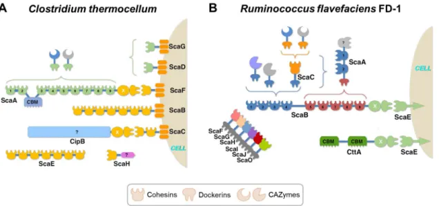
weight >3 MDa), termed the ‘cellulosome’ (Figure 1.3) (see references11–14 for a comprehensive
review on cellulosomes). Cellulosomes show different levels of complexity and are mainly localized at the cell surface12. These are known as integrating systems and are composed by one
or more scaffoldins, where CAZymes are integrated and brought to the vicinity of the substrate. The scaffoldin is a structural subunit composed of several cohesin modules that bind their binding
6
partners, the dockerin modules, present in CAZymes and other relevant proteins. Most of the scafoldins have in their modular structure a CBM from family 3a, that specifically binds to recalcitrant cellulosic substrates13. These multi-enzyme complexes can be attached to the
bacteria surface and display very complex and dynamic assemblies. One example is the cellulosome from Clostridium thermocellum, which has a primary scaffoldin (ScaA) comprising nine highly conserved type I cohesins, which allow the incorporation of different CAZymes and associated CBMs, through their type I dockerins (Figure 1.3A). To attach the scaffoldin subunit to the surface of the bacterial cell, membrane-associated proteins are bound to a type II cohesin12,15.
Bacteria of the genera Acetivibrio, Clostridium, Ruminococcus, Thermotoga16,17 and fungi of the
genera Neocallimastix, Piromyces and Orpinomyces12 are examples of anaerobic cellulolytic
microorganisms. The ecosystems where anaerobes are found to degrade plant polysaccharides to soluble sugars are as diverse as soils, sediments or water bodies. Recently, much attention has been given to the polysaccharide-degrading systems from anaerobic organisms that reside in the digestive tracts of invertebrates and vertebrates18,19. These offer novel systems for studying
carbohydrate recognition and are important for communication with the host, such as the human host, promoting health and nutritional benefits.
Figure 1.3. The anaerobic bacterial cellulosome. Schematic representation the architecture of the
cellulosomal assembly of (A) C. thermocellum and (B) R. flavefaciens FD-1. The colour code represents the
different specificities of cohesin-dockerin (Coh-Doc), that compose the various scaffoldins (Sca). The CAZymes modules, can additionally have appended CBMs. (Adapted from Bule et al., 201814).
Aerobic microorganisms secrete large quantities of CAZymes to the environment, organized in much simpler non-integrating systems. Bacteria from genera Bacillus, Micromonospora, Cellvibrio and Pseudomonas20 and fungi from genera Aspergillus21 are examples of aerobic cellulolytic
microorganisms. The enzymatic activities of the CAZymes are still complementary, showing strong synergy in the degradation of plant cell walls.
It is worth emphasizing in this section the recently identified lytic polysaccharide monooxygenases (LPMO) as key players in the first steps of plant biomass degradation19. LPMOs are
7
copper-dependent enzymes which cleave crystalline substrates by oxidizing the glycosidic bonds. By introducing chain breaks in insoluble polysaccharides, such as cellulose and chitin, and also in some hemicelluloses, LPMOs have the ability to enhance the activity of the glycoside hydrolases.
CAZymes and CBMs are classified into sequence-based families in the CAZy database (www.cazy.org)22. This database, with regular updates, is dedicated to classifying and analyse
genomic, structural and biochemical information concerning CAZymes and associated CBMs involved in the synthesis, modification and breakdown of oligo- and polysaccharides22. Currently
(as of March 2020), there are numerous different CAZymes and CBM families identified: 167 families of glycoside hydrolases (GHs), 110 families of glycosyl transferases, 40 families of polysaccharide lyases (PLs), 17 families of carbohydrate esterases (CEs), 16 families of auxiliary activities (including the 6 LPMO families), and 86 families of CBMs.
1.2.1 Carbohydrate-binding modules: the non-catalytic domains associated with
Carbohydrate Active enZymes
CBMs are a class of carbohydrate-binding proteins, defined as non-catalytic protein domains, with amino acid sequences ranging from 30 to 200 amino acids23,24. These modules were initially
defined as cellulose-binding domains (CBDs), as the first examples of CBMs mainly bound to crystalline cellulose25. However, these modules show a highly diverse range of ligand specificities,
between different families and even within the same family. Several characterized CBMs recognize non-crystalline cellulose, chitin, xylan, mannan, galactan, soluble α- and β-glucans and insoluble storage polysaccharides, such as starch and glycogen22.
The number of newly identified CBM sequences with putative carbohydrate binding is growing fast due to the exponential increase of sequence information derived from microbial genomics, metagenomics and transcriptomics data. Many of these proteins await elucidation and assignment of a carbohydrate-binding function26,27.
1.2.1.1 Classification of CBMs
Based on the conservation of protein fold, CBMs are divided into 7 different fold families. Most CBMs identified to date are classified in the β-sandwich family of protein folds. To provide additional functional relevance to the CBM classification, these modules have been grouped into three types: A, B, and C, according to the mode of interaction with the carbohydrates and the architecture of the binding site23,24.
CBMs from type A have a planar hydrophobic surface decorated by aromatic residues that interact
with flat crystalline polysaccharides, such as chitin or cellulose. This type of interaction is observed in the crystal structure of a CBM63-containing Bacillus subtilis expansin in complex with β1,4-D-linked cellohexaose28. Some type A CBMs have been reported to bind not only to
8
crystalline cellulose but also to soluble polysaccharides such as CBMs from family 2 and 3, which can also bind xyloglucans29, and CBM64 from Spirochaeta thermophila that binds a variety of
hemicelluloses30.
CBMs from type B or endo-type are classified as CBMs that bind to internal oligosaccharide
sequences. These CBMs exhibit a cleft or groove, that accommodates oligosaccharide chains with four or more residues, and show higher binding affinities with the increase of the oligosaccharide chain length.
One example is the highly thermostable family 4 CBM from Thermotoga maritima that binds β1,3-glucans and β1,3-1,4-glucans31,32 (Figure 1.4A). This CBM comprises the C-terminus of the
putative laminarinase Lam16A and the recognition of β1,3-D-linked laminarioligosaccharides
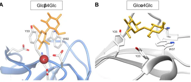
involves three tryptophan residues (W28, W58, and W99) and one tyrosine residue (Y23). One other example is the family 11 CBM from C. thermocellum, which recognizes mixed-linked β1,3-1,4-glucans32–34 (Chapter 4 of this Thesis is dedicated to the understanding of the molecular
determinants of this CBM specificity). This CBM is associated to the enzyme Lic26A-Cel5E, an enzyme that contains GH5 and GH26 catalytic domains that display β1,4-glucan and β1,3-1,4-glucan endoglucanase activity, respectively33.
CBMs from type C, or exo-type, are classified as CBMs that recognize the non-reducing end of an oligosaccharide sequence, binding in an optimal way to mono-, di- or trisaccharides, due to steric restriction in the binding site. Unlike the type B CBMs, type C do not contain the extended grooves in the binding-sites. Examples of this CBM type are the family 6 CBM from Bacillus
halodurans in complex with laminarihexaose35, the family 42 CBM from C. thermocellum, a
β-trefoil lectin that binds the arabinose side chains of complex hemicelluloses36, and family 13
CBM from Streptomyces lividans, another β-trefoil binding xylose or xylo-oligosaccharides37.
Remarkably, the family 6 CBM from Cellvibrio mixtus exhibits a type B cleft capable of recognizing β1,4-, β1,3-, and β1,3-1,4-linked glucose oligosaccharides, and a type C cleft that interacts with terminal residues of β1,4- and β1,3-linked glucose and also β1,4-xylose oligosaccharides38,39
(Figure 1.4B).
1.2.1.2 Functional Roles of CBMs
Although, in general, CBMs display low affinity towards their target ligands (in the µM-mM range)23, modular enzymes increase their avidity by containing multiple copies of CBMs,
enhancing the affinity for their target polysaccharide26. Although the mechanism by which CBMs
potentiate catalysis remains elusive26, the following four functional major roles are recognized for
CBMs23,24,26: 1) Targeting function, where CBMs target the joined catalytic modules to specific
regions on the carbohydrate substrate, such as reducing end, non-reducing end or internal polysaccharide chain, bringing the enzyme into close and prolonged contact with the target ligand; 2) Proximity effect, where CBMs increase the concentration of enzyme near its substrate, leading
9
Figure 1.4. Carbohydrate recognition by CBMs. (A) Type B protein-carbohydrate interactions illustrated
by 2 views of the 3D structure of family 4 CBM from Thermotoga maritima in complex with β1,3-D-linked laminarihexaose (PDB ID: 1GUI31). This type of CBMs displays a cleft arrangement in which the binding site accommodates glycan chains with four or more monosaccharide units. (B) Family 6 CBM from Cellvibrio
mixtus exhibits a type B cleft (in complex with β1,3-1,4-linked glucose tetrasaccharide (PDB ID: 1UZ0) and
a type C cleft (in complex with β1,4-linked glucose disaccharide) (PDB ID: 1UYX)38. Representations (not to scale) of individual 3D structures were done with program Chimera40 using the PDB atomic coordinates.
to a more rapid and efficient carbohydrate degradation; 3) Disruptive function, where CBMs act to disrupt the surface of tightly packed polysaccharides, such as crystalline cellulose fibres and starch granules, causing them to become more exposed to the catalytic module and, hence, increasing the degradation efficiency; and 4) Cell attachment, where CBMs adhere enzymes onto the surface of bacterial cell wall components, while exerting catalytic activity on an external carbohydrate substrate.
Several organisms possess CBMs that are not directly involved in plant cell wall degradation and have been found to perform other functions, acting in isolated or tandem forms. These modules can act as potential carbohydrate-sensors (or sensing domains) of the biomass availability in the extracellular medium, and examples are C. thermocellum CBMs from families 3 and 42 that trigger the expression of the enzymatic machinery specific for the degradation of the detected polysaccharides13,41. Additionally, certain CBMs, also known as Lysin motif domains or LysM
10
proteins acting as host immunity modulators, or be involved in the development of bacterial spores42.
Given the wide variety of ligand specificities, CBMs are an excellent model to study protein-carbohydrate recognition mechanisms. Additionally, these modules are interesting candidates for various applications in biotechnology, and some specific examples will be referenced in the next section.
1.2.2 Biotechnological applications of carbohydrate-binding proteins
Plant cell wall polysaccharides present major potential for biological and biotechnological applications, from paper and textiles industries, biotransformation of lignocellulosic materials into biofuels and other renewable products of biorefinery, and as sources of dietary fibres for both human and animal nutrition.In the past decades, the use of microbial and enzymatic systems to overcome the difficult deconstruction of such recalcitrant polysaccharides, yielding efficient and low-cost mechanisms, have gained importance43–45.
Not long after the identification of cellulosomes, their potential applications to biotechnology started to be explored46. Given their vast array of CAZymes and extreme habitat variability,
cellulosomes can be engineered as a multi-functional protein complex tool12,45,47. Chimeric
cellulosomes or designer cellulosomes have been used, for instance, as potential replacements or extensions of native cellulosomes to produce biofuels from cellulosic biomass48.
Additionally, each protein module in the cellulosome can be explored, individually or combined with free-acting CAZymes, for a wide spread of industrial and biotechnological applications47.
Synergistic actions between individual enzymes have been exploited to produce a combination of feed enzymes with the ability to degrade Chlorella vulgaris cell wall, with the purpose of improving the bioavailability of its valuable nutritional compounds for monogastric animal diets and facilitate the cost-effective use of microalgae by the feed industry44.
CBMs have also been largely explored for many biotechnological applications. Given their vast diversity, the ability to function autonomously in chimeric proteins and the controllable binding specificities so that the right solution can be adapted to an existing problem, make CBMs attractive candidates for a variety of applications49,50. From enhancers in biomass degradation51;
improvement of cellulose fibre properties for the paper and textile industries; functionalization of biomaterials in biomedicine; production, purification and immobilization of recombinant proteins; in food industry for the improvement of animal feed nutritional value; and as molecular probes for protein-carbohydrate interactions50.
As molecular probes, CBMs are valuable tools for the study of plant cell wall architecture. Recombinant CBMs have been used for characterizing native complex carbohydrates and engineered biomaterials50. CBMs from CAZy families 2a, 6, and 29 containing poly-histidine tags
11
such as crystalline cellulose, xylans, galactomannan and glucomannan52. Two recombinant
fluorescent CBM-probes consisting of a green fluorescent protein fused with a CBM3, that binds to both amorphous and crystalline cellulose, and mono-cherry fluorescent protein fused with a CBM17, that only binds to amorphous cellulose, have been used to reveal the surface accessibilities of amorphous and crystalline celluloses in Avicel53.
Given the increasing repertoire of CBMs, and their various potential and valuable applications, understanding the structural basis of their ligand specificity is of the most importance.
1.2.3 Clostridium thermocellum and Ruminococcus flavefaciens FD-1
The work developed in this Thesis focused on CBMs from Clostridium thermocellum (C. thermocellum) and Ruminococcus flavefaciens FD-1 (R. flavefaciens), two Gram-positive, anaerobic, cellulosome-producing cellulolytic bacteria that reside in different ecological niches, and of relevance in fields such as of bioprocessing and animal nutrition. C. thermocellum is a thermophilic bacterium54, found mostly in soils and hot springs and it is considered the most
efficient cellulolytic microorganism for the degradation of lignocellulosic biomass55.
C. thermocellum cellulosome (Figure 1.3A) was the first to be identified and characterized15 (see
section 1.2), and its complex architectural components have been extensively studied12,56,57. On
its turn, R. flavefaciens species are found in the digestive tracts of ruminants, other herbivorous animals and humans, and are among the most important ruminal cellulolytic bacteria, considered to be primarily responsible for plant cell wall biodegradation in the rumen58. Recent sequencing
of R. flavefaciens FD-1 genome has revealed one of the most complex cellulosomes (Figure 1.3B) identified to date, with one of the largest collection of cellulosome-associated proteins among known fibre-degrading bacteria14,27. Besides the numerous modular GHs, several CBMs were
identified to belong to known CAZy families and also putative CBM sequences27, some of which
have recently been classified into new families 75 to 8059.
Throughout the years, efforts were made to elucidate the molecular mechanism for the assembly of cellulosomes, enabling the identification of its different structural components. As such,
C. thermocellum CBMs have been widely studied and characterized, yielding a vast amount of
available information, with up to 90 CBMs from diverse families deposited in the CAZy database. Despite this, there are still numerous C. thermocellum CBMs that await elucidation and experimental validation. In contrast, given the more recent R. flavefaciens FD-1 genome sequencing, there is not much information available yet. Given the crucial roles of CBMs in plant cell wall biodegradation, the elucidation of the carbohydrate-specificities of the numerous assigned R. flavefaciens FD-1 CBMs and the understanding of mechanisms of carbohydrate recognition will promote the knowledge of this bacterium cellulolytic capabilities.
12
1.3 Methods for characterizing protein-carbohydrate interactions
Understanding the molecular basis for carbohydrate recognition by proteins and the relationship between structure and function are prerequisites for the development of future innovations in biological, biotechnological and industrial fields60. To this end, several state-of-the-art techniques
have been developed for the characterization of protein-carbohydrate interactions.
Characterization of protein-carbohydrate recognition often uses a combination of analytical methods, that can provide insights into the biological roles of each protein and their ligands, with structural studies, to understand the mechanisms of ligand-binding at molecular level. Carbohydrate microarrays61 allow to decode carbohydrate recognition by screening proteins for
ligand-binding and specificity. Other analytical methodologies, such as Isothermal Titration Calorimetry (ITC)62, MicroScale Thermophoresis (MST)60, Enzyme-Linked Immunosorbent Assay
(ELISA)63 and Affinity Gel Electrophoresis (AGE)62 can then be used to assess
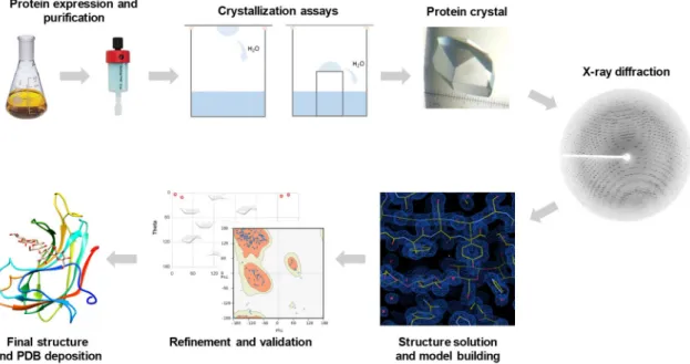
protein-carbohydrate affinity to target poly- and oligosaccharides. Structural characterization of the protein-carbohydrate recognition can be achieved by determining the molecular structures of protein-carbohydrate complexes, and X-ray crystallography64 and Nuclear Magnetic Resonance
(NMR) spectroscopy60 are the methods of choice, which can be used in a complementary manner.
High resolution data can also be integrated with lower resolution results from Cryo-Electron Microscopy (Cryo-EM) and Small Angle X-ray Scattering (SAXS) when characterizing large multi-component complexes65. Additionally, computational methodologies, such as Molecular
Dynamics (MD) and molecular docking66 are advantageous tools to complement the experimental
data.
In the next sections emphasis will be given to the methodological aspects and application of carbohydrate microarrays and X-ray crystallography, as these were the two major techniques applied to achieve the objectives of the proposed Thesis work plan.
1.3.1 Carbohydrate microarrays
The development of carbohydrate microarrays in the recent decades came to revolutionize the study of carbohydrate-protein interactions, satisfying the high demand for high-throughput methods to systematically array carbohydrate libraries and identify the specificity and biological role of carbohydrate-binding proteins67–73.
The main advantage of the microarray technology is that a wide diversity of carbohydrate probes can be immobilized on a microarray surface and simultaneously assessed for binding events, using only minute amounts of samples. This miniaturization feature of the microarrays, takes the most out of precious materials, both carbohydrates and protein analytes, while generating a large amount of information on a variety of carbohydrate-recognising systems74,75. Another important