Ana Paula Terrasso
Insert here an image
with rounded corners
modeling and drug discovery
Exploiting 3D differentiation of human stem cells
Oeiras
April, 2018
Dissertation presented to obtain the Ph.D degree in Sciences of
Engineering and Technology, Biomedical Engineering
modeling and drug discovery
Exploiting 3D differentiation of human stem cells
Ana Paula Barreto Terrasso
Dissertation presented to obtain the Ph.D degree in Sciences of
Engineering and Technology, Biomedical Engineering
Instituto de Tecnologia Química e Biológica António Xavier | Universidade Nova de Lisboa
modeling and drug discovery
Exploiting 3D differentiation of human stem cells
Ana Paula Barreto Terrasso
The work developed in this thesis was supervised by:
- Doctor Catarina Brito, Instituto de Biologia Experimental e
Tecnológica (iBET) e Instituto de Tecnologia Quimica e Biológica António
Xavier, Universidade Nova de Lisboa (ITQB-NOVA) (supervisor)
- Doctor Cláudia Almeida, CEDOC - Chronic Diseases Research Center,
Neural cell models for disease modeling and drug discovery: exploiting 3D differentiation of human stem cells
Copyright © 2018 by Ana Paula Barreto Terrasso
Instituto de Tecnologia Química e Biológica António Xavier Universidade Nova de Lisboa
From left to right: Dr. Duarte Barral, Dr. Júlia Costa, Dr. Luís Pereira de Almeida, Dr. Catarina Brito, Ana Terrasso, Dr. Cláudia Almeida, Dr. Dominik Paquet and Dr. Miguel Teixeira
Supervisors
- Doctor Catarina Brito, Instituto de Biologia Experimental e Tecnológica (iBET) e Instituto de Tecnologia Quimica e Biológica António Xavier, Universidade Nova de Lisboa (ITQB-NOVA), Portugal (supervisor)
- Doctor Cláudia Almeida, CEDOC - Chronic Diseases Research Center, NOVA Medical School, Universidade Nova de Lisboa, Portugal (co-supervisor)
Jury
- Doctor Dominik Paquet, Professor, Institute for Stroke and Dementia Research (ISD), University Hospital, Ludwig-Maximilians Universität Munich, Germany
- Doctor Luís Pereira de Almeida, Auxiliar Professor, Faculty of Pharmacy, University of Coimbra, Portugal
- Doctor Júlia Carvalho Costa, Principal Investigator, Instituto de Tecnologia Química e Biológica António Xavier, Universidade Nova de Lisboa (ITQB-NOVA), Portugal
- Doctor Duarte C. Barral, Principal Investigator, CEDOC - Chronic Diseases Research Center, Nova Medical School, Universidade Nova de Lisboa
- Doctor Miguel Teixeira, Cathedratic Professor, Instituto de Tecnologia Química e Biológica António Xavier, Universidade Nova de Lisboa (ITQB-NOVA), Portugal
Finantial support from :
Fundação para a Ciência e Tecnologia (FCT)
Ph.D grant PD/BD/52473/2014
iNOVA4Health (UID/Multi/04462/2013) financially supported by
FCT/MEC, through national funds and co-funded by FEDER under
PT2020
Tecnimede – Sociedade Técnico Medicinal S.A., the European
Regional Development Fund (FEDER) and the System of Incentives
for the Research and Technological Development (QREN) of the
Portuguese Government (project n.º 33913 – acronym PROiNEURO)
i
iii
Acknowledgements
I would like to express my gratitude to all the people who have contributed directly or indirectly to this thesis.
To my supervisor, Dr. Catarina Brito, for sharing this journey with me and for the constant support, guidance and motivation throughout these years. Thank you for the confidence, for the hours of scientific discussions and for the friendly conversation, and for always pushing me towards my goals and making me to potentiate my skills and grow as scientist. Also for stimulating such a challenging environment and the team spirit within the Advanced Cell Models Laboratory.
To my co-supervisor, Dr. Claudia Almeida, for introducing me to the field of neuronal intracellular trafficking, for all the fruitful scientific discussions and for inspiring me with your enthusiasm for research.
To Prof. Paula Alves, for the strong example of leadership and for transmitting us that excellence, rigor and hard work are fundamental for success. Thank you for giving me the opportunity to work in such an inspiring and excellent scientific environment at the Animal Cell Technology Unit.
To Dr. Augusto Filipe from Tecnimede - Sociedade Técnico-Medicinal, for the collaboration. For making available the compounds tested on the NT2 model and for always keeping the enthusiasm about our research.
To Dr. Eric Kremer, for all the support provided during our collaboration, for the fruitful discussions, for the knowledge shared about MPS VII and for receiving me in your lab.
To my colleagues in Advanced cell Models Lab, for being a truly team, for the discussion and critical suggestions and for the excitement shared. A special thanks to Catarina Pinto, Daniel Simão, Sofia Rebelo, Marta Estrada and Francisca Arez for the strong team spirit and for sharing this journey with me, for always being there, in good and not so good moments. Especially to Daniel, thank you for all the support, exchange of ideas and fruitful discussions. A thank to Marcos Sousa for the help with bioprocess operations. Also a thank to the rest of the people that
iv
contributed to the great atmosphere inside and outside the lab, especially to Marta Silva, João Sá, Mafalda, João Vidigal and Ana Oliveira. To all my colleagues, present and former members of the Animal Cell Technology Unit, for all the help provided and for creating such a great working environment, where we can continuously learn so much from all the different fields.
To Neuronal Trafficking in Aging Lab former and present members, especially to Tatiana Burrinha, for receiving me so well, for discussions and for the friendship.
Pessoalmente gostaria de agradecer,
A todos os meus amigos, por todo o apoio, por todos os momentos de descontração que passamos juntos e que me ajudam a seguir em frente.
Ao Paulo, pelo carinho, apoio e incentivo constantes, por toda a paciência e por estares sempre ao meu lado. Obrigado por acreditares sempre em mim e por me estimulares a querer sempre ir mais longe e melhor, por todos os momentos de descontracção e por toda a força que me deste.
À minha familia, que são a base de tudo. Ao meu irmão Zé Luis e aos meus pais, Ana Maria e António, sem vocês nada seria possível, obrigado por todo o amor, confiança e incentivo para seguir em frente, por estarem presentes e por todo o apoio incondicional. Obrigado por tudo o que me ensinaram, por serem um exemplo a seguir e por fazerem de mim aquilo que sou hoje.
v
Abstract
Neurological disorders are a major public health problem and are expected to rise dramatically together with the higher life expectancy and the shift towards an ageing society. Current therapeutic options can only ameliorate some of the symptoms and there are no effective treatments to target pathological mechanisms and stop disease progression. The human brain complexity hampers the understanding of the brain functioning at the molecular, cellular, and pathophysiological levels for many neurological disorders. This highlights the need for new brain models, which can contribute to unveil molecular mechanisms of neurological disorders, identify therapeutic targets and evaluate preclinically new therapies in a more adequate and predictive basis, withstanding its successful translation to the clinics. Despite their undeniable value, traditional animal models diverge from humans at biochemical and genetic levels. Moreover, 2D in vitro cell-based models do not mimic important aspects of brain cellular heterogeneity, architecture and microenvironment.
The main goal of this thesis was the development, validation and interrogation of 3D in vitro brain models for disease modeling and drug discovery. These models should include the presence of differentiated and functional human neurons and astrocytes, able to recapitulate the neuron-astrocyte interactions present in human brain, and mimic the endogenous microenvironment without the confounding effects of an exogenous matrix. To accomplish this, we took advantage of agitation-based culture systems to promote cellular aggregation and neural differentiation into neurospheroids. This strategy allows neural cells to establish cell and cell-extracellular matrix (ECM) interactions and generate complex 3D networks within the neurospheroids.
For drug screening applications, the human NTera2 (NT2) cell line was chosen to develop neurospheroids, since it provides an unlimited number of cells, with less expensive and time-consuming differentiation protocols. The development of a scalable and reliable bioprocess able to sustain the production of differentiated
vi
neurospheroids enriched in mature and functional human neurons and astrocytes is described in chapter 2. Toxicity assessment of prototypical neurotoxicants, combining endpoints based on neuronal- and astrocytic-specific gene expression and functionality in 3D, validated this brain cell model as a powerful tool to evaluate human neuronal and astrocytic toxicity. The potential of the NT2-derived neurospheroids for neuroprotection assessment was further validated. Implementation of an in vitro neuroprotection assay combining the human neuron-astrocyte co-culture neurospheroids with a cell viability endpoint is described in chapter 3. The neuroprotection assay setup was applied to several chemical and naturally-synthesized compounds and revealed several candidates with neuroprotective effect against an oxidant lesion. The robustness and simplicity of these assays enabled the use of complex 3D brain cell models as a platform to identify neurotoxic agents and to identify and validate drug candidates. This highlights the potential of 3D human brain cell models for drug testing, including both neurotoxicity and neuroprotection assessment.
For disease modelling, patient-specific induced pluripotent stem cells (iPSC) were used, as these can be differentiated in vitro into relevant somatic lineages with the genetic background of the patients. In chapter 4, the development of a Mucopolysaccharidosis type VII (MPS VII) human disease model, able to recapitulate the neuronopathic form of the disease is described. MPS VII is an orphan, ultra-rare, lysosomal storage disease (LSD) caused by a deficiency in β-glucuronidase enzymatic activity, which leads to an abnormal accumulation of glycosaminoglycans (GAGs) in the lysosomes of many tissues, including in the brain. For healthy and MPS VII iPSC-derived neural progenitor cells (NPC) 3D neural differentiation we took advantage of a strategy previously implemented in our laboratory using software-controlled perfusion stirred-tank bioreactors. The results demonstrated that neural differentiation of human MPS VII iPSC-NPC recapitulate major disease features, specifically GAGs accumulation. MPS VII neurospheroids interrogation revealed an upregulation in astrocytic GFAP gene expression and a
vii disturbance in MPS VII neuronal network activity and network functional connectivity, when compared with neurospheroids derived from iPSC of a healthy donor. This MPS VII human brain cell model can potentially contribute to elucidate the cellular processes responsible for disease progression and brain dysfunction and be applied for in vitro assessment of appropriate pharmacological treatments. The tools developed, namely the 3D neurospheroid culture system combined with the neuronal connectivity assay, have potential to assess neurological defects in other LSD and neurological disorders with variable phenotypes.
The work developed in this thesis provides simple and versatile in vitro approaches to generate human brain cell models for disease modeling and drug discovery that recapitulate human neuron-astrocyte interactions. Furthermore, the strategies described herein can be applied to other sources of neural cells or to iPSC from other neurological disorders and to evaluate different therapies in preclinical research.
ix
Resumo
As doenças neurológicas consituem um problema de saúde pública grave e prevê-se que a sua incidência aumente em paralelo com o aumento da esperança média de vida e o consequente envelhecimento da população. Os tratamentos disponiveis atualmente apenas aliviam alguns dos sintomas, não existem tratamentos efetivos, direccionados aos mecanismos patológicos e que impeçam a progressão das doenças. A elevada complexidade do cérebro humano dificulta a compreensão do seu funcionamento a nivel molecular, celular e patofisiológico. Isto demonstra a necessidade de desenvolver novos modelos celulares que contribuam para desvendar os mecanismos moleculares envolvidos no desenvolvimento e progressão das doenças neurológicas, identificar alvos terapêuticos e avaliar novas terapias de forma mais adequada e preditiva, garantindo o sucesso na sua translação para a clinica. Apesar da sua importância, os modelos animais usados tradicionalmente divergem dos humanos a nivel bioquimico e genético. Além disso, os modelos celulares bidimensionais (2D) não mimetizam aspectos importantes do cérebro humano, tais como a sua heterogeneidade celular, arquitectura e microambiente.
O objectivo principal desta tese é o desenvolvimento, validação e interrogação de modelos celulares de cérebro, com aplicação na descoberta de fármacos e em modelos de doenças neurológicas. Estes devem incluir neurónios e astrócitos humanos, diferenciados e funcionais, capazes de recapitular as interacções neurónio-astrócito existentes no cérebro humano e mimetizar o microambiente endógeno, sem a perturbação de uma matriz exógena. Foram usados sistemas de cultura agitados para promover a agregação celular e a diferenciação neural. Esta estratégia permite às células nos neuroesferóides estabelecer interacções célula-célula e célula-célula-matriz e gerar redes neuronais tridimensionais complexas.
Para aplicações em testes de fármacos, foi escolhida a linha celular Ntera2 (NT2), uma vez que permite obter um número ilimitado de células, com protocolos de diferenciação menos dispendiosos e demorados. O desenvolvimento de um
x
bioprocesso robusto e escalonavél, que permitiu a produção de neuroesferóides enriquecidos em neurónios e astrócitos humanos, maturos e funcionais, é descrito no capitulo 2. A avaliação da toxicidade de compostos neurotóxicos prototípicos, combinando endpoints baseados na expressão génica e na funcionalidade neuronal e astrocitica, permitiu validar este modelo como uma metodologia eficaz para avaliar toxicidade em neurónios e astrócitos humanos. A possibilidade dos neuroesferóides serem utilizados para avaliação de neuroprotecção foi também avaliada. A implementação de um ensaio in vitro para avaliar neuroprotecção, combinando a co-cultura de neurónios e astrócitos humanos em neuroesferóides e um endpoint de viabilidade celular, é descrita no capitulo 3. Este ensaio foi aplicado a diversos compostos, quimicos e sintetizados naturalmente e revelou vários candidatos com efeito neuroprotector contra uma lesão oxidativa. A robustez e a simplicadade destes ensaios potencia o uso de modelos celulares 3D mais complexos como plataforma para identificar compostos neurotóxicos e novos fármacos. Isto demonstra o potencial da aplicação de modelos celulares neurais para teste de fármacos, incluindo avaliação de neurotoxicidade e de neuroprotecção.
Para desenvolvimento de um modelo de doença, foram usadas células estaminais pluripotentes induzidas (iPSC), uma vez que estas podem ser diferenciadas in vitro em linhagens somáticas relevantes para o fenótipo da doença com os antecedentes genéticos dos pacientes. No capitulo 4 é descrito o desenvolvimento de um modelo celular humano que recapitula a forma neuronopática da doença Mucopolissacaridose tipo VII (MPS VII). A MPS VII é uma doença lisossomal de sobrecarga (DLS), orfã e ultra-rara, causada por uma deficiência na actividade do enzima β-glucuronidase, que leva à acumulação de glicosaminoglicanos (GAGs) nos lisosomas de diversos tecidos, incluindo no cérebro. Para diferenciação de células progenitoras neurais (NPC) derivadas de iPSC de um dador saudável e de outro com MPS VII, foi aplicada uma estratégia já implementada no laboratório, usando biorreatores de tanque agitado em perfusão.
xi Os resultados demonstraram que o modelo neural de MPS VII, derivado de iPSC-NPC humanas, recapitula as principais caracteristicas da doença, nomeadamente a acumulação de GAGs. Os neuroesferóides da doença revelaram uma sobreexpressão do gene astrocitico GFAP e distúrbios na funcionalidade da rede neuronal e na sua conectividade, quando comparados com os neuroesferóides derivados do dador saudável. Este modelo celular humano de MPS VII pode contribuir para elucidar os mecanismos celulares responsáveis pela progressão da doença e pela disfunção cerebral e ser aplicado na avaliação de tratamentos farmacológicos adequados para a doença. As metodologias desenvolvidas, nomeadamente o sistema de cultura de neuroesferóides, combinado com o ensaio de avaliação da funcionalidade e conectividade neuronal, podem ser aplicados na avaliação de defeitos neurológicos em outras DLS e doenças neurológicas com fenótipos variáveis.
O trabalho desenvolvido nesta tese fornece abordagens simples e versáteis para gerar modelos in vitro de células neurais humanas, com aplicação em ensaios pré-clínicos, que recapitulam as interações neurónio-astrócito presentes no cérebro humano. Para além disso, esta estratégia pode ser aplicada a outras fontes de células neurais ou a células de outras doenças neurológicas, para avaliar possiveis terapias em ensaios pré-clinicos.
xiii
Thesis publications
Terrasso AP, Pinto C, Serra M, Filipe A, Almeida S, Ferreira AL, Predroso P, Brito C, Alves PM. 2015. Novel scalable 3D cell based-model for in vitro neurotoxicity testing: combining human differentiated neurospheres with gene expression and functional endpoints. Journal of Biotechnology, 205, 82–92, doi: 10.1016/j.jbiotec.2014.12.011.
Terrasso AP, Silva AC, Filipe A, Pedroso P, Ferreira AL, Alves PM, Brito C. 2017. Human neuron-astrocyte 3D co-culture-based assay for evaluation of neuroprotective compounds. Journal of pharmacological and toxicological methods, 83, 72–79, doi: 10.1016/j.vascn.2016.10.001.
Bayó-Puxan N*, Terrasso AP*, Creyssels S, Simão D, Begon-Pescia C, Lavigne M, Salinas S, Bernex F, Bosch A, Kalatzis V, Levade T, Cuervo AM, Lory P, Consiglio A#, Brito C#, Kremer EJ# (*equal contribution; #co-senior authors). 2018. Lysosomal and network alterations in human mucopolysaccharidosis type VII iPSC-derived neurons. Journal of Cell Science. Under Revision.
xiv
Additional publications
Simão D, Silva MM, Terrasso AP,Arez F, Sousa MF, Mehrjardi NZ, Šarić T, Gomes-Alves P, Raimundo N, Gomes-Alves PM, Brito C. 2018. 3D differentiation of iPSC-derived NPC recapitulates human brain microenvironment. Stem Cell Reports. Under final
revision.
Figueira I, Tavares L, Jardim C, Costa I, Terrasso AP, ..., Santos CN. 2017. Blood– brain barrier transport and neuroprotective potential of blackberry-digested polyphenols: an in vitro study. European Journal of Nutrition, 1-18, doi: 10.1007/s00394-017-1576-y.
Figueira I, Garcia G, Pimpao RC, Terrasso AP, ..., Brito C, Santos CN. 2017. Polyphenols journey through blood-brain barrier towards neuronal protection. Scientific Reports 7(1), doi: 10.1038/s41598-017-11512-6.
Simão D, Terrasso AP, Teixeira AP, Brito C, Sonnewald U, Alves PM. 2016. Functional metabolic interactions of human neuron-astrocyte 3D in vitro networks. Scientific Reports, 6:33285, doi: 10.1038/srep33285.
Simão D, Arez F, Terrasso AP, Pinto C, Sousa MF, Brito C, Alves PM. 2016. Perfusion Stirred-Tank Bioreactors for 3D Differentiation of Human Neural Stem Cells. Methods in Molecular Biology, doi: 10.1007/7651_2016_333.
Materne EM, Ramme AP, Terrasso AP, Serra M, Alves PM, Brito C, Sakharov DA, Tonevitsky AG, Lauster R, Marx U. 2015. A multi-organ chip co-culture of neurospheres and liver equivalents for long-term substance testing. Journal of Biotechnology, 205, doi: 10.1016/j.jbiotec.2015.02.002.
xv
Table of contents
Chapter 1. Introduction ... 1 Chapter 2. Novel scalable 3D cell-based model for in vitro neurotoxicity testing: Combining human differentiated neurospheres with gene expression and functional endpoints ... 51 Chapter 3. Human neuron-astrocyte 3D co-culture-based assay for evaluation of neuroprotective compounds ... 81 Chapter 4. Lysosomal and network alterations in human mucopolysaccharidosis type VII iPSC-derived neurons ... 107 Chapter 5. Discussion and perspectives ... 151
xvii
List of figures
Figure 1.1: Drug discovery pipeline in the pharmaceutical industry.. ... 4 Figure 1.2: Different cell sources available for derivation of human neural cells.. ... 6 Figure 1.3: Major components of brain extracellular matrix (ECM).. ... 17 Figure 1.4: Stepwise degradation of glycosaminoglycans (GAGs) ... 30 Figure 1.5: Thesis aims and its discrimination by chapter.. ... 34 Figure 2.1: Neurospheroid culture ... 57 Figure 2.2: Neurotoxicity assays – dose-response curves. ... 58 Figure 2.3: Characterization of the neuronal population in neurospheroids. ... 63 Figure 2.4: Characterization of neurotransmitter phenotype of neuronal population in neurospheroids ... 65 Figure 2.5: Characterization of astrocytic population in neurospheroids. ... 66 Figure 2.6: Functional characterization of astrocytic population in neurospheroids ... 67 Figure 2.7: Neurotoxicity assays – functional and gene expression endpoints ... 69 Figure 3.1: Schematic workflow for addition of test compounds in neuroprotection assay over tBHP insult in 3D human neuron-astrocyte co-culture ... 88 Figure 3.2: Cell viability assays in 3D human neuron-astrocyte co-culture. Immunofluorescence microscopy of 3D human neuron-astrocyte co-culture ... 90 Figure 3.3: Idebenone neuroprotective effect over tBHP insult in 3D human neuron-astrocyte co-culture ... 91 Figure 3.4: Idebenone neuroprotective effect over the chloramphenicol insult in 3D human neuron-astrocyte co-culture. ... 92 Figure 3.5: Semi-automation of Presto blue cell viability endpoint ... 93 Figure 3.6: Neuroprotective effect of chemically synthesized test compounds over the tBHP insult in human neuron-astrocyte co-culture ... 94 Figure 3.7: Nicotinamide and Linezolid neuroprotective effect over the tBHP insult in 3D human neuron-astrocyte co-culture ... 95 Figure 4.1: Generation and characterization of human MPS VII iPSC ... 123
xviii
Figure 4.2: Characterization of human MPS VII iPSC-NPC and neurons in 2D cultures. ... 125 Figure 4.3: Schematic experimental workflow for differentiation of neural cells from control and MPS VII iPSC-NPC in 2D (top) and 3D (bottom) culture systems ... 126 Figure 4.4: Characterization of MPS VII neurospheroids ... 128 Figure 4.5: MPS VII neural cells recapitulate known disease features ... 130 Figure 4.6: Transmission electron microscopy (TEM) of MPS VII neurons... 132 Figure 4.7: Lysosome function alteration in MPS VII NPC and neurons ... 133 Figure 4.8: MPS VII neuronal activity and MPS VII neurospheroids calcium (Ca++) imaging analysis ... 136 Figure 5.1: Schematic representation of the major aims of the thesis and the achievements of each chapter (2-4) ... 153
List of tables
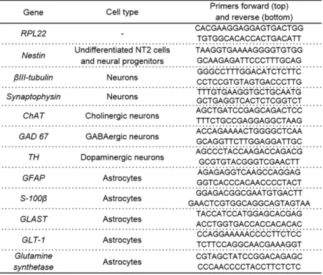
Table 2.1: List of primers used in NT2 neurospheroids qRT-PCR analysis ……….……62 Table 4.1: List of primers used in MPS VII cells qRT-PCR analysis………….……….120
xix
Abbreviations
2D Two-dimensional 3D Three-dimensional Acr Acrylamide AD Alzheimer diseaseASD Autism spectrum disorder
BDNF Brain-derived neurotrophic factor
BMP Bone morphogenic protein
cAMP Cyclic adenosine monophosphate CNS Central Nervous System
CSPGs Chondroitin sulfate proteoglycans
DIV Days in vitro
DMEM Dulbecco's Modified Eagle's Medium
EB Embryoid bodies
ECM Extracellular matrix EGF Embryonic growth factor ERT Enzyme replacement therapy ESC Embryonic stem cells
FBS Fetal bovine serum
FDA Fluorescein diacetate FGF Fibroblast growth factor FSG Fish skin gelatin
GABA γ-aminobutyric acid
GAD65/67 Glutamate decarboxylase 65/67
GAGs Glycosaminoglycans
GDNF Glia-derived neurotrophic factor GFAP Glial fibrillary acidic protein GLAST Glutamate aspartate transporter
xx Gln Glutamine GLT-1 Glutamate transporter 1 Glu Glutamate GS Glutamine synthase HTS High-throughput screening iN Induced neurons
iNSC Induced neural stem cells
iOPC Induced oligodendrocyte progenitor cells iPSC Induced pluripotent stem cells
LPS Lipopolysaccharides LSD Lysosomal storage diseases
MPS Mucopolysaccharidosis
MPS VII Mucopolysaccharidosis type VII
NE Neuroepithelial
NGF Nerve growth factor
NPC Neural progenitor cells NSC Neural stem cells
NT2 NTera2 clone D1
P/S Penicillin and Streptomycin PBS Phosphate buffered saline PCNA Proliferating cell nuclear antigen
PD Parkinson disease
PFA Paraformaldehyde
PGs Proteoglycans
PI Propidium iodide
PNNs Perineuronal nets PSC Pluripotent stem cells
qRT-PCR Quantitative real-time polymerase chain reaction
xxi
RG Radial glia
RPL22 Ribosomal protein L22
RT Room temperature
SD Standard deviation
SDS-PAGE Sodium dodecyl sulphate - polyacrylamide gel electrophoresis SFEBq Serum-free, floating embryoid body-like, quick aggregates
SHH Sonic hedgehog
tBHP Tert-butyl hydroperoxide TGF- β Transforming growth factor β TH Tyrosine hydroxylase
TLR4 Toll-like receptor 4 Tx-100 Triton X-100
vGluT1 Vesicular glutamate transporter β-gluc β-glucuronidase
r β-gluc Recombinant β-glucuronidase βIII-tub βIII-tubulin
β-tub β-tubulin
ChaT Choline acetyltransferase DAPI 4’-6-diamino-2-phenylindole
Syn Synaptophysin
PSD-95 Postsynaptic density protein 95 PVDF Polyvinylidene difluoride
VGluT1 Vesicular glutamate transporter 1
β-hex β-hexosaminidase
1
1.
CHAPTER 1
2
Table of contents
1. Neuroscience research: need for CNS modeling ... 3 2. Human cell sources for CNS modeling... 6 2.1. Primary cultures ... 7 2.2. Immortalized cell lines and in vitro differentiation of neural cells ... 8 3. Cell fate determinants: patterning and differentiation towards neural lineage ... ...14 4. Culture strategies for CNS modelling and drug screening... 18 4.1. Scaffold-dependent cultures ... 19 4.2. Scaffold-free cultures ... 21 5. Brain in vitro models for neuroscience research ... 23 6. Lysosomal storage diseases – Mucopolysaccharidosis type VII ... 28 7. Aims and Scope of the thesis ... 33 8. References ... 34
3
1. Neuroscience research: need for CNS modeling
Neurological disorders are a major public health problem and are expected to rise dramatically together with the increase in life expectancy and the shift towards an ageing society. Around 450 million people currently suffer from mental disorders and the forecasts indicate that by 2050 the number will triple (WHO Mental Disorders Fact Sheet, 2017). Neurological disorders represent significant economic and social burdens and high costs for healthcare systems, accounting for as much as 45% of the annual health budget in Europe (DiLuca et al., 2014; Harper, 2014; Rosemann, 2015). Current treatments for most neurological disorders only ameliorate some of the symptoms, alleviating patient’s mental impairment. Despite major and continuously growing investment, there are no effective disease-modifying treatments available to target pathological mechanisms and stop disease progression (Calcoen et al., 2015; Payne et al., 2015). Drug development pipeline have proven lengthy, costly and relatively unproductive process (Fig. 1.1) with a high number of late-stage failures and attrition rates around 90% (Calcoen et al., 2015; Hay et al., 2014). This is even worst for central nervous system (CNS) drugs, which are more likely to fail in Phase III drug development than non-CNS drugs, mainly due to lack of efficacy (Kesselheim et al., 2015). On average, the overall success rate is of 4.1%, but for example, in Alzheimer’s disease (AD) drug development, from 2002 to 2012, 413 clinical trials have been performed, mainly with drugs to improve cognition and disease-modifying small molecules or immunotherapies, with an overall success rate of only 0.4% (Calcoen et al., 2015; Cummings et al., 2014).
4
Figure 1.1: Drug discovery pipeline in the pharmaceutical industry. The different stages of drug
development process (drug discovery, preclinical research, clinical trials and regulatory approval); typically only 1 in 10 000 compounds are approved and the process costs more than 1 billion euro overall. 3D cell models can represent a complementary tool in preclinical research, offering increased predictive capacity, when compared to 2D cell-based assays. The use of these models can be parallel to animal models, reducing the dependency on animal models, costs and time. Adapted from Kunz-Schughart et al., (2004) and from http:// www.researchamerica.org/ advocacy-action/ issues-researchamerica-advocates/bench-bedside-drug-development-pipeline.
This highlights the need for novel preclinical models that enable new therapies to be developed and tested in a more adequate and predictive context, reducing the number of drugs that will fail in late-stages clinical trials (Fig. 1.1). To do so, the understanding of the pathophysiological bases for many neurological disorders is essential. However, the high degree of complexity of the human brain and its poor accessibility in vivo makes it difficult to investigate specific biochemical and cellular mechanisms. While of undeniable value, animal models and several in vitro cell-based models fail to precisely recapitulate the clinical pathophysiological state, hampering the relevance of preclinical studies using these models. This can be
5 accounted for the fact that animal and in vitro models do not mimic important aspects of the in vivo brain cellular composition, architecture and microenvironment. Animal models have been widely used to study genetic and pathological mechanisms of neurodegenerative diseases and to test new therapeutic drugs (Breslin and O’Driscoll, 2013; Fitzgerald et al., 2015). While mimicking brain tissue complexity, at biochemical and genetic levels there are differences between species and many human diseases resulting from complex interactions between genes and environmental factors can not be recapitulated in animals (Payne et al., 2015). Indeed, concordance between rodents and human toxicity can be very low with rodents being predictive of human toxicity in less than half of the cases (Olson et al., 2000). The transcriptional profile of AD mouse models is not similar to human AD (Hargis and Blalock, 2018) and these models do not reflect the extensive neuronal loss seen in the human condition (LaFerla and Green, 2012). Consequently, numerous drugs that demonstrated efficacy in the animal models have not shown efficacy when translated to the clinic. In fact, around 500 neuroprotective therapies that were successfully developed in rodent models of stroke, failed at some stage when translated to humans, with only one treatment being approved (O’Collins et al., 2006; Sena et al., 2010). Furthermore, human in vitro models for neurological disorders are commonly based on neuronal-like cell lines, mainly tumor-derived, but these do not recapitulate the cell heterogeneity of human brain and its microenvironmental features and are not genetically matched to the patient (Choi and Tanzi, 2012; Ross and Akimov, 2014). The recent advances in cell reprogramming technologies and pluripotent stem cell (PSC) neural differentiation strategies, allowing obtaining neural cells with the genetic background of the patient, provided unique opportunities to investigate important aspects of brain function, development and disease pathogenesis at cellular level. Therefore, given our poor understanding of the pathogenesis of complex diseases and the low predictive capacity of currently available animal and
6
in vitro models, it is clear that there is a high demand to develop brain cell models
that can withstand a successful translation of drugs to the clinics.
2. Human cell sources for CNS modeling
Human cell sources available for the derivation of neural cells for CNS modeling can be divided into two main categories: (1) primary cultures of human brain cells and (2) immortalized cell lines and in vitro differentiation of neural cells (Fig. 1.2).
Figure 1.2: Different cell sources available for the derivation of human neural cells. Neural
progenitor cells (NPC) or neural cells can be isolated directly from human fetal or adult brains. NPC are able to keep to some extent their self-renewal and multipotent capacities and can be differentiated into neural cells. Embryonic stem cells, isolated from blastocyst inner cell mass, and induced pluripotent stem cells, can be committed towards the neural lineage through neural induction protocols, generating neural progenitor cells, which can be further differentiated into neural cells. Somatic cells can be reprogrammed into induced pluripotent stem cells or directly reprogrammed into neural progenitor cells or terminally-differentiated neural cells. Immortalized cell lines are usually derived from tumor cells and some of these present ability to differentiate towards neural lineage into neural-like cells by addition of neurotrophic factors. Adapted from Conti and Cattaneo, (2010) and Jakel et al., (2004).
7
2.1. Primary cultures
Human brain cells isolated from fetus, can be maintained in vitro as primary cultures, including one or several brain cell types (Darbinyan et al., 2013; Mattson and Rychlik, 1990; Ray et al., 2014), or ex vivo as organotypic slice cultures, keeping the in situ cellular organization and three-dimensional (3D) tissue architecture within the slice (Humpel, 2015). The first cell culture of human neurons isolated from live adult brain was recently reported (Spaethling et al., 2017), with identification of 5 distinct cell types in culture: oligodendrocytes, microglia, neurons, endothelial cells and astrocytes and cell-type- and individual-specific transcriptional hierarchies were identified. Although it is a very valuable resource, the use of human brain tissue is limited due to the lack of availability and ethical concerns. Additionally, human brain tissue to study development and early defects in neurological disorders is not readily available, as brain biopsies from living patients are limited, and autopsy material is normally representative of later disease stages and limits sample quality (Darmanis et al., 2015). The use of terminally-differentiated cell types isolated from the CNS, as neurons, is also limited due to the impossibility of expansion in vitro. To overcome these issues, self-renewing multipotent neural stem cells (NSC) can be isolated from specific niches in fetal or adult brain tissue and expanded and terminally differentiated in
vitro into neurons and glia cells (Conti and Cattaneo, 2010; Simão et al., 2014).
Primary NSC have a limited self-renewal capacity in vitro due to the low level of expression of telomerase, which is required for continuous cell proliferation (Ostenfeld et al., 2000). Moreover, NSC differentiation potential, temporal development and phenotype varies with the tissue age, in vitro expansion and culturing method (Conti and Cattaneo, 2010; Zhang, 2006). Altogether, these aspects highlight the great limitations in the use of primary cell sources from CNS origin for in vitro modeling.
8
2.2. Immortalized cell lines and in vitro differentiation of
neural cells
Historically, the first hypothesis to overcome the difficulties associated with the use of primary cultures of brain cells was to establish immortalized cell lines. These can be from tumoral origin or immortalized in vitro by transfection of oncogenes. The advantages include being easy to obtain and allowing expansion of large numbers of cells. However, most of immortalized cell lines, as the widely used neuroblastoma cell lines, have the disadvantage of continuous proliferation, as they not generate post-mitotic cell-types and therefore do not allow the recapitulation of the neuronal development occurring in the CNS, as well as the organ-specific structural and functional features (Conti and Cattaneo, 2010; Gupta et al., 2012). The majority of the cell lines can be induced in vitro to differentiate in a more neuronal-like phenotype, usually by removal of specific growth factors or addition of neurotrophic factors. The human neuroblastoma cell line SH-SY5Y, which grows continuously as an undifferentiated cell population with neuroblast-like morphology, can be differentiated towards neuronal-neuroblast-like cells, with retinoic acid (RA) or specific neurotrophic factors, such as brain-derived neurotrophic factor (BDNF) (Shipley et al., 2016). SH-SY5Y cells have been used for neurotoxicity and neuroprotection testing in vitro and to understand the mechanisms of action of neurotoxic chemicals, such as okadaic acid and antidepressant drugs (Choi et al., 2011; Del Barrio et al., 2011). Also, Parkinson’s disease (PD) and AD-associated neuronal defects have also been modeled using neuroblastoma cell lines (Dodel et al., 2011; Fang et al., 2006; Presgraves et al., 2004). However, these models comprise immature neuronal-like cells and do not take into account the multiple cell types involved in the brain pathology and their interactions, which contribute to the disease.
Neural progenitor cells (NPC) can be immortalized in vitro, usually by transfection of oncogenes, to overcome its limited self-renewal capacity due to growth arrest and senescence. These immortal NSC lines can be expanded in vitro
9 and differentiated in neural cells. The human fetal NPC Lund mesencephalic cell line (LUHMES), immortalized by a tetracycline-controlled v-myc gene, can be differentiated into morphologically and biochemically mature dopamine-like neurons, following exposure to tetracycline, glial cell line-derived neurotrophic factor (GDNF) and dibutyryl cyclic adenosine monophosphate (cAMP). LUHMES were used for neurotoxicity testing and for PD modeling (Scholz et al., 2011; Zhang et al., 2014), either in co-culture with astrocytic cells (Efremova et al., 2015) or in a 3D culture setup (Smirnova et al., 2016), in an attempt to increase the level of mimicry of the human brain of the model. NPC immortalized in vitro by overexpression of the myc family transcription factors (ReNcell cells) were differentiated into the three neuronal lineages by removal of growth factors and addition of neurotrophic factors (Donato et al., 2007). A high-throughput screening assay for evaluation of chemical compounds effects was implemented using this ReNcell line (Breier et al., 2008) and differentiated neuronal cells were applied in AD modeling, based on overexpression of human b-amyloid precursor protein (APP) or APP and presenilin 1 (PSEN1), containing familial AD mutations (Choi et al., 2014). This 3D culture of differentiated neurons in Matrigel has been reported to recapitulate amyloid and tau pathologies with robust extracellular deposition of β-amyloid, and detection of aggregates of phosphorylated tau in neuronal cell soma and neurites (Choi et al., 2014). Notably, this was the first cell-based model able to recapitulate key features of AD pathophysiology in vitro, which have not been previously observed in mouse models and in in vitro 2D culture systems. The largely studied NTera-2 (NT2) cell line, a pluripotent cell line derived from a malignant embryonic carcinoma (Pal and Ravindran, 2006; Pleasure and Lee, 1993), upon exposure to RA, can be differentiated into neural cells, including neurons and astrocytes (Bani-Yaghoub et al., 1999; Goodfellow et al., 2011; Pleasure and Lee, 1993; Terrasso et al., 2015). NT2-derived neurons maintain a post-mitotic neuronal phenotype, with axons and dendrites elaborating processes and synaptic contacts and present different neurotransmitter subtypes, mainly glutamatergic and
10
GABAergic neurons (Coyle et al., 2011; Podrygajlo et al., 2009). NT2-derived astrocytic cells show phenotypic and functional markers, such as glial fibrillary acid protein (GFAP), the glutamate transporters GLAST and GLT-1 and the glutamine synthase (GS) enzyme (Goodfellow et al., 2011; Hill et al., 2012). NT2 cell line has been widely used to model specific neural functions (Goodfellow et al., 2011; Hill et al., 2012) and for neurotoxicity studies (Hill et al., 2008; Stern et al., 2013) due to its easy expansion in vitro and the simple neural differentiation protocol. In comparison with other cell lines, NT2-derived neural cells can attain a high degree of maturation, making them an useful human brain cell source. We promoted neural differentiation of NT2 cells as aggregates, generating neurospheroids that were mainly composed of functional neurons and astrocytes and were further applied for drug testing (Simão et al., 2016; Terrasso et al., 2017, 2015).
Several technological advances are nowadays allowing the derivation of human neural cells from different cell sources, such as embryonic stem cell (ESC) and induced pluripotent stem cells (iPSC). First human ESC lines were derived by Thomson and colleagues in 1998, providing a new starting point to attain recapitulation of tissue development in vitro and to generate a wide variety of cells types. A decade ago, reprogramming human somatic cells into iPSC by the introduction of four embryonic transcription factors (Oct4, Sox2, Klf4 and c-Myc) was firstly published by Yamanaka and colleagues (Takahashi et al., 2007). This technology opened the possibility of generating PSC with the genetic background of patients, which could then be induced to differentiate towards relevant somatic lineages, including the neural lineage (Chambers et al., 2009; Fernandes et al., 2015; Shi et al., 2012).
Initially ESC were differentiated towards the neural lineage via embryoid bodies (EB) formation (Dhara and Stice, 2008; Zhang et al., 2001), but more recently directed differentiation factors have been employed to differentiate ESC and iPSC into neural cells (Chambers et al., 2009; Shi et al., 2012). NPC can be efficiently generated from ESC and iPSC, in adherent culture, through inhibition of
11 bone morphogenetic protein (BMP) signaling by its antagonist noggin (Gerrard et al., 2005). RA and nerve growth factor (NGF) were found to be potent enhancers of neuronal differentiation, eliciting extensive outgrowth of processes and the expression of neuron-specific molecules (Schuldiner et al., 2001). Directed ESC or iPSC differentiation to enriched populations of specific neuronal subtypes can also be achieved. Derivation of midbrain dopaminergic neurons was induced by sequential application of the patterning molecules Sonic hedgehog (SHH), Fibroblast growth factor 8 (FGF8), BDNF, GDNF, transforming growth factor type β (TGF-β), dibutyryl cAMP and ascorbic acid (Perrier et al., 2004; Zhang and Zhang, 2010). Human iPSC-derived dopaminergic progenitor cells, differentiated by floor plate induction protocol, using BDNF, GDNF, cAMP and ascorbic acid, were grafted into the brain of a primate model of PD (Kikuchi et al., 2017). The authors reported that cells survived and functioned as mature midbrain dopaminergic neurons, extended dense neurites into the host striatum, indicating that human iPSC-derived dopaminergic progenitors are clinically applicable for the treatment of patients with PD (Kikuchi et al., 2017). Enriched populations of forebrain GABAergic neurons were obtained using purmorphamine and SHH interchangeably for neuroepithelia patterning (Ma et al., 2012). Human iPSC generated from Huntington’s disease patient were differentiated into striatal GABAergic neurons by combining morphogens and neurotrophins SHH, BDNF, dibutyryl cAMP and valpromide that contain the same CAG repeat expansion as the mutation in the HD patient (Zhang et al., 2010). The generation of a culture enriched in ESC-derived astrocytes was first reported by Gupta and colleagues (2012) by combining BMP-mediated Smad and LIF-mediated JAK-STAT signaling. These astrocytes promoted the protection of ESC-derived neurons against oxidative insults (Gupta et al., 2012). One year later, a chemically defined xeno-free medium was developed for fast generation of astrocytes from NSC derived from PSC (Shaltouki et al., 2013). Astrocytes differentiated by this culture method have been reported to display similar gene expression patterns, morphological characteristics and functional properties to
12
primary astrocytes (Shaltouki et al., 2013). Human iPSC from healthy individuals and patients with early-onset familial AD or late-onset sporadic form of AD were differentiated into astrocytes by addition of FGF2, ciliary neurotrophic factor and BMP2 (Jones et al., 2017). These astrocytes expressed functional markers, including GFAP, GLT-1 and GS, comparable to that of adult astrocytes in vivo, and AD astrocytes exhibited a pronounced pathological phenotype, with a significantly less complex morphological appearance than healthy astrocytes and aberrant expression and localization of GLT-1 and GS (Jones et al., 2017).
To shortcut the differentiation process from PSC, somatic cells can be directly reprogrammed into a terminally differentiated neural cell types, skipping all developmental precursor cell stages, or into an intermediate progenitor stage, such as multipotent NSC (Mertens et al., 2016; Pang et al., 2011). Payne and colleagues reported that a combination of four transcription factors (Brn2, Ascl1, Myt1l and NeuroD1) was enough to convert human fibroblasts into induced neurons (iN) that expressed multiple neuronal markers and matured to form synapses and generate action potentials. A minimal set of three transcription factors (Mash1, also known as Ascl1, Nurr1 and Lmx1a) was described to directly generate functional dopaminergic neurons from mouse and human fibroblasts (Caiazzo et al., 2011). Direct neuronal conversion, contrary to iPSC reprogramming, does not revert putative cellular aging markers. Therefore, these iN could be important tools to develop models of sporadic, late-onset neurodegenerative diseases, as aging is one of the most important risk factor for these disorders. Contrary, differentiation of neural cells from PSC recapitulates human neural development, potentially allowing studying neurodevelopmental diseases, impossible to address with iN (Mertens et al., 2016). For the generation of induced NSC (iNSC), human fibroblasts were directly reprogrammed with a single factor, Sox2 (Ring et al., 2012). Induced oligodendrocyte progenitor cells (iOPC) were generated by direct lineage conversion of mouse fibroblasts using different sets of three transcription factors, Sox10, Olig2 and Nkx6.2 (Najm et al., 2013) or Sox10, Olig2 and Zfp536 (Yang et al.,
13 2013). Embryonic and postnatal mouse fibroblasts were recently convert into astrocytes (iastrocytes) by three transcription factors involved in defining the astroglial cell fate, NFIA, NFIB, and SOX9 (Caiazzo et al., 2015). Small molecules can also be used to directly convert mouse and human fibroblasts into functional astrocytes without genetic manipulation (Tian et al., 2016). Direct neural reprogramming could be a more efficient strategy, in terms of time and costs than neural differentiation of iPSC. Still, iN are post-mitotic cells that cannot be expanded in vitro, which is a major disadvantage (Mertens et al., 2016). Thus, applications that require large numbers of neural cells may favor iNSC or iPSC-based strategies.
The widespread implementation of iPSC was accelerated by the knowledge accumulated with the ESC field and the fact that iPSC bypass the ethical concerns underlying the use of tissue from embryonic origin. With the recent advances in gene editing tools, such as CRISPR-Cas9, new strategies to precise genetic manipulations to introduce or to correct disease-associated mutations in iPSC are available, allowing accurate functional analysis (Doudna and Charpentier, 2014; Hockemeyer and Jaenisch, 2016). Recently, a set of isogenic stem cell lines containing PD-associated mutations in α-synuclein was generated by CRISPR-Cas9 gene editing (Arias-Fuenzalida et al., 2017). iPSC rapidly became a valuable tool for modelling human neurological disorders and for drug testing. For example, neurotoxicity of the chemotherapeutic drugs paclitaxel, vincristine and cisplatin was evaluated in iPSC-derived neurons that showed morphological disruption, decreased neurite outgrowth, decreased cellular viability and apoptosis (Wheeler et al., 2015). iPSC technology has also been widely applied for disease modeling, potentially contributing to elucidate early disease mechanisms and progression of neurological disorders. Indeed, disorders such as AD (Israel et al., 2012; Kondo et al., 2013), Down syndrome (Briggs et al., 2013), autism spectrum disorder (ASD) (DeRosa et al., 2010) and schizophrenia (Brennand et al., 2011) have been modeled using iPSC. Familial and sporadic AD iPSC-derived neurons showed phenotypes
14
relevant to the disease, such as accumulation of β-amyloid and phosphorylated tau protein (Israel et al., 2012; Kondo et al., 2013). Down syndrome iPSC-derived neural cultures showed a two-fold bias towards glial lineages and were up to two times more sensitive to oxidative stress-induced apoptosis than healthy neural cultures (Briggs et al., 2013). Schizophrenia iPSC-derived neurons showed reduced neuronal connectivity together with decreased neurite number, postsynaptic density protein 95 (PSD-95) levels and glutamate receptor expression (Brennand et al., 2011). Also, the neuronopathic forms of lysosomal storage diseases (LSD), such as mucopolysaccharidosis (MPS) IIIB (Lemonnier et al., 2011), MPS IIIC (Canals et al., 2015) and Pompe disease (Higuchi et al., 2014) were modeled employing human iPSC lines. The knowledge generated contributed to understanding disease mechanisms and to the treatment of these rare diseases (Borger et al., 2017). Further, this demonstrates that patient-specific iPSC can be used to model features of neurological disorders in vitro, for analyzing disease pathogenesis and evaluating drugs. Still, some limitations of iPSC-based disease modeling must be circumvent. These limitations include the time, costs and labor required for iPSC reprogramming and neural differentiation. Further, improve the maturity of neural cells differentiated from PSC is another of the challenges the field is facing. It is required to develop PSC neural differentiation strategies that allow obtaining fully mature and functional neural cells, which age in culture in order to mimic late-onset neurodegenerative diseases phenotypes.
3. Cell fate determinants: patterning and differentiation
towards neural lineage
In vivo, following implantation of the blastocyst and gastrulation, 3 distinct
germ-layers are generated: endoderm (that gives rise to internal organs), mesoderm (that gives rise to bone, muscle, heart and vasculature) and ectoderm (that gives rise to the nervous system and the skin). Neurodevelopment is spatiotemporally regulated and requires sequential and progressive restrictions in
15 cell fate from embryonic ectoderm. Three main events characterize early neurodevelopment: (1) neural induction through specification of the embryonic ectoderm to form the neural plate, mainly composed of neuroepithelial (NE) cells; (2) neurulation through serial morphogenetic transformations, including cell division, morphological changes and migration, to generate the neural tube along the developing embryo; and (3) neural patterning through expansion and division of the neural tube into functionally and spatially distinct neuraxial regions; NE cells originate radial glia (RG) cells, which maintain their epithelial characteristic (Price et al., 2011; Zirra et al., 2016). Further differentiated cell types, such as intermediate progenitors and terminally differentiated distinct neural subtypes, are generated from self-renewing asymmetric divisions of RG cells, through concerted molecular programs in specific regions of the nervous system. Once neurons are produced, they migrate outwards to their final locations, extend axons to target other neurons and begin forming the neural network (Kelava and Lancaster, 2017; Zirra et al., 2016).
Neurogenesis precedes gliogenesis, with radial glial serving as NSC substrate for both cell types and as scaffold for migration. Neurons and oligodendrocytes develop following a step-wise process: (a) stem cells are specified towards the neural lineage; (b) NSC migrate away from the germinal centers; (c) NSC exit cell cycle and (d) undergo terminal differentiation during which a given cell type initiates its physiological function (Molofsky and Deneen, 2015). Whether astrocytes follow the same pattern of development as neurons and oligodendrocytes has not yet been established (Molofsky and Deneen, 2015). However, it has become increasingly clear that the same patterning factors that control neuronal subtype generation also play an instructive role in astrocyte differentiation and regulate astrocyte subtype generation (Molofsky and Deneen, 2015).
During embryogenesis, germ-layer specification is highly dependent on TGF-β superfamily, which comprises Activin, Nodal, TGF-β and BMP families and signals
16
through a number of SMAD proteins downstream effectors (Pauklin and Vallier, 2015). Non-neural identities are promoted by BMP and Activin/Nodal signaling. Combined gradients of Nodal and BMP within the primitive steak control endoderm and mesoderm germ layer specification, whilst blocking neuroectoderm formation (Pauklin and Vallier, 2015; Zirra et al., 2016). BMP4 and its downstream effectors Smad1/5/8 interact with mesoderm regulators to repress endoderm markers, induced by Nodal and the downstream Smad2/3 that directly controls the transcriptional activity of a broad number of endoderm genes (Pauklin and Vallier, 2015). Furthermore, Smad1/5/8 and Smad2/3 bind to the same region of Nanog promoter, suggesting that these might compete to modulate the expression of key pluripotency markers.
Differentiation of PSC into neural cells requires the combination of mitogens and morphogens that mimic the in vivo developmental cues to specify cell identity. Insights from developmental neurobiology provided a conceptual framework to rationalize media composition for the directed differentiation of human PSC towards the neural lineage: the ectoderm produces BMPs to promote epidermal differentiation, while neural inducing regions antagonize BMPs to permit neural induction. Based on these mechanisms, Chambers and colleagues (2009) developed the most widely adopted approach for neural conversion of PSC, termed the dual-Smad inhibition protocol. The need for an intermediate EB stage is bypassed and the efficiency of generation of neural rosettes increased greatly by applying both Nodal and BMP4 antagonists in combination, for inhibition of downstream SMAD proteins. This protocol was further modified by addition of retinoids, improving the efficiency of specification towards a forebrain identity (Shi et al., 2012). Still, current approaches for directed differentiation of human PSC often fail to capture the dynamic and overlapping nature of the neurodevelopmental processes (Zirra et al., 2016). In vivo, the stem cell niche is regulated by multiple factors, such as the differentiating cell types and secreted signaling molecules, extracellular matrix (ECM) components, the 3D structural architecture of cells within the niche and
17 mechanical forces such as tension, rigidity and even fluid flow (Murrow et al., 2017). Nevertheless, the precise mechanisms by which individual components regulate the niche are still largely unknown.
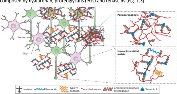
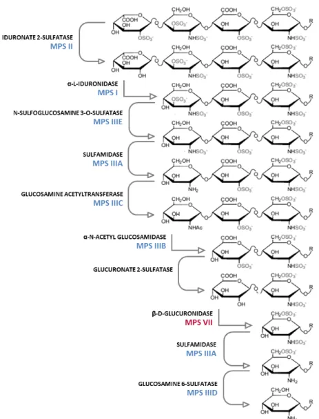
The role of extracellular cues, including ECM and other microenvironmental factors, in cell growth and differentiation is now beginning to be understood. The mechanical properties of the ECM, such as the stiffness or the elasticity, affect the adhesion and the interactions between the cells and the matrix, thus affecting cell morphology and resulting in the transduction of signals that can regulate cell differentiation (Guilak et al., 2009). Structurally, the ECM provides binding sites to neural cells and facilitates its organization into distinct CNS regions. Chemically, it is a source of diverse molecular signals that guide cellular growth, activity and survival. During CNS development, specific ECM components are dynamically regulated in a temporal and spatial manner to facilitate neurogenesis, neural cell migration and differentiation and axonal growth and guidance (Lau et al., 2013). The neural ECM of healthy brain tissue is a complex mixture of proteins, mainly composed by hyaluronan, proteoglycans (PGs) and tenascins (Fig. 1.3).
Figure 1.3: Major components of brain extracellular matrix (ECM). ECM components are mainly
arranged into perineuronal nets that are condensed around cell bodies and dendrites of neurons or diffused as the neural interstitial matrix between cells of the brain parenchyma. Adapted from Lau et al. (2013).
18
The chondroitin sulfate PGs (CSPGs) aggrecan, versican, neurocan and brevican are the primary organizational component of the brain ECM. These PGs bind tenascin glycoproteins, which act as linkers and provide binding sites to the cells (Hopkins et al., 2015; Lau et al., 2013). Hyaluronan is a negatively charged glycosaminoglycan (GAG), thereby attracting cations and charged proteins and increasing the hydration of brain tissue (Hopkins et al., 2015). Brain ECM is distributed within the perineuronal nets (PNNs) and the neural interstitial matrix, which consists of ECM components in the brain parenchyma that are not as tightly associated as in the PNNs (Fig. 1.3) (Lau et al., 2013). PNNs compose most of the brain ECM and are a condensed layer of a mesh-like matrix consisting mainly in PGs, tenascin r and link proteins, located around neuronal cell bodies and proximal dendrites. PNNs prevent random synapse formation and are thought to be critical for synaptic plasticity (Hopkins et al., 2015; Lau et al., 2013). Enzymatic degradation of CSPGs or destabilization of PNNs was shown to enhance neuronal activity and synaptic plasticity after CNS injury (Soleman et al., 2013).
4. Culture strategies for CNS modelling and drug screening
Typical strategies for culturing neural cells for CNS modelling rely on monolayer systems (2D). These, although easy to manipulate, fail to recapitulate the in vivo microenvironment and lack the structural architecture of the tissue, in many cases precluding the establishment of physiologically-relevant cell-cell and cell-ECM interactions (Breslin and Driscoll, 2013; Fitzgerald et al., 2015; Picollet-D’hahan et al., 2016). Contrasting with 2D, three-dimensional (3D) culture systems allow cells to establish a network of cell-cell and cell-ECM interactions in all planes, which affects cell morphology, polarity, cell signaling and gene expression (Pampaloni et al., 2007).
As mentioned previously, precise control of stem cell differentiation and tissue architecture is essential for development, organogenesis and tissue homeostasis. 3D culture methods enable the formation of self-organizing tissue-like structures
19 that recapitulate key aspects of the tissue of origin, such as the presence of multiple differentiated cell types, cell migration and activation of developmental gene expression programs (Murrow et al., 2017). This 3D environment can be relevant not only for neuronal maturation, functionality and signaling, but also for the penetration and action of drugs (Kim et al., 2004), potentially improving the outcome of cell-based assays in drug screening.
A vast number of distinct 3D culture strategies have been developed and employed in the last years. Nonetheless, several limitations need to be circumvent to take advantage of the full potential of 3D cultures. The 3D culture strategies can be divided in two main categories: (1) scaffold-dependent cultures, employing scaffolds based on natural or synthetic biomaterials as cell supportive matrixes and (2) scaffold-free cultures, in which cells were allowed to self-assemble into aggregates without an exogenous matrix.
4.1. Scaffold-dependent cultures
Aiming at recreating the in vivo cues such as mimicking tissue structure, mechanical and biochemical properties, a wide range of synthetic and biologically-derived materials have been used as scaffolds for 3D culture (Pampaloni et al., 2007; Picollet-D’hahan et al., 2016). These are mainly hydrogels and include biologically inert polymers, such as alginate, agarose or chitosan, or biologically active polymers, such as collagen or hyaluronan (Picollet-D’hahan et al., 2016). Biological active external cues (e.g. RGD adhesion domains) can also be incorporated in the scaffold in order to further enhance cell adhesion to the matrix, cell migration, cell differentiation and cellular functions (Guilak et al., 2009). For example, by controlling the density of the ligand ephrin-B2 along a soluble biopolymer, NSC differentiation was enhanced in vitro and in vivo, and ephrin-B1 conjugates strongly enhance PSC differentiation toward a dopaminergic phenotype (Conway et al., 2013).
Two main strategies can be followed for cell incorporation in the scaffold: (1) cells are directly seeded in a pre-formed scaffold or (2) cells are encapsulated
20
within the scaffold material, allowing them to spontaneously organize. Both strategies have already been applied for 3D culture of neural cells and generation of neural tissue-like structures (Irons et al., 2008; Lancaster et al., 2013). More recently, the field has been extended to more sophisticated scaffolding methods that include microfabrication, microfluidics and 3D bioprinting (Picollet-D’hahan et al., 2016). The combination of cellular supporting matrices and microfluidics technology, which controls physical and chemical conditions (e.g. temperature, gas tension, medium and soluble factors composition and precise control of fluid flow and shear stress) at microscale level, allows the generation of organ-on-chip models (Sackmann et al., 2014). These can potentially enhance the differentiation, function, and long-term survival of many cell types. The 3D bioprinting approaches allow creating complex and layered scaffolds able to sustain cell survival, proliferation and neural differentiation (Devillard et al., 2014; Lozano et al., 2015). Although presenting a great potential in recreating the natural physical and structural environment of living tissues, biologically active scaffold components can profoundly affect the properties of the culture and cell phenotype, which can lead to confounding effects specially when modeling disease. Further, scaffold-dependent culture methods are difficult to control in terms of diffusion of gases and nutrients (Kelava and Lancaster, 2017; Ranga et al., 2016; Serra et al., 2012). These methods are laborious, require technical knowledge and equipment and the analysis of most biological readouts is challenging, making difficult its applicability in high-throughput screenings (Hopkins et al. 2015).
Ideal 3D scaffolds should provide native spatial arrangement, adequate biomechanical properties and biocompatibility. The mechanical properties of the scaffold should match the CNS tissue characteristics, not affect, or affect positively, cell differentiation and ensure efficient neurite outgrowth and neural connectivity. Cell culture on hydrogel surfaces with variable moduli indicated that softer gels (100–500 Pa) favour neuronal differentiation while harder gels (1–10 kPa) favour glial differentiation (Saha et al., 2008). Brain tissue stiffness in vivo is decreased in
21 neurodegenerative disorders such as AD so, when modeling disease, stiffer scaffolds should be chosen to better mimic the pathological state (Franze et al., 2013). Still, the ideal 3D scaffold cannot be defined due to the lack of knowledge on disease-induced ECM changes.
4.2. Scaffold-free cultures
Scaffold-free methods take advantage of the capability of some cell types to self-assemble into aggregates, without the support of an exogenous scaffold. This allows cells to produce their own ECM without the confounding effects of an exogenous matrix, which can overlook the biochemical and biophysical contribution of the endogenous ECM factors.
Conditions in which the adhesive forces between the cells are greater than for the substrate are required for cells to aggregate. Cell aggregation can be facilitated by buoyancy or stirring. Thus, cell spheroids can be generated in static systems by forced floating methods, such as low-adherence culture surfaces and hanging-drop methods, or in agitation-based culture systems, such as shake flasks, spinner flasks, rotary wall vessels and stirred-tank bioreactors.
In low adherence surfaces and in the hanging drop method, no adherence cue is provided to the cells, cell aggregation is forced and is strongly dependent on cell-cell contacts. Depending on the heterogeneity of the initial cell-cell population and its aggregation capacity, some lack of reproducibility could exist and control of tissue size is difficult, while being also time-consuming and cumbersome aggregation methods (Picollet-D’hahan et al., 2016). Moreover, these systems provide low mass transfer properties and inherently it is difficult to maintain long-term cultures and the scalability of the system is very low, precluding many applications.
Agitation-based culture systems allow the generation of more homogenous cultures, presenting improved mass transfer properties over static culture systems. Cell aggregation is highly dependent not only on the cell type but also on system hydrodynamics. The geometry of the vessel, the stirring rate, and the type and size of the impeller are parameters that affect the culture hydrodynamics and,
22
therefore, are important to optimize when aiming at promoting cell aggregation and culturing aggregates (Kinney et al., 2011). The stirring creates shear, which may increase the number of cell-cell contacts and promote cell aggregation and aid in controlling the size of the aggregates (Kehoe et al., 2010). However, stirring can also subject the cells to shear stress, which can negatively impact cell physiology. To minimizing detrimental effects of shear stress, impellers able to suspend cells and mix the fluid gently and operating at lower stirrer speed were developed for PSC culture (Marks, 2003; Olmer et al., 2012).
Software-controlled stirred-tank bioreactors allow monitoring and controlling physical and chemical culture parameters, such as hydrodynamics, temperature, pH and dissolved oxygen. Furthermore, operation in a perfusion mode allows a stable flow of nutrients and differentiation/ neurotrophic factors, while clearing the cell debris and toxic byproducts at a controlled rate and maintaining the cells within the vessel (Simão et al., 2016). The major disadvantages of stirred tank bioreactors are the requirement for technical knowledge and specialized equipment, which is costly.
Efficient neuronal differentiation of PSC and NSC spheroids can be performed in scaffold-free 3D cultures, attaining increased neuronal differentiation efficiencies with reduced culture times, when compared to static 2D cultures (Chandrasekaran et al., 2017; Simão et al., 2014; Terrasso et al., 2015). We recently showed that iPSC-derived NSC cultured in a stirred suspension culture system self-assemble into aggregates, and differentiate into the three neural lineages, neurons, astrocytes and oligodendrocytes (Simão et al., 2016; Simão et al., 2018). Neurospheroids proteome and transcriptome dynamics revealed significant changes at cell membrane and ECM composition during neural differentiation, with an enrichment in PGs typical of brain ECM (Simão et al., 2018). These results demonstrate that iPSC-NSC 3D neural differentiation recapitulates extracellular space dynamics, with accumulation of endogenous ECM and secreted components and remodeling along differentiation.