resumo O quitosano é um policatião de origem natural que tem vindo a ser investigado como sistema não viral de vectorização de genes devido à sua biocompatibilidade e baixa toxicidade. No entanto, a sua baixa eficiência de transfecção tem dificultado o seu uso generalizado. Num estudo anterior mostrámos que a conjugação de resíduos de imidazol a cadeias de quitosano resulta numa melhoria da eficiência de transfecção do polímero. O principal objectivo deste estudo foi avaliar a aplicação de quitosano modificado com imidazol (CHimi) como vector para entrega de genes em medicina regenerativa, bem como encontrar novas vias para melhorar a eficiência. A expressão genética mediada por CHimi com dois graus de substituição -13% e 22% das aminas primárias do quitosano - foi avaliada em células 293T (células embrionárias humanas do epitélio do rim) por um período de 8 dias, usando o gene da β-Galactosidase (β-gal) como gene repórter. Os complexos de CHimi-DNA foram preparados numa razão molar de 18 entre aminas primárias e grupos fosfato. As células transfectadas com estes complexos apresentam um pico de actividade da β-gal às 72 horas pós-transfecção, verificando-se a expressão sustentada da proteína repórter durante todo o período de avaliação. Nestas condições a viabilidade celular não é comprometida. Quando se efectua um segundo tratamento das células com complexos à base de CHimi, a actividade de transfecção volta a aumentar, sem haver alterações na viabilidade celular. Verificou-se também que células transfectadas com estes vectores sobrevivem a um ciclo de congelação/descongelação, mantendo uma actividade de transfecção sustentada no tempo. Uma polietilenimina comercial (Escort V) foi usada como referência neste estudo. Apesar de os níveis de transfecção mediados por este vector serem duas ordens de grandeza mais elevados, a viabilidade celular decresce até aos 50% após cada tratamento.
De forma a investigar o processo de transfecção mediado por polímeros de CHimi, o tráfego intracelular destes complexos foi estudado por microscopia confocal de varrimento laser. Complexos de CHimi e DNA marcados com fluoróforos foram encontrados no citoplasma celular 2 horas depois da transfecção, sendo detectados até 48 horas pós-transfecção. Estes resultados podem em parte explicar a expressão sustentada de β-gal ao longo do tempo. Os complexos foram detectados no interior do núcleo 4 horas pós-transfecção. O DNA marcado com fluorescência não foi observado na forma livre em nenhum dos momentos analisados, enquanto que CHimi foi detectado num evento único no citoplasma. Num ensaio “cell-free” de transcrição/tradução in
vitro não foi detectada a síntese de proteína quando o DNA estava
complexado com CHimi, apesar de este ser expresso na ausência do polímero. Este conjunto de resultados sugere que, apesar dos complexos poderem ser encontrados no interior do núcleo rapidamente após a transfecção, a expressão genética parece depender da desintegração do complexo. O CHimi é um potencial candidato a vector para transporte de genes num cenário de regeneração. Este material medeia uma expressão proteica sustentada sem afectar a viabilidade celular. Com este sistema, as células toleram uma segunda adição de complexos, pelo que a administração repetida poderá ser potencialmente usada como estratégia para prolongar o efeito terapêutico de uma proteína de interesse. Em relação aos resultados de tráfego intracelular dos complexos, e considerando o perfil de expressão genética obtido, pode pôr-se a hipótese de que a expressão sustentada do gene resulta de um processo de libertação dependente do tempo. Neste sentido, ajustar a velocidade de degradação dos polímeros de CHimi pode ser usado como estratégia para melhorar o processo de expressão do gene tendo em vista o fim terapêutico pretendido.
abstract Chitosan is a polycation of natural origin, emerging in the non-viral gene delivery vectors scene due to its biocompability and low cytotoxicity. However, its low transfection efficiency has hampered its wide application so far. We have previously shown that grafting imidazole moieties into the chitosan backbone results in improved transfection efficiency of this polymer. The main goal of this study was to assess the application of imidazole-grafted chitosan (CHimi) as gene delivery vector in a regenerative medicine scenario and to find avenues to further improve its efficiency.
Gene expression mediated by CHimi with two degrees of substitution - 13% and 22% of chitosan primary amines - was assessed in 293T cells for periods up to 8 days, using the β-Galactosidase (β-gal) gene as reporter gene. CHimi-DNA complexes were prepared at a primary amine to phosphate groups molar ratio of 18. Cells transfected with the CHimi-based complexes have a peak of β-gal activity 72 hours post-transfection and show a sustained β-gal production for 8 days. During this time period cell viability is not impaired. When a second treatment with CHimi-based complexes is performed, transfection activity increases, without changes on cell viability. Additionally, cells transfected with CHimi-based vectors are able to withstand a freeze/thawing cycle, maintaining a sustained transfection activity. A commercially available polyethylenimine (Escort V) was used as a reference. Though transfection levels are two orders of magnitude higher, cell viability decreases up to 50% after each treatment. In order to investigate the transfection process mediated by CHimi-based vectors a study of the intracellular pathway of the complexes has been performed by confocal laser scanning microscopy. Complexes formed by fluorescently labeled CHimi and DNA were found inside the cell cytoplasm 2 hours after transfection and were detected up to 48 hours post-transfection. These results could explain in part the sustained gene expression over time. Complexes were detected inside cell nucleus since 4 hours post-transfection. Fluorescently labeled DNA in the free form was not observed at any of the time points analyzed. Free CHimi was detected in the cytoplasm in an atypical event. In a cell-free in vitro transcription/translation assay no protein production was detected when DNA was complexed with CHimi, though expressed when using plasmid DNA in the absence of CHimi. Taken together these results suggest that, though CHimi-based complexes can be detected inside cell nucleus promptly after transfection, gene expression is dependent on the complex disassembling.
CHimi is a potential candidate vector for gene delivery in a regenerative scenario. This material is able to mediate a sustained protein expression without impairing cell viability. In our system, cells can sustain another addition of the complexes suggesting that repeated administration could be used as a strategy to prolong the therapeutic effect. In view of the trafficking results and considering the gene expression profile, one can hypothesize that the observed sustained transgene expression is a time dependent release process. Thus, tuning the degradation rate of CHimi-based polymers could be a strategy to further improve the overall transgene expression process to fulfill the therapeutic end.
T
ABLE OF CONTENTSCHAPTER I – INTRODUCTION 1
1.GENERAL INTRODUCTION... 3
2.GENE DELIVERY SYSTEMS... 5
2.1. Viral gene delivery ... 5
2.2. Non-viral gene delivery... 6
3.INTRACELLULAR TRAFFICKING... 15
3.1. Cellular uptake... 15
3.2. Endosomal escape ... 18
3.3. Nuclear import ... 21
3.4. Beyond delivery ... 24
CHAPTER II – AIM OF THE THESIS 27 CHAPTER III – MATERIALS AND METHODS 31 1.MATERIALS... 33
1.1. Imidazole-grafted chitosan ... 33
1.2. Plasmid DNA ... 33
2.METHODS... 34
2.1. General procedures... 34
2.2. Gene expression studies ... 36
2.3. Intracellular trafficking... 40
2.4. Cell-free gene expression... 43
2.5. Statistical data analysis ... 45
CHAPTER IV – RESULTS 47 1.GENE EXPRESSION STUDIES... 49
1.2. Freeze/thaw ... 53
1.3. Re-transfection studies ... 54
2.INTRACELLULAR TRAFFICKING... 58
2.1. Fluorescence microscopy ... 58
2.2. Cell-free gene expression... 63 CHAPTER V – DISCUSSION 67 CHAPTER VI – CONCLUDING REMARKS AND FUTURE WORK 75 CHAPTER VII – REFERENCES 79
I
NDEX OF FIGURESFigure 1: General structure of a cationic lipid... 8
Figure 2: Example of a neutral lipid - DOPE. ... 8
Figure 3: Example of other lipids used in gene delivery. ... 9
Figure 4: Poly(L-lysine) (PLL) chemical structure. ... 10
Figure 5: Chemical structure of poly(ethylenimine) (PEI). ... 11
Figure 6: Synthesis scheme of PAMAM cascade dendrimers. ... 12
Figure 7: Chemical structure of chitosan ... 13
Figure 8: Chemical structure of imidazole-grafted chitosan... 14
Figure 9: Biological barriers to gene delivery. ... 15
Figure 10: Multiple portals of entry in mammalian cells. ... 16
Figure 11: The proton sponge effect. ... 19
Figure 12: Schematic drawing of the pCMV-SPORT-βgal and pCMV-GFP constructs with the respective restriction sites. ... 34
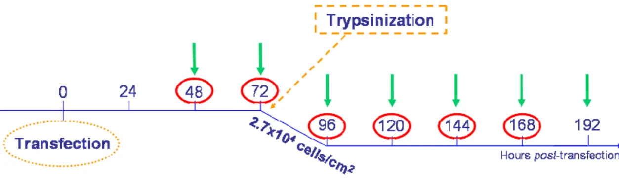
Figure 13: Experimental design for long term gene expression evaluation. ... 37
Figure 14: Experimental design to evaluate long term gene expression after re-seeding cells at 2.7x10⁴ viable cells/cm2... 37
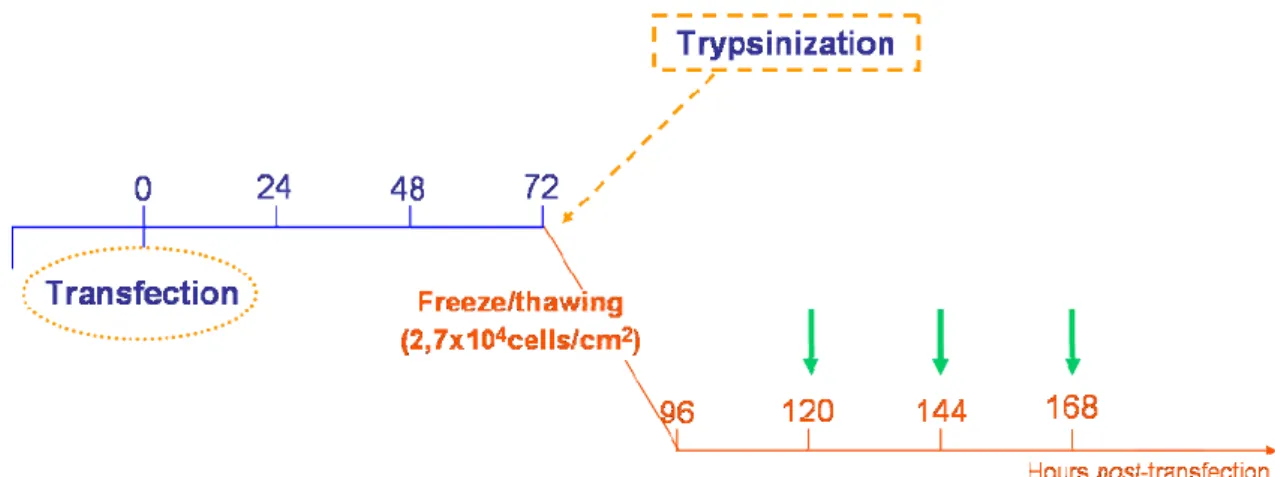
Figure 15: Experimental design for evaluation of transfection activity after freeze/thaw process. .. 38
Figure 16: Experimental design for re-transfection studies... 39
Figure 17: Molecular structure of 5(6)-Carboxy-X-rhodamine N-succinimidyl ester (ROX)... 41
Figure 18: Schematic drawing representing the covalent bonding between DNA and Label IT® reagent... 42
Figure 19: Transfection activity as function of time of cultures treated with CHimi2- and CHimi3-based vectors.. ... 49
Figure 20: β-gal activity and the total protein content of CHimi-based vectors transfected cells as a function of time. ... 50
Figure 21: Transfection activity as a function of time of cells transfected with PEI (Escort V).. ... 51
Figure 22: β-gal activity and the total protein content of PEI (Escort V) transfected cells. ... 51
Figure 23: Evaluation of β-gal specific activity over time... 52
Figure 24: Cell viability as a function of post-transfection time.. ... 53
Figure 25: Effect of a freeze/thawing cycle on β-gal specific activity as a function of time in culture after transfection. ... 54
Figure 26: Evaluation of β-gal specific activity as a function of time after re-transfection with CHimi2 and CHimi3, comparing to non-re-transfected cells.. ... 55
Figure 27: Experimental procedure performed as re-transfection control.. ... 55
Figure 28: Transfection activity of cultures transfected only once (solid line) or two times (dashed line) with (A) CHimi2 and (B) CHimi3. ... 56
Figure 29: Evaluation of β-gal specific activity after re-transfecting cells with PEI-based vectors... 56
Figure 31: CLSM images obtained 2 hours after transfecting 293T cells with CHimi2ROX-DNACy5
complexes... 60 Figure 32: CLSM images of 293T cells 4 hours after transfection with CHimi2ROX-DNACy5
complexes... 61 Figure 33: CLSM images obtained 6 hours after transfecting 293T cells with CHimi2ROX-DNACy5
complexes... 61 Figure 34: CLSM images of 293T cells 24 hours after being transfected with CHimi2ROX-DNACy5
complexes... 62 Figure 35: CLSM images of GFP positive 293T cells 48 hours after transfection with CHimi2
-DNACy5 complexes... 63ROX Figure 36: Gel electrophoresis of CHimi2-based complexes after incubation in phosphate buffer
(0.1 M, pH 7.8), 90 minutes at 30 °C. ... 64 Figure 37: Effect of CHimi-pTTR complexes on the in vitro transcription/translation assay of
I
NDEX OFT
ABLESTable 1: Examples of current gene therapy clinical trials using viral-vectors ... 6 Table 2: Conditions tested in the in vitro transcription/translation assay. ... 44 Table 3: Zeta potential, average size (Z-Average) and polydispersity index (PdI) of CHimi2-DNA
L
IST OF ABBREVIATIONS β-gal – β-GalactosidaseAIDS – Acquired immunodeficiency syndrome ATP – Adenosine triphosphate
BCA – Bicinchoninic acid BSA – Bovine serum albumine CHimi – Imidazole-grafted chitosan
CHimi1 – Chitosan with a degree of substitution of 5% CHimi2 – Chitosan with a degree of substitution of 13% CHimi2ROX – CHimi2 fluorescently labeled with rhodamine
CHimi3 – Chitosan with a degree of substitution of 22% CLSM – Confocal laser scanning microscopy
CMV – Cytomegalovirus DA – Degree of N-acetylation
DAPI – 4'-6-Diamidino-2-phenylindole
DMEM – Dulbecco's Modified Eagle's Medium DMSO – Dimethyl sulfoxide
DNA – Deoxyribonucleic acid
DNACy5 – DNA Fluorescently labeled with Cy5
DS – Degree of substitution
E. Coli – Escherichia coli
EDC – 1-Ethyl-3-[3-dimethylaminopropyl]carbodiimide hydrochloride FT-IR – Fourier Transform Infrared Spectroscopy
GFP – Green fluorescent protein GPC – Gel permeation chromatography MES – 2-(N-morpholino)ethanesulfonic acid Mw – Average weight molecular weight NPC – Nuclear pore complex
N/P – Amine to phosphate molar ratio NHS – N-hydroxysuccinimide
NLS – Nuclear localization sequences ONP – o-nitrophenyl-β-pyranoside
ONPG – ortho-nitrophenyl-β-D-galactopyranoside PBS – Phosphate buffered saline
PDL – Poly(D-lysine) PEG – Poly(ethylene glycol) PEI – Poly(ethylenimine)
PLL – Poly(L-lysine)
PAMAM – Polyamidoamine dendrimers PNS – Peripheral nervous system
pTTR – Plasmid encoding for TTR protein RNA – Ribonucleic acid
RNAi – RNA interference
ROX – 5(6)-Carboxy-X-rhodamine N-succinimidyl ester SCID – Severe combined immunodeficiency
SD – Standard deviation siRNA – Short interfering RNA SV40 – Simian Virus 40 TTR – Transthyretin
1.
G
ENERAL INTRODUCTIONConceptually, gene therapy appeared in the early seventies [1]. The idea rapidly spread out due to the enormous potential in a vast range of applications. In a broad sense, gene therapy attempts to provide to the cells of a patient the genetic information required for producing a protein that in turn will have a therapeutic effect, to correct or modulate a disease [2]. The premise is the use of genes as pro-drugs to induce in vivo the production of therapeutic proteins [3], using the “patient’s own cells as mini-bioreactors” [4]. This approach circumvents limitations of direct recombinant protein administration like low bioavailability, systemic toxicity or high cost of manufacturing [3].
In a first stage, gene therapy was thought to be applied to monogenic diseases, like cystic fibrosis [5], but with the culmination of the Human Genome Project and the development of highly sensitive techniques, new targets have been identified [6]. Gene therapy is currently studied as strategy to treat, or modulate diverse health disorders, such as cancer [7], AIDS [8] cardiovascular [9] or neurological diseases [10]. Additionally, gene therapy has been applied in a regenerative medicine scenario as a mean to induce the production of proteins, such as growth factors, to promote the repair and/or functional recovery of an injured tissue or organ, either as a therapeutic tool by itself or in combination with cellular therapies and/or tissue engineering [11]. RNAi gene therapy is a recent approach and is particularly in fashion since the Nobel Prize in Medicine for RNAi research of Fire and Mello in 2006 [12]. RNAi strategy differs from classical gene therapy scheme once it introduces short interfering RNA (siRNA) in order to inhibit a specific protein expression, instead of promoting it. Although it induces a transient effect, RNAi is very promising in the gene therapy field [13], namely in cancer therapies [14]. Indeed, siRNA is being tested in ten of the ongoing gene therapy clinical trials [15].
The first registered gene therapy clinical trial dates from 1989 and in 2006 more than a thousand have been performed [15]. Very optimistic reports were published [16]. However, in 1999 the first drawback to gene therapy put a chill on the high expectations. A patient involved in a clinical trial for ornithine transcarbamylase enzyme deficiency died after multiple organ failure due to intrahepatic infusion of an adenoviral vector [4]. Since then, gene therapy has had cyclically its ups and downs. Promising results published in the first years after the beginning of a French trial for X-linked several combined immunodeficiency (SCID) initiated in 1999 – the so called “bubble boy” disease – brought back the optimism [17, 18]. However, up to now, four of the 10 patients involved in the trial developed leukemia [19] and one of them died in the past year [20]. This adverse event is consequence of the random insertion of the retrovirus vector in the proximity of an oncogene that leads to an aberrant expression of the “therapeutic protein” – IL- 2Rγ – and malignant cell expansion [21]. Another setback in gene therapy clinical trials was reported very recently with a patient involved in a trial for arthritis. The causes are under investigation, but suspicions point to an adverse reaction caused by a second injection of the adeno-associated virus, used as gene vehicle [22]. This would be the first fatality in a trial not studying a life-threatening disease.
The recurrent setbacks limited gene therapy progress and concerns about viral vectors safety were highlighted. Viruses were the first choice for the transport of genes due to their high efficiency. They are naturally gene-delivery vehicles and as sophisticated products of evolution they are very skillful [23]. Therefore, viral vectors represent 70% of the ongoing gene therapy clinical trials [15]. Some of the viral vectors systems tested in clinical trials will be briefly described during this review. The tribulations occurred in the trials, the potential oncogenicity and high immunogenicity after repeated administration led to a revaluation of the use of viral vectors for therapy [20]. Although several efforts are being made in order to increase viruses’ safety [24], significant expectations shifted to non-viral vectors. So far, the non-viral approach is used in approximately 30% of the gene therapy clinical trials, but the relative percentage is increasing over time [15].
The primary challenge for the non-viral approach is delivery [25]. Non-viral gene delivery systems are comparatively less efficient, what hampers its wide application [26]. However, important attempts are being made in order to close the gap [27]. A broad literature has been published in the last few years with different strategies and formulations for increasing non-viral gene delivery efficiency and target cell populations. This will be reviewed as well. However, the strategy to get better non-viral gene delivery systems seems to depend on a more detailed knowledge about the way vectors interact with cells and which are the rate-limiting steps for delivery. The design of efficient and functional non-viral systems relies on the better understanding of the cell barriers and the mechanism involved in the intracellular trafficking of these gene carriers [26].
The present work has been performed in the framework of a project that aims at the development of safe and effective biomaterial-based delivery systems of therapeutic genes to promote neuroregeneration in the peripheral nervous system (PNS). It is proposed the use of chitosan in the design of novel polycations to serve as carriers of DNA for specific delivery of genes to PNS cell populations. The basic concept behind the approach is the modification of chitosan, a natural polymer with known low cytotoxicity, in order to develop a gene carrier with higher affinity for nervous system cells, improved trafficking into the cell and into the nucleus, as well as, in the cell cytoplasm. One of the strategies that were explored was the improvement of endosome escape potential of the vector. It was proved that the introduction of imidazole groups in the chitosan backbone increased the transfection efficiency of chitosan by enhancing the endosomal escape of the vectors [28]. The main goal of the present work was to further assess the application of these materials as gene delivery vectors for application in a regenerative medicine scenario.
In view of the above, the strategy that was followed was to study in vitro the intracellular mechanisms occurring in imidazole-grafted chitosan mediated transfection as a way to further understand the action of these vectors and, ultimately find new avenues to improve gene delivery efficiency.
2.
G
ENE DELIVERY SYSTEMSThe greatest hurdle to gene therapy application is the development of non-toxic and efficacious gene delivery systems. The ideal gene delivery vector should assist DNA transport to the nucleus of the target cell, leading to appropriate transfection efficiency (normally defined as the percentage of treated cells that express the therapeutic gene or transgene) and suitable “therapeutic protein” production. Additionally, it should guarantee low toxicity and immunogenicity, be biodegradable and stable [29].
Gene delivery systems are usually divided in two categories: recombinant virus and non-viral vectors.
2.1. Viral gene delivery
The use of recombinant viruses as vehicles to transport genes was inspired in their capability to deliver their genetic material in host nuclei to initiate expression of its own genome using host machinery. For therapeutic purposes, the transgene can be assembled in the viral genome and viruses’ innate mechanism of infection will assist the transgene transport into the cell nucleus [29]. To construct a virus-based vector for gene delivery applications, genes encoding viral components essential for propagation should be separated or removed to prevent reconstruction by recombination into productive viral particles. These genetic components are replaced by the therapeutic gene [24].
Adeno-, retro-, lenti-, adeno-associated or herpes simplex viruses are examples of some engineered viruses for gene delivery applications that have been tested in gene therapy clinical trails (see Table 1). These viruses can be divided in two categories: integrating and non-integrating. In integrating vectors as retrovirus and adeno-associated virus a long-life expression of the therapeutic gene is expected, whereas non-integrating virus (adenovirus or herpes simplex virus) trigger a transient expression.
Retrovirus vectors were the first being developed and used in clinical trials due to its relatively simple and effective design. They tend to establish a chronic infection but are unable to infect non-dividing cells [24]. Lentiviruses are part of the retrovirus family, but as they rely on active transport of their genetic material into cell nucleus, they are able to infect also non-dividing cells [24]. The wide tropism (range of cells that can be productively infected by a virus) of lentiviruses can difficult cell targeting, but the unspecific integration in host genome is the most important disadvantage of the viruses from the retroviruses family.
The main challenge in adenovirus-mediated infection is how to make expression persist [30]. These are non-integrating viruses, but they are very efficient in a large variety of tissues. Additionally, most adults had already been exposed to adenovirus what could compromise a gene therapy strategy due to pre-existing immunity [30]. Even though, adenovirus vectors are the most used vectors in ongoing gene therapy clinical trials [15], due to their great potential [31].
As the name suggests, adeno-associated virus require helper virus (as adenovirus) to mediate productive infection. These vectors carry limited size DNA inserts, but have wide tropism, being able to infect both dividing and non-dividing cells. Although adeno-associated viruses are generally classified as integrating viruses, these vectors can also promote episomal gene expression [24]. Herpes simplex based vectors can not integrate in host genome, but the virus persists after primary infection in state of latency. An important advantage of herpes simplex vectors is their capability to accommodate large DNA inserts. Herpes simplex vectors show wide tropism, but in vivo they are neurotropic [10] and therefore, particularly promising for neuronal gene delivery [32]. Even though, in the actual clinical trials scene they are mainly tested in the treatment of cancer diseases [15].
Table 1: Examples of current gene therapy clinical trials using viral-vectors (information available at [15]) Virus-based vector Disease Category Phase Date
Iniciated/approved
Administration
route Country Total number
HIV infection Infectious diseases I 2003 intramuscular Belgium Metastatic prostate cancer Cancer diseases III 2006 ― Netherlands Cystic Fibrosis Monogenic disease I 2005 Intranasal USA Operable High grade glioma Cancer diseases I 2005 Intratumoral Belgium Cystic Fibrosis monogenic disease I/II ― Intrabronchial France Prostate cancer Cancer diseases III 2007 Intratumoral USA Head and neck cancer Cancer diseases II 1997 ― UK End Stage Renal Disease Cardiovascular diseases III 2007 Perivascular collagen
collar device USA Class II-IV stable angina Cardiovascular diseases II 2005 Intramyocardial Belgium Colorectal cancer Cancer diseases II 2006 Intratumoral USA Glioblastoma multiforme Cancer diseases I 2005 ― Germany Glioblastoma Cancer diseases III 2006 Intratumoral UK Non-small cell lung cancer Cancer diseases I ― ― China Severe combined immune deficiency due to
adenosine aeaminase (ADA) deficiency Monogenic disease I/II 1999
In vitro - bone marrow transplantation France Metastatic melanoma Cancer diseases I/II 2003 Intravenous Italy HIV infection Infectious diseases I 1997 Intravenous Australia Malignant melanoma Cancer diseases I/II ― In vitro - Subcutaneous Scotland HIV infection Infectious diseases I/II 2004 In vitro - Intravenous USA CD 19+ Leukemia and Lymphoma Cancer diseases I 2006 In vitro - Intravenous USA
32 169 7 35 146 Lentivirus Adeno-associated virus Adenovirus
Herpes simplex virus
Retrovirus
The high efficiency of viral vectors was the main impelling force for its widespread testing on clinical trials. Important developments were achieved over recent years [33], however the risk of insertional mutagenesis or immunological responses, and the problems that occurred in clinical trials held back viral gene transfer progress. While viral gene delivery research slowed down, non-viral gene delivery had a boost [34], towards the design of “artificial viruses” [35].
2.2. Non-viral gene delivery
Non-viral vectors were presented as an alternative to viral vectors to mediate the process of gene delivery. Nevertheless, despite the intensive research during the past decade, non-viral vectors still do not compare with viral vectors in terms of efficiency [36]. Therefore, the bulk of non-viral research is focused on improving vectors efficiency in the delivery of genes.
Non-viral gene delivery involves liposomal and polymeric vectors, as well as, direct DNA injection. The first case is a chemical approach in which lipids or polymers are used to complex and transport DNA into the cell. The direct DNA injection has been shown to lead to gene expression, but high levels of expression are limited to exposed tissues such as skin or muscle [37]. Naked DNA is unsuitable for systemic administration due to fast clearance by the mononuclear phagocyte system and degradation by serum nucleases that can eliminate DNA within 30 minutes [38]. Consequently, physical/mechanical methods are frequently applied in order to improve direct DNA delivery.
2.2.1. Physical/mechanical methods
Physical methods apply physical fields in order to compromise cell membrane integrity, inducing permeability to DNA molecules. Electroporation applies electric forces; sonication uses ultrasound waves; laser irradiation induces membrane permeability in the site of the beam impact by local thermal effect; magnetofection uses DNA-coated magnetic beads and an external magnetic field is applied in order to concentrate particles in the target cells [39]. Physical methods for gene transfer are normally well tolerated and no significant toxicity is reported [39]. However, its application seems to be limited to exposed or surgically exposed tissues [2] or for genetic modification ex vivo. The “physical approach” to gene delivery is relatively new and the poor efficiency or the lack of in
vivo specificity, as well as the expensive equipment required, are issues still to be improved.
Microinjection and the gene gun are mechanical methods used to improve naked gene transfer. Microinjection uses the simpler approach: with a glass needle the genetic material is injected in the cell cytoplasm or inside the nucleus. Up to 100% of the recipient cells can be transfected with this method, but it is very meticulous and cells must be transfected one by one. Furthermore, this method is limited to in vitro or ex vivo applications [39]. With the gene gun, cells or tissues can be transfected by shooting accelerated gold particles coated with DNA. The major application of this method is DNA vaccination, where limited expression is needed in exposed tissues as skin and muscle [40]. The gene gun is being tested in a number of clinical trials [15], but the major barrier to its wide application is the shallow penetration of particles in biological tissues [41]. Even though, physical and mechanical methods have already demonstrated potential in gene transfer and whether independently, or in reasonable combination with other non-viral vectors, a place in gene delivery seems to be assured [39].
2.2.2. Liposomes
Cationic liposomes were introduced as gene delivery vectors by Felgner in 1987. He first used 2,3-bis(oleoyl)oxipropyl trimethylammonium chloride (DOTMA) and dioleoylphosphatidylethanolamine (DOPE) liposomes in order to assist DNA delivery into cells [42]. DOTMA is a cationic lipid with a hydrophobic moiety that ensures the assembly into bilayer vesicles and an amino head group to bind phosphate DNA groups (Figure 1). DOPE is a neutral lipid (Figure 2) typically called “helper”
lipid, because it increases gene delivery efficiency of cationic liposomes [43]. Cholesterol has been used as an alternative helper lipid [44]. The presence of DOPE in cationic liposomes formulations is thought to induce endosome destabilization, facilitating DNA escape from these vesicles and, ultimately, leading to an enhanced gene expression [45]. DOTMA/DOPE liposomes are nowadays commercially available as the transfection reagent, Lipofectin®.
Figure 1: General structure of a cationic lipid.
O N H3 P O O O H O O C O O C +
Figure 2: Example of a neutral lipid - DOPE.
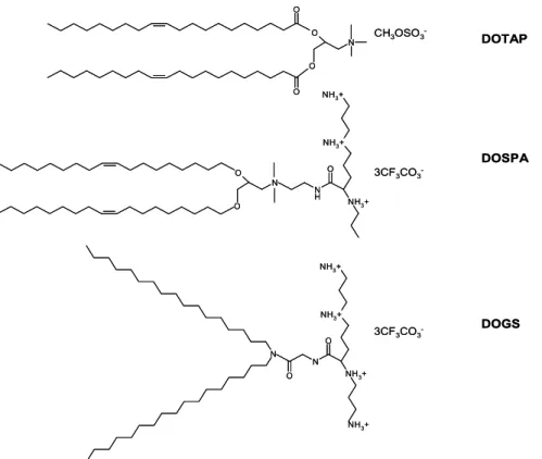
In liposome-mediated gene delivery an electrostatic interaction is established between the permanently positive charge of the cationic lipid and the negatively charged DNA, making “transportable” condensed units – lipoplexes – able to be internalized by the cell. A positive charged lipoplex is essential to allow cell binding of the particles and subsequent endocytosis [46]. Based in Felgner strategy, several lipids were synthesized and used as liposomes for gene delivery (see Figure 3), such as DOTAP (1,2-dioleoyl-3-trimethylammonium-propane), that efficiently protects DNA from serum DNases degradation [47] or multivalent cationic lipids (DOSPA – 2,3-dioleyloxy-N-[2(spermine-carboxamido)ethyl]-N,N-dimethyl-1-propanaminium trifluoroacetate – or DOGS – di-octadecylamidologycylspermine) that exhibit improved efficacy compared to monovalent lipids, but also higher toxicity [48].
O O N O O DOTAP CH3OSO3 -DOSPA 3CF3CO3 -O N O N H O NH3+ NH3+ NH3+ N N O O NH3+ NH3+ NH3+ NH3+ DOGS 3CF3CO3 -O O N O O DOTAP CH3OSO3 -DOSPA 3CF3CO3 -O N O N H O NH3+ NH3+ NH3+ N N O O NH3+ NH3+ NH3+ NH3+ DOGS 3CF3CO3
-Figure 3: Example of other lipids used in gene delivery.
The clinical applications of cationic liposomes are mainly focused on cancer or cystic fibrosis [49] and represent about 8% of the gene therapy clinical trials [15]. However, cationic liposome-mediated gene delivery has some critical limitations, including the lack of reproducibility in the fabrication process, colloidal instability, significant toxicity after repeated administration [23], lack of targeting [40] and limited applicability by intravenous route due to impaired ability to go beyond the vasculature [49]. Aiming at circumvent these limitations, several authors are working on the improvement of biological properties of lipidic gene carriers. The incorporation of poly(ethylene glycol) (PEG) in the lipoplex structure intends to increase the particle circulating time and reduce unspecific interactions with serum components [44]. The binding of saccharides aims at enhancing gene delivery efficiency and also improve the lipoplex storage stability [44]. In a different approach, conjugation with transferrin was used to enhance gene expression by means of receptor-mediated endocytosis [50] and also to target transferrin-expressing tumor cells [51]. Formulations that expose cell-surface receptor binding peptides have also shown promising results on targeting upper airways epithelial cells [52].
2.2.3. Cationic polymers
As cationic lipids, polymers bearing groups which are protonated at physiological pH have been used as gene carriers. Cationic polymers can establish electrostatic interactions with DNA and form condensed particles – polyplexes – shielding DNA from nucleases activity.
The use of polymers shows important advantages for gene delivery. Polymers can be specifically tailored to a proposed application by choosing the appropriate molecular weight, controlling physicochemical properties and coupling cell specific target moieties [53].
Both synthetic and natural polymers have already been tested as gene carriers and will be reviewed in the next sections.
2.2.3.1. Synthetic polymers
Synthetic polymers as “off-the-shelf” materials form the basis for much of the non-viral gene delivery literature. However, these polymers face significant problems such as toxicity or limited biodegradability. Three of the major synthetic polymers used as gene carriers are described in the following paragraphs.
2.2.3.1.1. Poly(L-lysine)
Poly(L-lysine) (PLL) (Figure 4) was one of the first cationic polymers to be intensively studied as gene carrier. A large variety of molecular weights [54] and conjugation with specific ligands such as folate [55] or histidine [56] have already been tested. With an optimum ratio between PLL amino groups and DNA phosphate groups, small complexes can be produced, capable to enter the cell. However, these complexes tend to aggregate [2] and accumulate in the endosome. The end result is a rather high toxicity [57], also attributed to the low degradation rate of PLL [58].
Although several efforts has been made to overcome PLL-based vectors low transfection efficiency and cytotoxicity [57], in the actual scene of gene delivery it looks unlikely that PLL-based polyplexes will find some clinical application [23]. Its low transfection efficiency is attributed to the poor escape from endosomal vesicles, as will be described in more detail afterwards.
NH CH CO C H2 CH2 C H2 CH2 N H2 x
Figure 4: Poly(L-lysine) (PLL) chemical structure.
2.2.3.1.2. Poly(ethylenimine)
In 1995, Boussif and colleagues [59] introduced poly(ethylenimine) (PEI) as a potential gene carrier. Transfection experiments with this synthetic polymer were promising from the beginning, and PEI became the gold standard in non-viral gene delivery [60].
PEI can occur as branched (bPEI) or linear (lPEI) morphological isomers, depending on the linkage between the repeating ethylenimine units (Figure 5). The branched isoform contains primary, secondary and tertiary amines, each with the potential to be protonated. This results in a high positive charge density polymer, which can effectively buffer a wide pH range [61]. Moreover, bPEI can condense DNA in small complexes, protecting it from serum nucleases degradation [47]. However, the high density of positive charges that confers particular properties to bPEI results in a rather high toxicity, which is one of the major limiting factors for its in vivo use [53].
CH2 x N CH2 CH2 y CH2 NH C H2 CH2 N H2 CH2 x CH2 NH a) b) A) B) CH2 x N CH2 CH2 y CH2 NH C H2 CH2 N H2 CH2 x CH2 NH a) b) A) B)
Figure 5: Chemical structure of poly(ethylenimine) (PEI). A) linear; B) branched
The linear isoform of PEI was introduced in gene delivery studies more recently and promising results were published. Some authors reported improved transfection efficiency and less toxic effects on lPEI-mediated gene delivery, comparing to bPEI [62, 63]. However, for these materials – lPEI and bPEI – cytotoxicity and transfection activity seem to be dependent on the molecular weight [62, 64], the degree of branching of the polymer [63] as well as some experimental conditions, like DNA compactation [59] or the cell type studied [65].
Despite the superior transfection efficiency mediated by PEI-based vectors, the high positive charge hampers cell targeting on account of several unspecific interactions. To overcome this lack of targeting ability of PEI-based vectors some authors are working to achieve specificity by conjugation with specific ligands like transferrin (in order to target tumors) [66] or epidermal growth factor (to enhance uptake by epithelial cells) while masking the charge surface in PEG-shielded particles [67].
One auspicious strategy recently developed is to make PEI-based vectors biodegradable as mean to reduce the toxicity, while maintaining the high transfection efficiency. Possible strategies are crosslinking low molecular weight PEIs [68] or to link PEI with β-cyclodextrin [69]. The degradation products are thought to be easier eliminated by the cell [69]
2.2.3.1.3. Dendrimers
Chemistry similar to that described for PEI could be found in dendrimers. Starburst® polyamidoamine dendrimers (PAMAM) are synthetic and highly branched, spherical polymers with a large number of amines in the perimeter of the molecule (Figure 6). The size and surface charge are controlled by varying the number of synthetic steps (“generations”) and cytotoxicity seems to be dependent on these parameters [70]. Dendritic polymers gained popularity because of their versatility and simplicity in transfection [40]. These cationic polymers mediate relatively high
transfection efficiency in a wide variety of cell lines, including primary cells that are usually more difficult to transfect [71]. Introducing arginine residues to the dendritic surfaces increases gene delivery efficiency, comparing with that of native PAMAM [72].
Figure 6: Synthesis scheme of PAMAM cascade dendrimers (adapted from [70]).
Recent advances on dendrimers research include a better understanding on the role of dendrimers chemistry in vivo and the development of biodegradable chemistries (see [73] for a review).
There is no agreement regarding dendrimers toxicity. Several authors did not ascribe toxicity to PAMAM [71, 74], but some damaging effect seems to occur, especially in the case of dendrimers with higher degrees of positive charges on the surface, like the ones resulting from the grafting of arginine moieties [72]. Issues related to the toxicity associated with the positive charge of dendrimers must be solved to introduce such systems in the clinic [73].
2.2.3.2. Natural polymers
There are only a small number of cationic polymers of natural origin available. Nevertheless, natural polymers present striking properties for gene delivery applications such as biocompability and minimal cytotoxicity. Chitosan is one of the most studied naturally derived polymeric gene carriers.
2.2.3.2.1. Chitosan
Chitosan is an aminopolysaccharide poly [β-(1–4)-2-amino-2-deoxy-D-glucopyranose] comprised of N-acetyl glucosamine and glucosamine units (Figure 7). This polymer is biodegradable, biocompatible, cheap, non-toxic and tightly condenses DNA, forming polyelectrolyte complexes
suitable for gene delivery [75]. Reduced amounts of chitosan can be found in some fungi. However chitosan major source is obtained by alkaline deacetylation of quitin, naturally occurring in crustacean shells, squids or insects.
O N H CO C H3 O H OH O O NH2 O H OH O x y
Figure 7: Chemical structure of chitosan constituted by x) repeating units of N-acetyl-D-glucosamine and y) repeating units of D-glucosamine.
Chitin can be distinguished from its deacetylated derivative chitosan by the latter solubility in dilute aqueous acid solutions. Solubility of chitosan depends on its degree of acetylation (DA) defined as the molar percentage of acetyl units per mol of chitosan [76]. β-(1–4) linkages between D-glucosamine residues of chitosan are specifically cleaved by chitosanase, produced by some bacteria. However, chitosan can also be hydrolyzed by several other enzymes such as lysozyme, pectinase, cellulases, hemicellulases, lipases, and amylases, among others [77]. The in vivo degradation of chitosan was soon taken as an interesting property for several applications, because biodegradability is critically related to safety issues [78].
Mainly due to its high biocompatibility, chitosan has been proposed as an alternative to other non-viral vectors, however the transfection activity is rather low comparing with commercial available liposomes [79-81] or PEI [82, 83]. Nevertheless, there is a broad interest on chitosan-based vectors, and several efforts are being made towards improving chitosan transfection efficiency. Many strategies have been tested as hydrophobic modification with 5β-cholanic acid [84], chitosan thiolation [85], grafting with PEI to increase transfection while maintaining the low toxicity [82] or binding specific ligands, like transferrin [81], lactose [79] or folate [86] to enhance cell uptake. The transfection experiments with chitosan are strongly influenced by the DA [80] and molecular weight of the polymer, as well as by the stoichiometry of the chitosan–DNA complex, serum concentration and pH of the transfection medium [87].
2.2.3.2.2. Imidazole-grafted chitosan
Grafting imidazole moieties to a polymeric backbone has been first proposed for PLL as a mean to improve the transfection efficiency of these vectors [56]. It was suggested that due to its pKa (~6) imidazole could increase the polymer buffering capacity in the endosomal/lysosomal pH range, improving vectors escape from endosomal vesicles. Furthermore, some authors proposed that once imidazole is part of histidine aminoacid it could induce better biocompatibility of these systems [88].
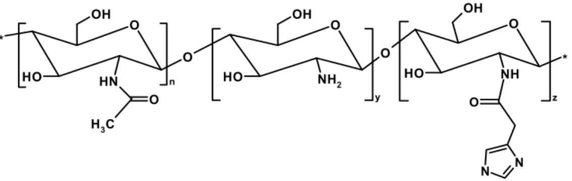
In a previous study, we showed that grafting imidazole moieties to a chitosan backbone (Figure 8) results in the improvement of the transfection efficiency mediated by this polymer [28]. Three degrees of substitution with imidazole were studied: 5% (CHimi1), 13% (CHimi2) and 22% (CHimi3), at various N/P molar ratios (ratio between primary amine groups of chitosan to phosphate groups of DNA). Transfection efficiency of imidazole-grafted chitosan (CHimi) was tested in 293T cells. It was shown that the transfection efficiency increases up to an N/P molar ratio of 18, being higher in CHimi2 and CHimi3 mediated transfection. In terms of particle size, zeta potential or DNA protection there is no significant differences between imidazole-grafted and the unmodified chitosan at the higher N/P molar ratios, suggesting that the increase in transfection efficiency is caused by the incorporation of imidazole moieties. Additionally, no toxic effects were attributed to CHimi based polymers [28]. Therefore, as CHimi-mediated transfection shows great potential for gene delivery, a more detailed study on the mechanisms underlying the gene delivery promoted by these new vectors is required.
N N O O NH O OH O H * N H O O OH O H NH 2 n C H3 O O y OH O H * z
Figure 8: Chemical structure of imidazole-grafted chitosan constituted by (n) units of N-acetylated monomer, (y) units of the deacetylated monomer and (z) units of the imidazole-grafted monomer.
From mechanical strategies to tailored polymers, several efforts are being made in order to create efficient strategies to deliver DNA into the cell nucleus. However, non-viral gene delivery vectors have still a long way to go in order to get closer to viruses efficiency. The first approach to improve transfection efficiency was essentially focused on testing different vector compositions. Some of these strategies were reviewed in the previous paragraphs. More recently, many authors turned their attention to the understanding of the molecular mechanisms underlying transfection and the interactions of non-viral vectors with cells. The premise is that the future non-viral systems design will probably benefit from the attained know-how in such studies.
3. I
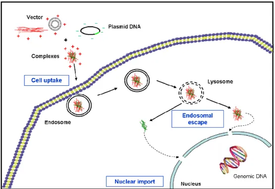
NTRACELLULAR TRAFFICKINGAs discussed in the previous paragraphs, liposomes and cationic polymers are currently being explored as carriers to transport genes into the nucleus of cells. In this process they must overcome several biological barriers toward an efficient non-viral gene delivery (Figure 9). These barriers include binding to the cell surface and cross the membrane, escaping lysosomal degradation, and get into the nucleus [89]. The understanding of these barriers is critical for the development of more effective gene vehicles [90].
Figure 9: Biological barriers to gene delivery.
3.1. Cellular uptake
The cell membrane is the first barrier for non-viral gene delivery. In the absence of a carrier molecule, the highly anionic nature of DNA hinders its passive diffusion through the hydrophobic cell membrane. Association of DNA with cationic lipids or polymers greatly reduces the net negative charge of the particles, increasing the cellular uptake [36]. Even though, most lipoplexes and polyplexes cannot readily cross the cell membrane due to their large size and, in case of polyplexes, also due to the hydrophilic nature. In a first stage, DNA-carrier complexes interact with glycosaminoglycans or specific cell receptors on cell membrane and are thereafter internalized into endosomal vesicles [91]. Endocytosis has been established as the main mechanism for non-viral vectors internalization [26].
Complex physico-chemical properties, as size [92, 93], or the surface charge density determine the interactions with glycosaminoglycans presented on the cell surface. Ultimately, these properties are critical in the uptake process as well as in the overall transfection efficiency [87]. Several studies have been performed in order to correlate physico-chemical properties and polyplexes uptake. Some authors established a size requirement below 100 nm [94] to allow endocytosis of DNA-polymer complexes. However, this is not a well defined boundary, since nanoparticles with a mean size of 150-300 nm are also assumed as suitable for gene delivery [95]. Conversely, some authors suggested that particle size is not a restriction for cellular uptake [87], since Ishii and co-workers obtained good transfection results with 5-8 µm chitosan-DNA complexes [96]. It is important to note that it could be difficult to establish a maximum size for complexes uptake by cells because DNA-polymer suspensions are often polydisperse suspensions. Furthermore, some issues about endocytosis are yet to be fully understood [97] what could explain this apparent lack of agreement.
3.1.1. Endocytic pathways
The three main endocytic pathways discussed in this review are clathrin-mediated endocytosis, caveolar-mediated endocytosis and macropinocytosis (Figure 10).
Phatocytosis (particle-dependent) Macropinocytosis (> 1µm) mediated Clathrin-endocytosis (~120 nm) Caveolin-mediated endocytosis (~60 nm) Clathrin- Caveolin-independent endocytosis (~90 nm) Pinocytosis Phatocytosis (particle-dependent) Macropinocytosis (> 1µm) mediated Clathrin-endocytosis (~120 nm) Caveolin-mediated endocytosis (~60 nm) Clathrin- Caveolin-independent endocytosis (~90 nm) Pinocytosis
Figure 10: Multiple portals of entry in mammalian cells (adapted from [98]).
Clathrin-mediated endocytosis is the best characterized and the most common pathway. It occurs constitutively in all mammalian cells being responsible for the uptake of several essential nutrients. For a long period it was referred as “receptor-mediated endocytosis”, but is now clear that the title was unsuitably applied, because other pinocytic pathways also involve receptor-ligand interactions [98, 99]. In clathrin-mediated endocytosis the internalized material follows an endolysosomal pathway, pursuing from early to late endosomes, and ultimately ending in the lysosomal compartment. The early endosome represents the first stage to which clathrin-coated vesicles deliver their content. The early endosomes traverse to the perinuclear cytoplasm on microtubules tracks, where they fuse with late endosomes or lysosomes. Along this route, the pH drops from
6.0-6.8 in early endosomes to approximately 5 in lysosomes [100]. This pH reduction combined with high concentration of lysosomal enzymes aims at degrading the internalized material [100].
In caveolar-mediated endocytosis, particles are internalized in primary endocytic vesicles that fuse, to larger, more complex tubular membrane organelles – “caveosomes” – which maintain a neutral pH [101]. These organelles are transported through microtubules to the perinuclear region and their content is delivered in the endoplasmatic reticulum [101]. It has been described that the Simian Virus 40 (SV40) can activate a signaling cascade to activate caveolae-localized surface receptors and trigger its own uptake [101]. Calveolar-mediated endocytosis of DNA-carrier complexes is still extremely unexplored and some authors believe that it is unlikely to significantly contribute to constitutive endocytosis, because caveolar vesicles are slowly internalized [26] and generally considered small in size [26, 98]. However, it is important to notice that caveolae constitutes 10-20% of cell surface in endothelial cells [98], where this endocytic pathway could be a relevant portal to enter into the cell [26].
Macropinocytosis has been recognized as an alternative endocytic pathway for some time, but its relevance remained elusive in many cellular processes [102], as in polyplexes uptake. Macropinosomes are formed by actin-driven ruffling of plasma membrane which is dependent on membrane cholesterol content [103]. The fusion between macropinosomes and lysosomes occurs only in macrophages [102]. However, some reports suggest that in non-phagocytic cells macropinosomes can also acidify utilizing a vacuolar-type H+-ATPase and electrogenic Cl- shunt
mechanism, similar to the one described for endosomes [104]. Furthermore, it was reported that the pH reduction is slighter than in late endosomes or lysosomes [92]. Macropinocytosis has been taken into account in gene delivery studies very recently [92, 93, 104], and its specificities may compromise some endosomal escape strategies, as will be pointed out in the section 3.2.
A recent study from Rejman and co-workers with latex beads intended to differentiate the endocytic pathway of particles according to their diameter. They found out that particles up to 200 nm are internalized in clathrin-coated pits, whereas bigger particles enter non-phagocytic cells by macropinocytosis or caveolar mediated endocytosis [93]. This was a very surprising result because caveolar vesicles typically appear as rounded plasma membrane invaginations of 50±80 nm [98, 101]. However the most striking result of this study is to point out macropinocytosis as an alternative pathway to mediate bigger particles uptake in non-phagocytic cells. This issue can help to explain Ishii and colleagues results [96] and challenge the concept that the optimal vector should form small complexes with plasmid DNA [88, 94, 95].
Another remarkable point that should be considered when analyzing endocytosis and its role on gene delivery is the cell cycle. The membrane tension varies in the different phases of the cell cycle. During mitosis, it significantly increases, inhibiting deformation required for invagination [105] and therefore limiting the internalization of particles by endocytic pathways.
3.1.2. Receptor-mediated endocytosis to enhance uptake
Grafting specific ligands to target a specific cell population can also improve transfection efficiency by promoting cellular uptake via receptor-mediated endocytosis. Transferrin-conjugated polymers are thought to be internalized via transferrin receptor that is overexpressed in cells with high metabolic activity. Mao and colleagues developed three schemes to conjugate transferrin with chitosan vectors and they achieved a three-fold increase in transfection in human embryonic kidney 293 cells [81]. In contrast, Simões and colleagues showed that transferrin-conjugated lipoplexes may enter cells even in the presence of free transferrin, what suggests that conjugation of transferrin is not increasing complexes uptake [50]. The authors proposed that the large size of the conjugate can hinder the interaction ligand-receptor, impairing endocytosis via transferrin-receptor. Other ligands such as integrins [106], sacharides [79] or growth factors [107] have also potential to enhance gene delivery efficiency by means of receptor-mediated endocytosis. However, the success of a targeting strategy depends on the conjugation chemistry, the length of spacer between ligand and complex, the ligand-receptor binding strength, and the number of targeting ligands per complex [23]. Additionally, targeting can also be hampered in high positive charge density particles due to unspecific interactions [66].
Sometimes, conjugation of specific ligands gets irreproducible results, so the efficient cell-specific targeting requires careful optimization of the various parameters that affect cell-surface binding [23]. When the strategy is to promote receptor-mediated endocytosis it is important to keep in mind that the success of targeting and the transfection upgrading will primordially depend on the finite number of receptors available on cell surface for binding and internalization [36].
3.2. Endosomal escape
For long time authors believed that all the endocyted complexes followed the endolysosomal pathway, but with recent considerations about macropinocytosis and caveolar-mediated endocytosis [92, 93] new concerns arose. Even though, after cell uptake via any endocytic pathway, the complexes are limited to endocytic vesicles with no access to the cytosol or to the nucleus. The escape from this vesicles is critical for efficient transfection [26], since cellular uptake does not seem to be the limiting step for either polyplexes or lipoplexes-mediated gene delivery [91]. Therefore, several authors are focused on developing strategies to promote endosomal escape of the complexes and they are mainly taken into consideration the endolysosomal pathway.
3.2.1. The flip-flop mechanism
There are some proposed mechanisms for endosomal escape in lipidic carriers. The most accepted is probably the flip-flop model proposed by Xu and co-workers [108]. They propounded
that, after being internalized by the cell, lipid-DNA complexes initiate destabilization of the endosome membrane what results in flip-flop of anionic lipids on the cytoplasmatic face of the membrane. The anionic lipids laterally diffuse into the complex and form charge-neutralized pairs with the cationic lipids. Ultimately, the membrane is destabilized and the DNA released into the cytoplasm [108].
Alternative theories suggest that DNA can be released from the lipoplex due to charge neutralization with anionic macromolecules, or membrane destabilization by pH-sensitive lipids like DOPE [109].
3.2.2. The proton sponge effect
For polyplexes the endosomal escape discussion is mainly focused on works based on the gold standard bPEI. The high transfection efficiency of this polymer is attributed to its buffering capacity within the physiological pH range. According with the so-called “proton-sponge” hypothesis first proposed by Behr [110], cationic polymers with the ability to buffer endosomal pH induce osmotic swelling of endosomes due to excessive proton and chloride accumulation with secondary water movement (see Figure 11). Buffering capacity in the endosomal pH range has also been proven in poliamidoamine dendrimers. In contrast, no chloride accumulation was detected with PLL [104]. The amino groups of PLL have a pKa around 10 and are all protonated at physiological pH what explains the lack of buffering capacity of this polymer. PLL-DNA complexes are sequestered for several hours in endosomal vesicles what makes endosomal escape the main limiting factor in PLL-driven gene delivery [56].
Figure 11: The proton sponge effect (adapted from [111]).
Chitosan has limited buffering capacity in the endosomal acidic pH range 4.5-5.5 [81, 112]. The pKa of chitosan primary amino groups is around 6.3, what limits its buffering capacity in more acidic pH [112]. Studies with bafilomycin A1 – a specific endosomal proton pump inhibitor - report no influence in chitosan mediated transfection, suggesting that this polymer could not induce a proton sponge effect [83]. According to this concept and because the cellular toxicity and side
effects of lysosomotropic agents like chloroquine make it impractical for in vivo gene delivery some authors are focused in designing new vectors with buffering capacity [88].
Despite the widespread acceptance of the proton sponge hypothesis, there are some reports that challenge this theory. Godbey and co-workers found no lysosomal involvement in PEI-DNA complexes routing to the nucleus [113]. Their results are based on confocal images through time of fluorescent-labeled lysosomes and complexes. PEI-DNA complexes have been found in the nucleus 4 hours post-transfection [114] and no changes were detected in lysosomal pH [113]. However, in Behr proton sponge hypothesis, PEI-induced osmotic rupture occurs in endosomes, previous to fusion with lysosomes, as explained by Akinc and colleagues, who further confirmed Behr’s theory [115]. Also in disagreement with Godbey and colleagues, fluorescent and electron microscopy results obtained by Bieber demonstrate that PEI-DNA complexes accumulate in lysosomal compartment [116]. Nevertheless they also challenge the endolysosomal swelling and suggest that PEI-DNA complexes could induce enough local membrane damage to trigger the release of the complexes into the cytoplasm [116]. In accordance with both theories, some research with linear PEI (lPEI) showed that smaller complexes could escape rapidly from lysosomes via proton sponge mechanism, whereas larger complexes reach the cytoplasm via membrane damage [62]. To all this discussion we can add the recent report of Rejman and colleagues [117] that suggests that PEI-mediated transfection occurs after caveolar-mediated endocytosis of PEI-DNA complexes. This report indirectly confronted the proton sponge effect and its relevance for PEI transfection efficiency, because caveolar vesicles have neutral pH and do not fuse with late endosomes [101].
It is now clear that there is a broad lack of accordance in literature about cell transfection mechanisms. A clear explanation can only be achieved with standardized protocols as well as a deeper knowledge about specific features of each endocytic pathway and the specificity of each inhibitor recurrently used in this kind of studies.
3.2.3. Imidazole containing polymers
As referred in section 2.2.3.2.2, imidazole group of histidine has a pKa around 6.0 and turns cationic in a slightly acidic medium (like in endosomal vesicles). Therefore, it owns some buffering capacity in the endosomal pH range and could induce vesicular escape of complexes based on imidazole containing polymers by the “proton sponge” mechanism [53]. Midoux and co-workers describe significant improvement in PLL-mediated transfection efficiency after grafting histidine to the polymer [56]. Complexes based on histidylated PLL were smaller, had lower surface charge, reducing non-specific interactions [118] and enhanced cell viability [119].
The homopolymer of histidine has also been tested for gene delivery. However, since polyhistidine is insoluble in aqueous solution at pH> 6, it was conjugated with gluconic acid to impart solubility at physiologic pH [88]. This conjugated polymer showed moderate transfection ability; still, the study
confirms the advantage of imidazole containing polymers as an initial basis for construction of biocompatible gene delivery vehicles [88].
A similar approach is found with urocanic acid-grafted polymers. Urocanic acid, bearing an imidazole ring, was grafted to water soluble chitosan as mean to improve endossomal escape of chitosan-based complexes through the proton sponge mechanism [120]. As previously referred, chitosan amino groups have a pKa value around 6.3, but very limited buffering capacity is found [81, 112]. On the other hand, imidazole ring of urocanic acid has a pKa of 6.9. This slight difference could help urocanic acid-modified complexes to protonate in endosome in a faster and tighter way, probably leading to proton sponge effect [120]. Modified complexes shown stronger compaction, smaller size and improved transfection results, while cell viability is maintained [120].
We have recently proved that grafting imidazole moieties to a chitosan backbone results in improved transfection efficiency [28]. This improvement seems to be consequence of the polymer buffering capacity, since transfection activity is significantly reduced in the presence of bafilomycin A1 (unpublished data). Our own results further corroborate that the incorporation of imidazole moieties represents a promising strategy to enhance polymers transfection efficiency while maintaining cell viability [53].
3.2.4. Fusogenic peptides
Other strategies are being developed in order to overcome the endosome bottleneck to gene delivery. One important approach is adapted from viruses that use short amino acid sequences with fusogenic properties. It was revealed that these sequences have particular features. There is a conserved hydrophobic side chain with several residues of glutamic acid. The carboxylic groups in the side chain of this amino acid protonate when the pH drops and the conformation of the peptide changes from random coil to α–helix. The helix is amphipatic with the hydrophobic residues on one side what can disturb the lysosomal membrane leading to vesicle content release [121]. However, the use of protein structures like fusogenic peptides has important disadvantages such as low stability of peptides, high costs for peptide synthesis and immunogenic potential of these structures [53].
3.3. Nuclear import
Nuclear entrance has been presented as the Achilles’ heel of non-viral gene delivery [122]. After escaping from endosomal vesicles, DNA should transverse the cell cytoplasm to the proximity of cell nucleus and, ultimately, get in. Indeed, in most cell types a fundamental limitation to gene expression in currently used non-viral systems seems to be the inability of DNA to migrate from the cytoplasm to the nucleus [123].
3.3.1. From the cytoplasm to the nucleus proximity
The cell cytoplasm is composed of a network of microfilament and microtubule systems and a variety of subcellular organelles bathing in cytosol. In addition, cytosol has a high protein concentration, resulting in a significant molecular crowding which limits the diffusion of molecules, such as polyplexes [60]. Hence, after endosomal escape, is not expected that cationic polyplexes or even DNA can diffuse freely. Furthermore, low mobility in the cytoplasm makes plasmid DNA an easy target to cytoplasmatic nucleases [53], limiting its availability.
Very little data is published on transport of nucleic acid through the cytoplasm to within proximity of the nuclear pore. However, some studies reported the importance of DNA size on diffusion. DNA of 100 bp could achieve high mobility and enhanced transfection efficiency, whereas for larger fragments (>1000 bp) the diffusion is remarkably reduced [124], suggesting the existence of transport mechanisms other than diffusion. Polymer-DNA complexes transport seems also to be an energy dependent process. Suh and colleagues showed that bPEI-DNA complexes are transported into the perinulear region by an energy-dependent mechanism [122]. This mechanism could be only part of cationic complexes transport, but there is evidence on microtubule network involvement [122] and association to motor proteins such as dynein [125].
3.3.2. Complex disassembling
Concerning the nuclear import, one question that needs to be addressed is whether the DNA disassembles from the liposome or polymer prior to or after nuclear entry. For liposome-mediated gene delivery Xu and colleagues postulate that during transfection, DNA is released from the lipid in the cytoplasm and is then trafficked uncoated by an inefficient mechanism into the nucleus [108]. The disassembling of DNA from the lipid prior to entering the nucleus was thought to be critical for the ultimate expression of the gene, because when lipid-DNA complexes were directly injected in the nucleus the transgene expression was reduced [126]. In this way, the incorporation of lipids such as DOPE or cholesterol, which favors the DNA release from lipid formulations, is common strategy to enhance gene delivery [36]. However, the opposite theory has also been proposed by Cornellis and colleagues, who recently showed that lipoplexes microinjected into the nucleus can also promote gene expression and that the cytoplasmatic dissociation of DNA from the lipoplex is not the limiting step for lipofection [127].
The intracellular mechanisms occurring after polymeric vectors-mediated transfection has been slightly explored and the published studies are mostly focused on PEI or PLL-based complexes. Pollard and co-workers proposed that cationic polymers (bPEI and PLL) could promote gene accessibility to the nucleus without complex disassembling [126].
Nuclear localization of bPEI was demonstrated by fluorescence labeling in immortalized endothelial cells [114]. bPEI enters the cell nucleus alone or assembled with DNA [114] by diffusion, rather than by some active transport mechanism [128]. It was suggested that, through interaction with
anionic lipids from the cell or endosome membrane, complexes are covered by a lipidic coat that promotes fusion with nuclear envelope and DNA is released inside nuclei [114]. The complex disassembling is point of disagreement and some authors proposed that DNA is released by competition with genomic DNA [126], or rRNA highly concentrated in the nucleolus [129]; whereas other reports support that there is no DNA release, since transcription occurs efficiently, even with DNA tightly complexed with the polymer [116].
As discussed in section 2.2.3.1.2, linear PEI (lPEI) shares the chemical formula with branched PEI (bPEI) but the different structure confers remarkable differences in intracellular trafficking. lPEI complexes disassemble in cytoplasm which is correlated with the earlier detectable gene expression relative to bPEI [130]. However, to low molecular weight lPEI polymer, alternative mechanisms had been revealed by confocal laser scanning microscopy. lPEI-based complexes can reach the nucleus intact; some of these polyplexes may dissociate inside the nuclei, but disassembling can also occur in the cytosol and, in that conditions, DNA or polymer reach the nucleus on their own [62].
Experiments to determine transfection mechanism of PLL complexes are quite difficult since these polyplexes are mostly entrapped in the endosomal vesicles and no nuclear localization is detected except after lysosome destabilization induced by agents such as chloroquine. Even though, in long incubation experiments in vitro, the dissociation of DNA and the polymer can be proved [130]. In chitosan-mediated transfection no significant dissociation of the complexes in the cytoplasm was detected [87]. Chitosan can be found inside the cell nucleus [96], where the disassembling is supposed to occur [87]. It was also proposed that the presence of the polymer in the nucleus does not interfere with the gene expression [87], however there are no evidences attesting these theories.
3.3.3. The nuclear membrane
The nuclear envelope is the final obstruction to plasmid DNA entry into the nucleus. The transport of macromolecules through the nuclear membrane is crucial for the metabolism of eukaryotic cells [109]. In consequence, there are specific mechanisms to allow and control substances access into the nucleus. The nuclear envelope is a double membrane perforated by nuclear pore complexes (NPC). The NPC allows passive diffusion of molecules up to 50 kDa. Larger molecules should contain specific sequences – nuclear localization sequences (NLS) – that are able to mediate nuclear import and transverse the nuclear pore in an energy-dependent manner [60]. The size scale of the NPC make it unlikely transporter for DNA, but some studies report DNA entry in intact nuclei [131] and in reconstituted nuclei extracted from Xenopus laevis eggs [132].
Besides passive diffusion and energy-dependent transport, a third process can allow particles to get into the nucleus. When cells undergo mitosis, the nuclear envelope breaks down and the
![Table 1: Examples of current gene therapy clinical trials using viral-vectors (information available at [15])](https://thumb-eu.123doks.com/thumbv2/123dok_br/15917920.1093484/21.892.106.767.421.768/table-examples-current-therapy-clinical-vectors-information-available.webp)
![Figure 11: The proton sponge effect (adapted from [111]).](https://thumb-eu.123doks.com/thumbv2/123dok_br/15917920.1093484/34.892.266.653.705.957/figure-proton-sponge-effect-adapted.webp)

![Figure 18: Schematic drawing representing the covalent bonding between DNA and Label IT ® reagent (available at [146])](https://thumb-eu.123doks.com/thumbv2/123dok_br/15917920.1093484/57.892.119.427.319.650/figure-schematic-drawing-representing-covalent-bonding-reagent-available.webp)
