2016/2017
Catarina Pedrosa Martins da Costa
Emerging Genetic Alterations Linked to
Male Infertility: X-Chromosome Copy
Number Variation and Spermatogenesis
Regulatory Genes’ Expression
Mestrado Integrado em Medicina
Área: Genética Médica Tipologia: Dissertação
Trabalho efetuado sob a Orientação de: Doutora Susana Fernandes E sob a Coorientação de: Professor Doutor Alberto Barros
Trabalho organizado de acordo com as normas da revista: Annals of Reproductive Medicine and Treatment Catarina Pedrosa Martins da Costa
Emerging Genetic Alterations Linked to
Male Infertility: X-Chromosome Copy
Number Variation and Spermatogenesis
Regulatory Genes’ Expression
DEDICATÓRIA
Type of Article: Original Research Article
1
2
Title: Emerging genetic alterations linked to male infertility: X-chromosome Copy 3
Number Variation and Spermatogenesis regulatory genes’ expression 4
5
Authors: Catarina Costa1, Maria João Pinho1,2, Alberto Barros1,2,3, Susana Fernandes1,2 6
1 Department of Genetics, Faculty of Medicine of University of Porto, Porto, Portugal 7
2i3S – Instituto de Investigação e Inovação em Saúde, University of Porto, Porto, 8
Portugal. 9
3Centre for Reproductive Genetics A Barros, Porto, Portugal. 10
Corresponding author: Susana Fernandes, Department of Genetics, Faculty of
11
Medicine of University of Porto, Al. Prof. Hernâni Monteiro, 4200 - 319 Porto, 12
PORTUGAL . Telephone: +351 22 551 36 47 E-mail: [email protected] 13 14 15 16 17 18 19
Title: Emerging genetic alterations linked to male infertility: X-chromosome Copy 20
Number Variation and Spermatogenesis regulatory genes’ expression 21
Abstract
22
The etiopathogenesis of primary testicular failure remains undefined in 50% of cases. 23
Most of these idiopathic cases probably result from genetic mutations/anomalies. Novel 24
causes, like Copy Number Variation and gene expression profile, are being explored 25
thanks to recent advances in the field of genetics. Our aim was to study Copy Number 26
Variation (CNV) 67, a patient-specific CNV related to spermatogenic anomaly and 27
evaluate the expression of regulatory genes AKAP4, responsible for sperm fibrous sheet 28
assembly, and STAG3, essential for sister chromatid cohesion during meiosis. One 29
hundred infertile men were tested for CNV67 with quantitative PCR (qPCR). Quantitative 30
real-time PCR was performed to evaluate gene expression patterns of the two mentioned 31
genes in testicular biopsies from 22 idiopathic infertile patients. 32
CNV67 deletion was found in 2% of patients, with the same semen phenotype described 33
in previous studies. Expression levels of AKAP4 and STAG3 were downregulated in 34
infertile patients when compared to control group (p<0.05). 35
Resulting data reinforce the role of CNV67 in male infertility etiology. Its frequency is 36
significantly higher in oligo/azoospermic men and evidence indicates consistency of 37
phenotype. Downregulation of AKAP4 and STAG3 cellular transcript levels was observed 38
in the testicular biopsies, suggesting that the gene expression is altered, contributing to 39
unsuccessful sperm production. 40
As one continues to better understand about the genetics of male infertility, there will be 41
undoubtedly a shift towards better diagnosis and treatment for those patients presenting 42
idiopathic infertility. 43
Keywords: Male Infertility; Spermatogenesis; DNA copy number variations; Gene 44 expression. 45 BACKGROUND 46
An estimated 15% of couples are infertile, not achieving a clinical pregnancy after 1 year 47
of unprotected intercourse, with a great impact on the individual, couple and society [1, 48
2]. Male reproductive dysfunction is the sole or contributory cause of infertility in half of 49
the couples [3, 4], with abnormalities of sperm number (azoospermia, oligozoospermia), 50
motility (asthenozoospermia) and morphology (terathozoospermia) being frequently 51
diagnosed [5, 6]. 52
53
Male infertility can be clinically divided in three main categories: acquired, congenital 54
and idiopathic, when no cause is identified [6, 7]. The idiopathic group still represents 55
50% of the cases of primary spermatogenic failure in humans [2, 8] and presently, due to 56
the lack of pathophysiological understanding, no specific treatment is offered [9]. Most 57
of the underlying causes are thought to be genetic [6, 10, 11], mainly due to 58
spermatogenesis defects [3, 12], correlated, or not, with environmental factors. 59
Spermatogenesis is a highly complex process controlled by several regulatory genes 60
which assure the correct maturation steps, from spermatogonia to sperm [3]. These men 61
are otherwise usually healthy, suggesting that any genes involved must either be only 62
expressed or be functionally required for spermatogenesis [3]. 63
64
During the last years, novel tests and diagnostic tools have been employed to identify rare 65
genetic mutations and polymorphism with putative direct or indirect effects on 66
spermatogenesis. The declining cost and increased power of whole-genome sequencing 67
studies, including evaluation of the increasingly important intergenic regions of the 68
genome, is leading to nascent paths of research and likely indicate that, in the future, such 69
studies will be used on daily-basis [13]. Likewise, genetic testing of Copy Number 70
Variation and spermatogenesis’s regulatory genes expression may reveal the etiology of 71
idiopathic patients and, consequently, increase the likelihood of successful paternity and 72
reduce potential risks to the progeny [13, 14]. 73
74
Copy Number Variation (CNV) has raised a considerable interest among scientific and 75
medical communities. CNV is conventionally defined as a DNA segment, 1 kb or longer, 76
that is present in a variable number of copies in the genome, between individuals [15]. 77
Since the first comprehensive CNV map of the human genome, in 2006, several diseases 78
have been linked to CNVs, mainly due to disruption of functional elements (either genes 79
or regulatory elements). In fact, it is well established that Y chromosome CNVs in the 80
AZF region are linked to spermatogenic impairment and are routinely analysed for 81
genetic male infertility diagnosis [9, 16]. These unbalanced quantitative variants can be 82
classified into gains (increased number of DNA copies compared to reference genome) 83
and losses (reduction or deletion compared to reference genome) [15]. 84
85
Recently, high-resolution X-chromosome specific array-comparative genomic 86
hybridization (aCGH) identified CNVs which could be related with male infertility [17]. 87
X chromosome genes are particularly tempting because men are hemizygous for the X-88
genes. Since compensation by a normal allele is impossible, it is more likely that a 89
mutation may affect the fertility of an individual [6, 10]. From the reported CNVs, 90
CNV67 was one of the most promising candidates, resembling AZF deletions of the Y 91
chromosome [16, 17]. 92
CNV67 deletion was exclusively found in infertile patients at a frequency of 1.1% 94
(p<0.01), ranging patient’s phenotypes from azoospermia due to Sertoli-Cell-Only 95
Syndrome (SCOS) to oligozoospermia. It is localized in Xq28 and is likely to be 96
maternally inherited [16]. It has been suggested that CNV67 deletion linked to 97
spermatogenic failure may be related to highly duplicated genes of X-Cancer Testis 98
Antigen (CTA) family, the most represented linked testis specific family. In fact, X-99
CTA genes comprise 10% of all X-linked genes and are expressed specifically in testis. 100
[18]. In particular, CNV67 deletion may remove the melanoma antigen family A, 9B 101
(MAGEA9B), expression levels in spermatocytes and in some tumour cell lines. It may
102
also affect chromosome X open reading frame (CXorf40A), situated at < 1Mb from the 103
deletion and regulation elements of Heat Shock Transcription Factor Family, X-Linked 104
1/2 (HSFX1/2) [16, 17]. 105
Gene expression profile can be used as a basis for identification of candidate genes that 106
contribute to male infertility [19-21]. To date, genetic studies in mice have identified more 107
than 200 genes that are specifically or preferentially involved in the complex regulation 108
of fertility and some are specifically expressed in the germ line [6, 10, 11, 19]. AKAP4 109
and STAG3 are strong candidate genes for male infertility [7, 22]. 110
The A-kinase anchor protein 4 (AKAP4), an X-linked member of the AKAP gene family, 111
encodes the most abundant protein of the spermatozoon’s fibrous sheet, a cytoskeletal 112
structure surrounding the region of the principal piece of sperm flagellum [23]. AKAP4 113
anchors cAMP-dependent protein kinase A (PKA) to the sperm fibrous sheet, which is 114
essential for sperm capacitation, playing a central role in the regulation of normal sperm 115
motility [11, 24, 25]. In fact, studies have shown that in AKAP4-deficient mice, though 116
sperm count was not reduced, they were immotile, resulting in male infertility [6, 11]. 117
Furthermore, another study verified no detection of AKAP4 immunolabeling in man with 118
0% sperm mobility [19]. Therefore, AKAP4 is likely required for the structural and 119
functional integrity of the fibrous sheath [25]. 120
Stromalin 3 (STAG3) is a component of all meiosis-specific cohesion complexes, a large 121
ring-shaped proteinaceous structure which tethers sister chromatids, providing cohesion 122
to the structure [22, 26]. Its deletion has been related to a Premature Ovary Failure (POF). 123
Interestingly, STAG3-deficient male mice display a severe defect in synapses and 124
premature loss of centromeric cohesion during the early stages of prophase I, which 125
causes an arrest during the zygotene-like stage, leading to infertility [22, 27]. 126
127
The aim of this study is to explore these emerging genetic alterations by quantifying the 128
copy number variation of CNV67 in a group of infertile men and consolidate the 129
pathophysiology which links CNV67 to male infertility. In addition, the expression of 130
spermatogenesis regulatory genes AKAP4 and STAG3 will be evaluated in infertile men 131
testicular biopsies and correlated with the (in)fertility status. 132
133
MATERIALS AND METHODS
134
This study includes two distinct analyses – CNV67 screening and expression profile of 135
AKAP4 and STAG3. Each analysis design will be explained separately.
136
CNV67 screening analysis
137
Patient samples
138
Peripheral blood samples were collected from 100 Portuguese idiopathic infertile men, 139
with different grades of spermatogenic impairment - 44 azoospermic (AZO), 47 severe 140
oligozoospermic (SOZ), 4 oligozoospermic (OZ) and 5 normozoospermic (N) men (Table 141
1). Infertile patients were selected on the basis of a comprehensive andrological 142
examination including medical history, semen analysis, scrotal ultrasound, and hormonal 143
and genetic analysis. Patients with abnormal karyotype or Y chromosome microdeletion 144
were excluded. Normal controls were fertile normozoospermic volunteers. 145
Genomic DNA (gDNA) extraction
146
Peripheral blood (3–5 mL) was collected through vein puncture from all participants. 147
High molecular weight DNA was isolated using a salting out method. 148
Quantitative PCR (qPCR)
149
The number of copies of CNV67 on each sample was determined by Quantitative PCR 150
(qPCR). TaqMan® probes were designed by the manufacturer (Applied Biosystems, 151
Foster City, USA) and were chosen to target specific regions. Hs03323870_cn was 152
selected for the target CNV67 (labeled with FAM) and Hs03323870 was selected for 153
RNase P (labeled with VIC) and used as the reference gene. Reactions were performed in 154
triplicate in a final volume of 20 µL according to the manufacturer’s instructions. Briefly, 155
the components of the reaction mix were: 4 µL genomic DNA, 10 µL 2X TaqMan® 156
Genotyping Master Mix, 1 µL 20X TaqMan® Copy Number Assay, 1 µL 20X TaqMan® 157
Copy Number Reference Assay (RNase P) and 4 µL nuclease-free water. qPCR was 158
carried out on a StepOnePlus™ Real-Time PCR System (Applied Biosystems). The 159
thermal cycling conditions were as follows: Initial enzyme activation for 10 minutes at 160
95°C, 40 cycles were performed, each one consisting of 15 seconds at 95°C and 60 161 seconds at 60°C. 162 163 Data Analysis 164
Applied Biosystems CopyCallerTM Software v2.0 was used to determine the copy number 165
status of each target region, and calculations were performed according to the maximum-166
likelihood algorithm of the software. Raw copy value (RCV) represents a non-integer 167
number of copy calculated, whereas predicted copy number (PCN) is defined as an integer 168
number of copy determined by the algorithm (0, 1, 2, or 3+). As CNV67 is located on X-169
chromosome, normal females will display PCN of 2 and normal males PCN equal to 1. 170
In the case of male alteration, Copy Number (CN) gain is defined as PCN higher than 1, 171
and PCN of 0 is regarded as CN loss. 172
AKAP4 and STAG3 expression
173
Patient samples
174
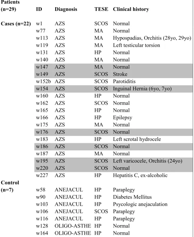
Testicular samples were collected from 22 idiopathic infertile men with AZS (used as 175
cases). Seven men with secondary infertility were used as controls. Testicular biopsies 176
were obtained to confirm the clinical diagnosis (diagnostic biopsy) or for sperm retrieval 177
(Testicular Sperm Extraction: TESE) to intracytoplasmic sperm injection (treatment 178
biopsy). Patients with abnormal karyotype or Y chromosome microdeletion were 179
excluded. Clinical information of each sample is shown in Table 2. 180
Each sample was divided into three aliquots: one was reserved for histological analysis, 181
the second (100-200mg) was processed for sperm extraction and the third (10mg) was 182
immediately transferred to a 1.5mL tube with mRNA later® solution (Ambion®, Foster 183
City, USA) and stored at -80ºC for further gene expression studies. 184
185
RNA isolation and reverse transcription (RT) reaction
186
After thawing the frozen pellets, cells were lysed on ice with 1000μL of TriPure Isolation 187
Reagent (Roche Diagnostics, Indianapolis, USA) and passed several times through a 188
syringe and needle. The total mRNA was then extracted according to the associated 189
protocol. At the end, RNA pellet was resuspended in 50μL of diethylpyrocarbonate 190
(DEPC)-treated RNase-free water (Promega, Wisconsin, USA) and incubated for 1h on 191
ice. RNA was then quantified in a Biotech Photometer UV 1101 (WPA, Cambridge, UK). 192
1µg of mRNA in a total volume of 10μL was reverse transcribed to complementary DNA 193
(cDNA) using qScriptTM cDNA SuperMix (Quanta, BiosciencesTM, Gaithersburg, USA), 194
with random hexamers as the priming method and according to the manufacturer’s 195
instructions. 196
Gene expression analysis by quantitative real-time PCR (qRT-PCR)
197
TaqMan® Gene Expression Assays were used for both targeted experimental genes 198
(AKAP4 - Hs00275849_m1 and STAG3 - Hs00429370_m1) All TaqMan® probes were 199
labeled with FAM dye and were purchased from Applied Biosystems. RNA 18S 200
Ribosomal (18S) was used as the housekeeping gene and TaqMan® Gene Expression 201
Assay was also utilized. 202
RNA expression levels were analysed by qRT-PCR on a StepOnePlus™ Real- Time PCR 203
System (Applied Biosystems). qRT-PCR was performed in a volume of 10μL, using 2μL 204
of cDNA, 2.5µL of Nuclease Free-water, 5µL of 2xKAPA probe MasterMix (Kappa 205
Biosystems, Boston, Massachusetts, USA) and 0,5µL of 20X TaqMan® Gene Expression 206
Assay for each gene, using a Fast Protocol according to manufacturer instructions. 207
Briefly, after initial enzyme activation for 2 minutes at 50°C and 20 seconds at 95°C, 40 208
cycles were performed, each one consisting of 3 seconds at 95°C and 20 seconds at 60°C. 209
Standard curves were performed with five points, in duplicates. Each PCR for relative 210
quantification was run in triplicate (technical replicates) and all genes were run together 211
with a negative control. 212
Data analysis and statistics
213
Data was analyzed using REST 2009 (Relative Expression Software Tool), which is a 214
standalone software tool that estimates up and downregulation for gene expression studies 215
(http://www.qiagen.com/rest). The purpose of this software is to determine whether there 216
are significant differences between samples and controls, while taking in account issues 217
of reaction efficiency and reference gene normalization. The obtained hypothesis test 218
P(H1) represents the probability of the alternate hypothesis that the difference between 219
the sample and control groups is due only to chance. Real time PCR-negativity was 220
defined by the absence of amplified product after 40 cycles and because REST software 221
uses Ct values and reaction efficiency for calculations instead of relative expressions 222
values, we proposed that the value of the last cycle of amplification (Ct = 40 cycles) 223
should correspond to the value of absence of relative expression. Wilcoxon Signed Rank 224
Test was used for the statistical analysis (StatView for Windows) with the significance 225
level set at p < 0.05. 226
227
RESULTS AND DISCUSSION
228
CNV67 screening analysis
229
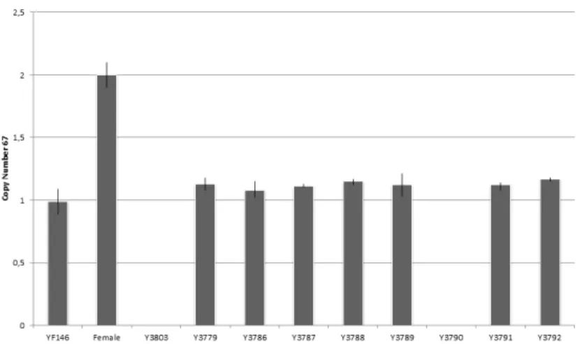
In order to screen CNV67 deletion, 100 samples were studied from infertile men with 230
different sperm phenotypes and concentration, as previously described, by RT-PCR 231
(Table 1). Two individuals – Y3790 and Y3803 – were found to have deletion (0 copies) 232
of CNV67 (2%). Y3790 was azoospermic, diagnosed with SCOS after biopsy and Y3803 233
presented a severe oligozoospermia (2 x10⁶ sperm/mL) (Fig. 1). Our data supports the 234
sperm phenotypes related to CNV67 deletion (azoospermia in a clinical context of SCOS 235
or oligozoospermia). Moreover, the findings indicate a significantly higher frequency in 236
our Portuguese population, even though our sample was quite small when compared to 237
previous studies [16, 17]. Whether the observed deletion is directly responsible for the 238
altered sperm phenotype (either affecting gene expression or regulatory elements) or is 239
related to increased genomic instability remains uncertain [17]. 240
AKAP4 and STAG3 expression
241
Quantification of testicular mRNA levels of genes expression was carried out by qRT-242
PCR in individuals showing spermatogenic failure. Two spermatogenesis related genes - 243
AKAP4 and STAG3 – were analyzed in 20 and 22 testicular biopsies samples,
244
respectively. Clinical and pathologic information on the cases and controls are presented 245
in Table 2. 246
Analysis of the qRT-PCR results was completed by using REST 2009 software. The 247
findings are summarized in Table 3. For reference gene normalization, 18S housekeeping 248
gene was used. Sample expression ratios were calculated with REST software using the 249
following formula: 250
Relative Expression = Concentration of Gene of interest ÷ Geometric mean
251
(concentration of reference gene 1, concentration of reference gene 2,…)
252
With the use of this software, the up or downregulation for each gene expression was 253
estimated comparing cases with controls. Results indicated that the AKAP4 and STAG3 254
were downregulated with statistical significance (p<0.05) in the case group compared to 255
the control group (Table 3; Fig. 2). 256
Interestingly, 6 cases (w147, w149, w154, w176, w186, w195) did not express AKAP4 257
and w220 patient did not express STAG3 (see Table 2). It has been demonstrated that the 258
reduction of gene expression in spermatogenic failure patients could not be exclusively 259
attributed to a decreased number of germ cells, but the contribution of the reduced cellular 260
expression should be also taken in account [19]. 261
AKAP4 encodes a protein involved in fibrous sheet assembly and its regulation [28], and
262
is exclusively expressed in germ cells, during the post-meiotic phase of spermatogenesis 263
[25, 29]. The findings of this study on AKAP4 expression exhibit a statistically significant 264
difference with downregulation in the case group. Interestingly, no expression was 265
detected in several SCOS patients. However, the 2 oligo-asthenozoospermic [23] patients 266
used as controls expressed AKAP4, contradicting the data published in a previous study. 267
Similarly, downregulation of STAG3 was found in the case group. The STAG3 encodes a 268
predominant STAG protein component of cohesin complexes in primary spermatocytes, 269
participating in the telomere attachment to the nuclear periphery, telomere maintenance, 270
chromosome pairing, chromosome synapses and maintenance of sister chromatid 271
cohesion [27]. This protein is exclusively expressed in meiosis. 272
273
Gene expression profiles can be used as a basis for identification of candidate genes that 274
contribute to spermatogenic impairment. One must emphasize that an inherent problem 275
in investigating testicular expression changes is the cellular complexity of the organ [21]. 276
Here we analyzed the transcriptional changes in a complete organ, with distinct germ cell 277
types. One advantage is that we revealed complex transcriptional changes related to the 278
whole testis during germ cell differentiation. The same point has the inherent 279
disadvantage, compared with isolated cell fractions, as we cannot directly identify the 280
locus of expression change. Furthermore, whether the observed differential expression 281
profiles represent the cause or consequence of spermatogenic impairment remains to be 282
elucidated. This data should be useful in delineating the patterns of gene expression 283
involved in male germline, which may contribute to understanding male infertility. 284
CONCLUSION
285
Classic male infertility tests, like karyotyping, Y chromosome microdeletions and FISH 286
analysis at somatic and germ cell levels, are no longer sufficient to investigate the 287
potential contribution of genome disorders on male infertility. A wide range of molecular 288
methods are required for better understanding of male infertility causes and, therefore, 289
increase the potential offer for a better treatment for infertile patients [30]. 290
Novel genetic alterations have been identified which may be of potential clinical 291
relevance in the etiology of male infertility in the medium term, like Copy Number 292
Variation (CNV). Of all CNVs related to male infertility, X-CNV67 was one of the most 293
interesting ones [16], with a consistent phenotype and significant frequency. It is likely 294
that rare single nucleotide polymorphisms (SNPs) and CNVs, although they are rare on 295
an individual basis, collectively they can contribute to explain a significant number of 296
cases of male infertility that are currently classified as idiopathic. [13]. 297
298
The present study also explores gene expression profile as an emerging genetic alteration 299
with implications in male infertility. Therefore we assessed the expression profile of 300
regulatory genes AKAP4 and STAG3 on infertile men testicular biopsies. Our data reports 301
altered expression of germ-line regulatory genes, providing an initial glimpse into the 302
complex regulatory network controlling germ line development. Further analyses in 303
larger series are required to better understand the biological implications of these 304
differences. 305
306
Although the importance of diagnosing genetic factors is fully recognized, the diagnostic 307
workup of infertility in men is still limited to a few genetic tests [17]. Genetic testing 308
allows clarifying an obscure infertility diagnosis and help to prevent miscarriage and 309
iatrogenic transmission of genetic defects to the offspring through Assisted Reproduction 310
Techniques (ART) [3, 4]. Therefore, we believe that efforts should be made in order to 311
identify potential genetic causes of infertility and, in this way, aid couples to make 312
informed decisions, optimize genetic testing and provide therapeutic targets [2, 31]. 313
314
In conclusion, our findings merit further investigation in order to elucidate the potential 315
of CNV67 in routine fertility workup and the role of AKAP4 and STAG3 in male 316
infertility. 317
318
Ethics approval and consent to participate - The local Ethical Committees of the
319
Faculty of Medicine of University of Porto/ Centro Hospitalar S. João approved the study. 320
Informed consent was obtained from the patients before being included in this study 321
during their reproductive medical treatment. 322
Acknowledgements - We would like to thank the patients involved for their participation,
323
Nuno Barros and Joaquina Silva for providing patients samples and their clinical data, 324
and Sofia Dória for help on statistical analysis. 325
326
REFERENCES
327
1. Zegers-Hochschild F, Adamson GD, de Mouzon J, Ishihara O, Mansour R, Nygren K, et 328
al. International Committee for Monitoring Assisted Reproductive Technology (ICMART) and 329
the World Health Organization (WHO) revised glossary of ART terminology, 2009. Fertility and 330
Sterility. 2009;92(5):1520-4. 331
2. Esteves SC. A clinical appraisal of the genetic basis in unexplained male infertility. 332
Journal of human reproductive sciences. 2013;6(3):176-82. 333
3. Elliott DJ, Cooke HJ. The molecular genetics of male infertility. Bioessays. 334
1997;19(9):801-9. 335
4. McLachlan RI, Krausz C. Clinical evaluation of the infertile male: new options, new 336
challenges. Asian Journal of Andrology. 2012;14(1):3-5. 337
5. Cooper T, Castilla JA. WHO LABORATORY MANUAL FOR THE EXAMINATION AND 338
PROCESSING OF HUMAN SEMEN. Journal of Andrology. 2009;30:9-. 339
6. Stouffs K, Tournaye H, Liebaers I, Lissens W. Male infertility and the involvement of the 340
X chromosome. Human Reproduction Update. 2009;15(6):623-37. 341
7. Krausz C. Male infertility: Pathogenesis and clinical diagnosis. Best Practice & Research 342
Clinical Endocrinology & Metabolism. 2011;25(2):271-85. 343
8. Lee JY, Dada R, Sabanegh E, Carpi A, Agarwal A. Role of Genetics in Azoospermia. 344
Urology. 2011;77(3):598-601. 345
9. McLachlan RI, O'Bryan MK. State of the Art for Genetic Testing of Infertile Men. Journal 346
of Clinical Endocrinology & Metabolism. 2010;95(3):1013-24. 347
10. Zheng K, Yang F, Wang PJ. Regulation of Male Fertility by X-Linked Genes. Journal of 348
Andrology. 2010;31(1):79-85. 349
11. Stouffs K, Lissens W. X chromosomal mutations and spermatogenic failure. Biochimica 350
Et Biophysica Acta-Molecular Basis of Disease. 2012;1822(12):1864-72. 351
12. Krausz C, Giachini C. Genetic risk factors in male infertility. Archives of Andrology. 352
2007;53(3):125-33. 353
13. Hotaling J, Carrell DT. Clinical genetic testing for male factor infertility: current 354
applications and future directions. Andrology. 2014;2(3):339-50. 355
14. Wosnitzer MS. Genetic evaluation of male infertility. Translational andrology and 356
urology. 2014;3(1):17-26. 357
15. Fanciulli M, Petretto E, Aitman TJ. Gene copy number variation and common human 358
disease. Clinical Genetics. 2010;77(3):201-13. 359
16. Lo Giacco D, Chianese C, Ars E, Ruiz-Castane E, Forti G, Krausz C. Recurrent X 360
chromosome-linked deletions: discovery of new genetic factors in male infertility. Journal of 361
Medical Genetics. 2014;51(5):340-4. 362
17. Krausz C, Giachini C, Lo Giacco D, Daguin F, Chianese C, Ars E, et al. High Resolution X 363
Chromosome-Specific Array-CGH Detects New CNVs in Infertile Males. Plos One. 2012;7(10). 364
18. Fratta E, Coral S, Covre A, Parisi G, Colizzi F, Danielli R, et al. The biology of cancer testis 365
antigens: Putative function, regulation and therapeutic potential. Molecular Oncology. 366
2011;5(2):164-82. 367
19. Bonache S, Algaba F, Franco E, Bassas L, Larriba S. Altered gene expression signature of 368
early stages of the germ line supports the pre-meiotic origin of human spermatogenic failure. 369
Andrology. 2014;2(4):596-606. 370
20. Chalmel F, Lardenois A, Evrard B, Mathieu R, Feig C, Demougin P, et al. Global human 371
tissue profiling and protein network analysis reveals distinct levels of transcriptional germline-372
specificity and identifies target genes for male infertility. Hum Reprod. 2012;27(11):3233-48. 373
21. Feig C, Kirchhoff C, Ivell R, Naether O, Schulze W, Spiess AN. A new paradigm for 374
profiling testicular gene expression during normal and disturbed human spermatogenesis. 375
Molecular human reproduction. 2007;13(1):33-43. 376
22. Llano E, Gomez-H L, Garcia-Tunon I, Sanchez-Martin M, Caburet S, Luis Barbero J, et al. 377
STAG3 is a strong candidate gene for male infertility. Human Molecular Genetics. 378
2014;23(13):3421-31. 379
23. Moretti E, Scapigliati G, Pascarelli NA, Baccetti B, Collodel G. Localization of AKAP4 and 380
tubulin proteins in sperm with reduced motility. Asian Journal of Andrology. 2007;9(5):641-9. 381
24. Turner RMO, Johnson LR, Haig-Ladewig L, Gerton GL, Moss SB. An x-linked gene 382
encodes a major human sperm fibrous sheath protein, hAKAP82. Journal of Biological 383
Chemistry. 1998;273(48):32135-41. 384
25. Hu Y, Yu H, Pask AJ, O'Brien DA, Shaw G, Renfree MB. A-kinase anchoring protein 4 has 385
a conserved role in mammalian spermatogenesis. Reproduction. 2009;137(4):645-53. 386
26. Hopkins J, Hwang G, Jacob J, Sapp N, Bedigian R, Oka K, et al. Meiosis-Specific Cohesin 387
Component, Stag3 Is Essential for Maintaining Centromere Chromatid Cohesion, and Required 388
for DNA Repair and Synapsis between Homologous Chromosomes. Plos Genetics. 2014;10(7). 389
27. Ayobami Ward JH, Matthew Mckay, Steve Murray and Philip W. Jordan. Genetic 390
interactions between the meiosis-specific cohesion components, STAG3, REC8 and RAD21L. 391
3G. 2016;6:1713-24. 392
28. Pereira R, Oliveira J, Ferraz L, Barros A, Santos R, Sousa M. Mutation analysis in 393
patients with total sperm immotility. Journal of Assisted Reproduction and Genetics. 394
2015;32(6):893-902. 395
29. Miki K, Willis WD, Brown PR, Goulding EH, Fulcher KD, Eddy EM. Targeted disruption of 396
the Akap4 gene causes defects in sperm flagellum and motility. Developmental Biology. 397
2002;248(2):331-42. 398
30. Benkhalifa M, Montjean D, Belloc S, Dalleac A, Ducasse M, Boyer P, et al. Emerging 399
molecular methods for male infertility investigation. Expert review of molecular diagnostics. 400
2014;14(1):37-45. 401
31. Dunn TM, McGovern MM, Bar-Chama N, Kardon NB. Genetic evaluation of male factor 402
infertility. Am J Hum Genet. 1999;65(4):A214-A. 403 404 405 406 407 408 409 410 411 412 413 414 415 416 417 418 419 420 421 422 423 424
FIGURE SECTION 425 426 427 428 429 430 431 432 433 434
Fig. 1 – Copy Number Variation 67 deletion of infertile man Y3790 and Y3803. Whiskers 435
refer to maximum and minimum of copies calculated. As CNV67 is located on X-436
chromosome, normal females will display Predicted Number of Copies (PCN) of 2 and 437
normal males PCN equal to 1. In the case of male alteration, Copy Number (CN) gain is 438
defined as PCN higher than 1, and PCN of 0 is regarded as CN loss. YF154 – Fertile man 439 control. 440 441 442 443 444 445 446 447 448 449
450 451 452 453 454 455 456 457 458 459
Fig. 2 – Expresion levels of AKAP4 and STAG3 in testicular samples. cDNA expression 460
was normalized using a housekeeping gene (18S). Boxes represents the interquartile 461
range, or the middle 50% of observations. The dotted line represents the median gene 462
expression. Whiskers represent the minimum and maximum observations. Significance 463
differences between groups are represented as: * p<0.05. The data was analysed by Mann-464 Whitney-Wilcoxon test. 465 466 467 468 469 470 471 472 473 474 * *
Table Section
475 476 477 478 479 480 481 482 483 484 485 486 487 488 489 490 491 492 493 494 495 496 497 498 499 500 501 502 503 504 505 506 507 508 509Table 1 – Clinical description of the study population Patient’s semen phenotype (n=100) Azoospermic Severe oligozoospermic (5x10⁶/mL) Oligozoospermic (5-15x106/mL) Normal 44 47 4 5
510 511 512
Table 2 - Clinical description of the study population Patients
(n=29) ID Diagnosis TESE Clinical history
Cases (n=22) w1 AZS SCOS Normal
w77 AZS MA Normal
w113 AZS MA Hypospadias, Orchitis (28yo, 29yo)
w119 AZS MA Left testicular torsion
w131 AZS HP Normal
w140 AZS MA Normal
w147 AZS MA Normal
w149 AZS SCOS Stroke
w152b AZS SCOS Parotiditis
w154 AZS SCOS Inguinal Hernia (6yo, 7yo)
w160 AZS HP Normal
w162 AZS SCOS Normal
w165 AZS HP Normal
w166 AZS HP Epilepsy
w175 AZS MA Normal
w176 AZS SCOS Normal
w183 AZS HP Left scrotal hydrocele
w186 AZS SCOS Normal
w187 AZS MA Normal
w195 AZS SCOS Left varicocele, Orchitis (24yo)
w220 AZS SCOS Normal
w227 AZS HP Hepatitis C, ex-alcoholic
Control
(n=7) w58 ANEJACUL HP Paraplegy
w90 ANEJACUL HP Diabetes Mellitus
w103 ANEJACUL HP Psycologic anejaculation
w106 ANEJACUL SCOS Paraplegy
w116 ANEJACUL HP Paraplegy
w128 OLIGO-ASTHE HP Normal
w164 OLIGO-ASTHE HP Normal
ANEJACUL – anejaculation; AZS –azoospermia; HP- hypospermatogenesis; MA –maturation arrest; OLIGO-ASTHE – oligo-asthenozoospermia SCOS – Sertoli-Cell-Only Syndrome; Shaded samples had no expression for at least one of the studied genes.
513 514 515 516 517 518 519 520 521 522
Table 3 – AKAP4 and STAG3 expression results Gene Type Rxn
Effic.
Expression Std. Error 95% C.I. P(H1) Result
18S REF 0.9558 1.000
AKAP4 TRG 0.9346 0.011 0.000 - 0.645 0.0-62.299 0.007 DOWN STAG3 TRG 0.9829 0.050 0.001 - 2.076 0.0-114.426 0.038 DOWN
REF – Reference. TRG – Target. Rxn Effic. – Reaction efficiency. Std. Error – Standard Error. 95% C.I. – 95% confidence interval. P(H1) - Probability of alternative hypothesis that difference between sample and control groups is due only to chance.
AGRADECIMENTOS
À Doutora Susana Fernandes, por toda a sua ajuda e orientação ao longo da elaboração deste trabalho, pela disponibilidade sempre demonstrada e pela forma construtiva como sempre me orientou, contribuindo para o meu enriquecimento pessoal, académico e científico.
À Doutora Maria João Pinho, pelo seu apoio na análise dos resultados obtidos no decurso deste trabalho. Agradeço também a disponibilidade sempre presente para questões relacionadas com o trabalho laboratorial.
Ao Professor Doutor Alberto Barros, pela orientação e enriquecimento científico, assim como por me ter proporcionado as condições necessárias para a elaboração desta tese de mestrado.
À Doutora Sofia Dória, por ter contribuído para a análise estatística deste trabalho e palavras de incentivo.
Um agradecimento extensivo a todo o pessoal do Serviço de Genética da Faculdade de Medicina da Universidade do Porto, que invariavelmente me apoiou no decorrer deste trabalho, prontificando-se sempre em auxiliar-me em questões técnicas e metodológicas. Agradeço ainda a grata oportunidade de debater as diversas dúvidas que se me foram deparando, e pelas palavras de incentivo e confiança.
À Dr.ª Joaquina Silva e Dr. Nuno Barros, por me disponibilizarem as amostras dos pacientes e respetiva informação clínica.
Aos meus amigos Ana Catarina Costa, Janete Guimarães, Maria Alves e Miguel Barbosa, que me acompanharam ao longo de todo o percurso deste trabalho com palavras de incentivo em especial nas fases de maior adversidade.
Aos meus pais, irmã e avós, por todos os bons ensinamentos que me transmitiram e me incentivaram sempre a dar o meu melhor. Agradeço todo o carinho e apoio demonstrados incondicionalmente, o que foi indispensável esta tese fosse possível. Deste modo, aqui fica um agradecimento especial.
Anexo 1
Nor as da Revista A als of Reproductive Medici e a d Treat e tReferentes ao trabalho intitulado EMERGING GENETIC ALTERATIONS LINKED TO MALE INFERTILITY: X-CHROMOSOME COPY NUMBER VARIATION AND SPERMATOGENESIS REGULATORY GENES’ EXPRESSION
Bringing Excellence in Open Access
Manuscript Formating Guidelines
Cover Letter: Corresponding author details with their affiliation(s) (Name, Surnames if any,
Department, University, State/province and Country) must be mentioned below the Title. The corresponding author should be starred in the authors list. Corresponding author should be provided with complete affiliation, contact number and E-mail address.
Word Limit for the Manuscripts
Research Article: Articles provide the clear description of new findings of chief importance. The
word limit is 5000 or fewer words excluding references and legends.
Review Article: Articles provide the systematic insights of latest advancements and current
happenings. The word limit is 5000 or fewer words excluding references and legends.
Mini Review: Articles provide the organized insights that are of broad interest. The word limit is
1500-2500 or fewer words excluding references and legends.
Perspectives/Commentaries: Articles provide discussions of suitable data analyses or of
thematic concerns to emerge from current scientific meetings. The word limit is 1500-2500 or fewer words excluding references and legends.
Short Communication: These articles communicate the clear description of the new findings
briefly. The word limit is 1500-2500 or fewer words excluding references and legends.
Editorial: Editorials convey views on any theme relevant to the Journal's concerns that contain
no more than 1200 Words excluding References.
Letters: Letters could be of two types Opinion Letters and Letter to the Editor. Both are written
by the Editors that convey any view/perspectives on current research interests or problems. The word limit is 1200 Words excluding References. There should be no figures and tables in this type of Manuscript.
Title: Title must be precise, self-explanatory and short. Abbreviations need to be avoided to the
most possible. Except the conjunctions, prepositions and articles rest of the title must be presented in title case. The Font Size of the Title should be Times New Roman 15, bold and centered.
Abstract: Each manuscript must contain an abstract of no more than 300 words for all
Research/Review and ground articles. An abstract need to be concise, informative, self explanatory of the work and must be free from citations.
Keywords: For review and research articles keywords remain mandatory. Keywords should be
precisely picked from the manuscript which is most commonly used in the article.
Introduction: This section should lay a strong background for the study leaving the readers to
understand the purpose and need for the study. This sector should contain citations for mentioned statements from the supporting papers.
Materials and Methods: This section should provide detailed procedures if the techniques are
new and if they are applied from well established procedures, they should be cited. This sector can have multiple sub sections as per the number of methods and methodologies used. Anyhow none of the techniques should be exactly copied with same data mentioned.
Results and Discussion: This sector should be describing the results and interpretations of the
above experiments. There could be multiple subheadings or described in a single paragraph. None of the content/data should be copied.
Conclusions: This should be clearly explaining the author thoughts, highlights and limitations of
the study.
Acknowledgements: Author could provide the grant details if any or express his gratitude
towards his interest.
Funding Acknowledgement: Author could provide the grant details if any or express his
gratitude towards his interest.
Figure Illustrations and Table Formatting
Research is effectively communicated in your figures & tables such as graphs, illustrations, diagrams or other visuals and tables that play significant role. Figures must be submitted only in high-resolution TIFF, PNG, JPEG or EPS formats. There is no limitation for the number of figures and tables for the author to submit in a Manuscript. Composite figures must be labeled A, B, C, etc. Figure legends are mandatory and each legend should not cross 100 words. It is important that figure numerical denotations must be mentioned in the text of Manuscript. Tables submitted must be in Word (.doc), Excel (.xls), and PPT formats only. Each table must contain a brief title of no more than one sentence, placed above the table with the table number. The legend and annotations should be placed below the table. Annotations may be used to explain abbreviations. All Tables submitted must be cell bases, editable and short. Avoid using color shades in the Table. Usage of special characters such as Asters must be denoted below with the text explaining the denotation. Do not use any hyperlinked text in the tables. Table legends must not cross 100 words.
Reference Formatting
Reference Style: JSciMed Central uses Vancouver referencing style that uses a numerical
approach. It refers to sources cited in the text must in numerical order according to their order of appearance. If the same piece of work is cited more than once then same citation number should be used. Each reference number should be enclosed by square brackets e.g. [2].
References format: Author(s) last name, Title, Journal Short Name, Year, Volume: Page
numbers.
1. Journal Article with less than 6 authors: Schneider LS, Dagerman KS, Insel P. Risk of death with atypical antipsychotic drug treatment for dementia: meta-analysis of randomized placebo-controlled trials. JAMA. 2005; 294: 1934-1943.
2. Journal Article with more than 6 authors: Rose ME, Huerbin MB, Melick J, Marion DW, Palmer AM, Schiding JK, et al. Regulation of interstitial excitatory amino acid concentrations after cortical contusion injury. Brain Res. 2002; 935: 40-46.
3. Book Reference: Gilstrap LC 3rd, Cunningham FG, VanDorsten JP. Operative obstetrics. 2nd edn. New York: McGraw-Hill. 2002.
4. Chapter in a Book Reference: Misra NC, Misra S, Chaturvedi A. Carcinoma gallbladder. In: Johnson CD, Taylor I, editors. Recent advances in surgery. London: Churchill Livingstone. 1997; 69-87.
5. Conference proceedings: Harnden P, Joffe JK, Jones WG, editors. Germ cell tumours V. Proceedings of the 5th Germ Cell Tumour Conference, 2001 Sep 13-15. Leeds, UK. New York: Springer, 2002.
6. Dissertation: Borkowski MM. Infant sleep and feeding: a telephone survey of Hispanic Americans [dissertation]. Mount Pleasant (MI): Central Michigan University. 2002. 7. General Link Reference: National Hospice and Palliative Care Organization.
JSciMed Central functions a double peer-review system, where the reviewers' and authors names are not disclosed for unbiased peer review system. All the published articles are permanently archived under a Creative Commons Attribution License 4.0.
All the published manuscripts are subjected to Article processing Charges where the author or author institution can pay a fee to the publisher at publication time, the publisher thereafter making the material available 'free' at the point of access (the 'gold' open access model). The author may contact the editorial office to know more details on the Processing charges.