BIOLOGICAL PREDICTORS OF SURVIVAL
IN LYMPHOMA AND MECHANISMS UNDERLYING
FOLLICULAR LYMPHOMA TRANSFORMATION INTO
DIFFUSE LARGE B CELL LYMPHOMA
Pedro Farinha
A PhD thesis in Medicine in the specialty of Anatomic Pathology Faculdade de Ciências Médicas - Universidade Nova de Lisboa
Abstract
Cancer biomarkers provide an opportunity to identify those patients most at risk for disease recurrence, predict which tumours will respond to different therapeutic approaches and ultimately define candidate biomarkers that may serve as targets for personalized therapy. New biomarkers are especially needed in the management of lymphoid cancers. At present, these tumours are diagnosed using a combination of morphologic, phenotypic and molecular features but prognosis and overall survival are mostly dependent on clinical characteristics. In most lymphoma types, these imprecisely assess a significant proportion of patients, in particular, those with very poor outcomes. Follicular lymphoma (FL) is the second most common lymphoma subtype worldwide. It is typically an indolent disease with current median survivals in the range of 8-12 years, but is usually fatal when it transforms into an aggressive high-grade lymphoma, characteristically Diffuse Large B Cell Lymphoma (DLBCL). Morphologically and functionally it recapitulates the normal cells of the germinal center with its survival dependency on non-malignant immune and immune-related cells. Informative markers of transformation immune-related to the intrinsic biology of FL progression are needed. Within this thesis two separate approaches to biomarker discovery were employed. The first was to study the
global expression of genes (‘genomics’) obtained using high-throughput, whole-genome-wide approaches that offered the possibility for discovery of new genetic abnormalities that might represent the important biological mechanisms of transformation. Gene signatures associated with early events of transformation were found. Another approach relied on hypothesis-driven concepts focusing upon the microenvironment, rich in several non-malignant cell types. The immunoarchitectural studies of macrophages, regulatory T cells and microvessel density on diagnostic biopsies of uniformly treated FL patients significantly predicted clinical outcome and, importantly, also informed on the risk of transformation. Techniques that enabled the use of routine formalin fixed paraffin embedded diagnostic specimens from the pathology department archives were preferentially used in this thesis with the goal of fulfilling a rapid
Resumo
Table of Contents
Abstract / Resumo 2
Table of Contents 6
List of Abbreviations 15
Acknowledgements 17
Co-Authorship Statement 19
Dedication 24
Section 1: Introduction 25
1.1 Lymphoma 25
1.1.1. Definition 25
1.1.2. Classification 26
1.1.3. Therapy 27
1.1.4. Prognosis 27
1.2 Follicular Lymphoma 28
1.2.1. Introduction 28
1.2.2. Germinal Center 29
1.2.3. t(14,18) & Genomic Mutations 30
1.2.4. Crosstalk between Malignant & Non-malignant cells 31
1.2.5. Transformation 35
1.2.6. Prognostic Markers in FL 37
1.3 Other Lymphomas 38
1.3.1. Diffuse Large B Cell Lymphoma 38
1.3.2. Hodgkin Lymphoma 39
1.3.3. Mantle Cell Lymphoma 40
1.3.4. Primary Mediastinal Large B Cell Lymphoma 41 1.4 Genotype & Phenotype Assessment in Cancer 41
1.4.1. Gene Expression Profiling (GEP) 41
1.4.4. Tissue Quality & Fixatives 45
1.4.5. Tissue Microarray 47
1.5 Hypothesis & Specific Aims 49
Aim 1 49
Aim 2 49
Aim 3 50
Aim 4 50
1.6 List of Figures 51
1.7 References 53
Section 2: Mechanisms of Follicular lymphoma transformation into
Diffuse Large B Cell Lymphoma 68
2.1 Introduction 68
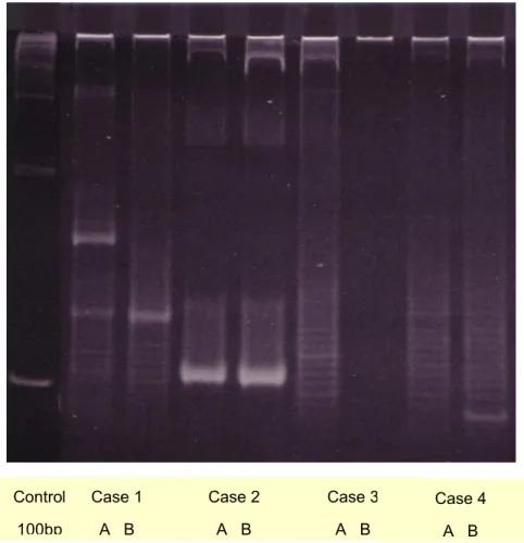
2.2 Experiment 1: Comparison of Gene Expression Profiling and
Genomic Abnormalities Using Paired Fresh Frozen Samples 68
2.2.1. Material & Methods 68
2.2.1.1. Patients & Sample Selection 68
2.2.1.2. Affymetrix U133 Plus 2.0 Array 69
2.2.1.3. CGH: BCCA Array 70
2.2.2. Results & Discussion 71
2.2.2.1. GEP 71
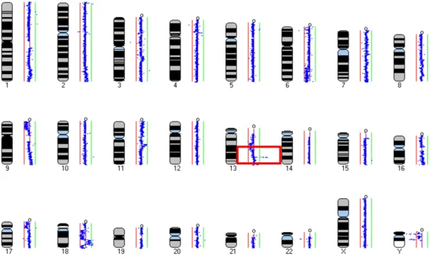
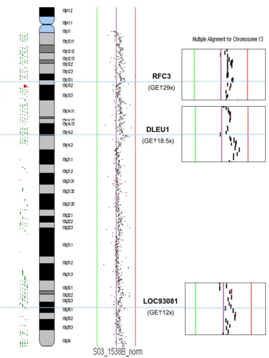
2.2.2.2. aCGH 72
2.2.2.3. Correlation GEP vs. aCGH 73
2.2.3. Conclusion 74
2.3 Experiment 2: Gene Expression Profiling of Composite Follicular and Diffuse Large B-cell Lymphoma Using Formalin Fixed Paraffin
Embedded Tissue Samples 75
2.3.1. Introduction 75
2.3.2. Material & Methods 76
2.3.2.1. Patients & Sample Selection 76
2.3.2.2. Composite FL and DLBCL 77
2.3.2.3. GEP: Agilent 44K Whole Human Genome 60mer 77
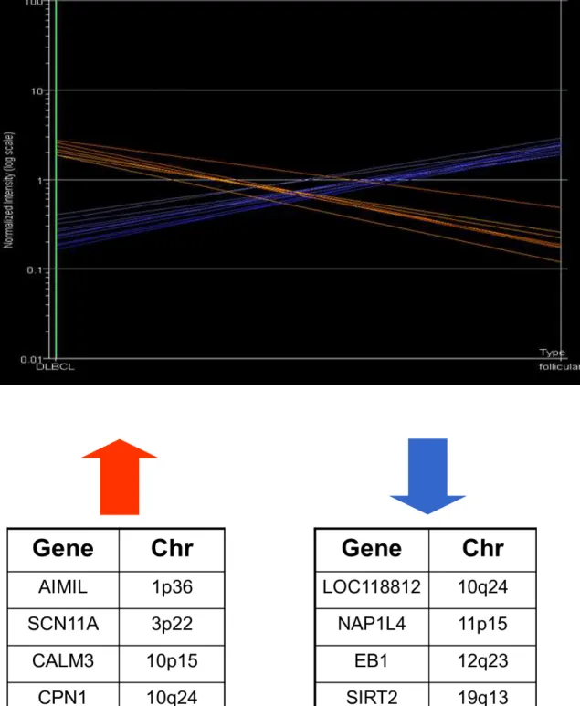
2.3.3. Results 78
2.3.4. Discussion 80
2.3.5. Conclusion 82
2.4 List of Tables 83
2.5 List of Figures 85
2.6 References 109
Section 3: New Biological Predictors of Survival and Transformation in Follicular Lymphoma: the Microenvironment 114
3.1 Introduction 114
3.2 Aim 115
3.3 Material & Methods 115
3.3.1. Patients & Sample Selection 115
3.3.2. Tissue Microarray & Immunohistochemistry 116
3.3.2.1. Malignant B Cells 117
3.3.2.2. Follicular Dendritic Cell (FDC) 118
3.3.2.3. Macrophages 118
3.3.2.4. T Cells 119
3.3.2.5. Microvessel Density & Follicle Size 120 3.3.3. Data Analysis Statistics and Survival Analysis 120
3.4 Results 121
3.4.1. Clinical Characteristics & Outcome 121
3.4.2. Pathology Variables 121
3.4.2.1. Malignant B Cells Markers 122
3.4.2.2. FDC Marker 122
3.4.2.3. Macrophages Markers 122
3.4.2.4. T Cell Markers 123
3.4.2.5. Microvessel Density 125
3.4.3. Multivariate Analysis 125
3.5 Discussion 126
3.5.1. Neoplastic FL B Cells 127
3.6 Conclusions 134
3.7 List of Tables 135
3.8 List of Figures 140
3.9 References 164
Section 4: Microenvironment in Follicular Lymphoma:
Monocyte & Macrophage Interactions in Patients with Follicular
Lymphoma Harboring a t(14;18): Is There a Clonal Relationship? 175
4.1 Introduction 175
4.2 Aim 177
4.3 Material & Methods 178
4.3.1. Patients & Samples Selection 178
4.3.2. FICTION Technique 178
4.3.2.1. Paraffin Samples 179
4.3.2.2. Touch Imprint Samples 179
4.3.2.3. Paraffin & Touches Antigen Retrieval 180
4.3.2.4. Immunofluorescence Staining 180
4.3.2.5. Fluorescence In Situ Hybridization 180
4.3.3. Image Capturing and Analysis 181
4.4 Results 181
4.4.1. Paraffin Samples – Pilot Study 181
4.4.2. Touch Imprints Samples 182
4.5 Discussion 183
4.6 Conclusion 187
4.7 List of Tables 188
4.8 List of Figures 190
4.9 References 205
Section 5: New Biological Predictors of Survival in DLBCL 209
5.1 Introduction 209
Section 5.1: Strong TP53 Expression is an Independent Predictor
of Outcome in de novo DLBCL Treated with either CHOP or R-CHOP 210
S5.1.3 Material & Methods 212
S5.1.3.1. Patients & Sample Selection 212
S5.1.3.2. Tumour Specimens – FFPET Samples 213
S5.1.3.3. DNA Extraction 214
S5.1.3.4. Roche’s AmpliChip p53 Assay 214
S5.1.3.5. Chip Design 215
S5.1.3.6. Data Analysis of Microarray Signals 216
S5.1.3.7. Statistical Analysis 216
S5.1.4 Results 217
S5.1.5 Discussion 218
S5.1.6 Conclusion 220
S5.1.7 List of Tables 222
S5.1.8 List of Figures 224
S5.1.9 References 237
Section 5.2: Addition of Rituximab (R) to CHOP Improves Survival in
Non-GCB Subtype of DLBCL 242
S5.2.1 Introduction 242
S5.2.2 Aim 244
S5.2.3 Materials & Methods 244
S5.2.3.1. Patients & Sample Selection 244
S5.2.3.2. Tumour specimens FFPET Samples 245
S5.2.3.3. Statistical Analysis 245
S5.2.4 Results 246
S5.2.5 Discussion 247
S5.2.6 Conclusion 249
S5.2.7 List of Tables 250
S5.2.8 List of Figures 254
S5.2.9 References 267
Section 5.3: Prognostic Significance of BCL6 Rearrangements
detected by Fluorescence in situ Hybridization in DLBCL 272
S5.3.1 Introduction 272
S5.3.3.2. FISH and IHC on TMAs 273
S5.3.3.3. Statistical Analysis 274
S5.3.4 Results 274
S5.3.5 Discussion 276
S5.3.6 Conclusion 278
S5.3.7 List of Tables 279
S5.3.8 List of Figures 281
S5.3.9 References 283
Section 5.4: MYC Aberrations are Associated with a Poor Prognosis in Diffuse Large B Cell Lymphoma Patients Treated with
R-CHOP Chemotherapy 287
S5.4.1 Introduction 287
S5.4.2 Aim 288
S5.4.3 Materials & Methods 288
S5.4.3.1. Patients & Sample Selection 288
S5.4.3.2. Tissue Microarray & Immunohistochemistry 288
S5.4.3.3. Cytogenetic Analysis 289
S5.4.3.4. Determination of DLBCL Cell of Origin Subtypes 289
S5.4.3.5. Statistical Analysis 289
S5.4.4 Results 290
S5.4.4.1. Cytogenetics, IHC and Cell of Origin 290 S5.4.4.2. Impact on survival of MYC rearrangements in R-CHOP treated
patients 291
S5.4.4.3. Risk of CNS relapse in R-CHOP treated patients with a MYC
rearrangement at diagnosis 291
S5.4.5 Discussion 292
S5.4.6 Conclusion 294
S5.4.7 List of Tables 295
S5.4.8 List of Figures 297
S5.4.9 References 300
Section 6: New Biological Predictors of Survival in Hodgkin
6.2 Aim 303
6.3 Material & Methods 304
6.3.1. Patients & Sample Selection 304
6.3.2. Gene Expression Analysis 305
6.3.3. Immunohistochemical Analysis 305
6.3.4. Data Analysis 306
6.3.4.1. Predictive Models 306
6.3.4.2. Statistical Analysis 308
6.4 Results 308
6.4.1. Gene Expression Analysis 308
6.4.2. Immunohistochemical Analysis 310
6.5. Validation Study 311
6.5.1. Material & Methods 311
6.5.2. Results 313
6.6 Discussion 315
6.7 List of Tables 318
6.8 List of Figures 329
6.9 References 344
Section 7: New Biological Predictors of Survival in Mantle
Cell Lymphoma: The Microenvironment 349
7.1 Introduction 349
7.2 Aim 349
7.3 Material & Methods 349
7.3.1. Patients & Sample Selection 349
7.3.1.1. Cohort 1 350
7.3.1.1.1. Patients & Samples Selection 350
7.3.1.1.2. Monoclonal Antibodies 350
7.3.1.1.3. Cell Preparation 351
7.3.1.1.4. Flow Cytometric Analysis 351
7.3.1.1.5. Gene Expression Analysis 351
7.3.2. Data Analysis 353
7.4 Results 353
7.4.1. Cohort 1 353
7.4.2. Cohort 2 356
7.5 Discussion 358
7.6 Conclusion 361
7.7 List of Tables 362
7.8 List of Figures 371
7.9 References 379
Section 8: New Biological Predictors of Survival in Primary
Mediastinal Large B Cell Lymphoma: The Microenvironment 385
8.1 Introduction 385
8.2 Aim 386
8.3 Material & Methods 386
8.3.1. Patient characteristics 386
8.3.2. Tissue Microarray Construction 387
8.3.3. Immunohistochemistry 387
8.3.4. Scoring 387
8.3.5. Data Analysis 387
8.4 Results 388
8.5 Discussion 288
8.6 Conclusion 391
8.7 List of Tables 292
8.8 List of Figures 395
8.9 References 403
Section 9: Conclusions 407
9.1 Summary 407
9.2 Discussion & Conclusions 408
9.4 References 419
Resumo alargado 429
List of abbreviations
Abbreviation Definition
BCCA – CLC British Columbia Cancer Agency Centre for Lymphoid Cancer
CTAG Centre for Translational and Applied Genomics CHLC Centro Hospitalar Lisboa Central
IPO Instituto Português de Oncologia
LLMPP Leukemia/Lymphoma Molecular Profiling Project
NHL Non-Hodgkin lymphomas
IPI International Prognostic Index
FLIPI Follicular Lymphoma Prognostic Index
LDH Lactate dehydrogenase
IPS International Prognostic Score
BCR B cell Receptor
TCR T cell receptor
SHM Somatic hypermutation CSR Class-Switch Recombination GEP Gene Expression Profiling
FFPET Formalin Fixed Paraffin Embedded Tissue
TMA Tissue microarray
IHC Immunohistochemistry
FISH Fluorescence In-Situ Hybridization
FICTION Fluorescence-Immunophenotyping and Interphase Cytogenetics as a Tool for Investigation Of Neoplasms CGH Comparative Genomic Hybridization
aCGH Array Comparative Genomic Hybridization BAC Bacterial Artificial Chromosome
bp Base pairs
CCD Charge Coupled device
cDNA complimentary DNA
Kb Kilobase pairs
FDC Follicular Dendritic Cells
TAM Tumour Associated Macrophages LAM Lymphoma Associated Macrophages MVD Microvessel Density
Tregs Regulatory T cells
FOXP3 Forkhead box P3
EBV Epstein-Barr virus
GCB Germinal Centre B cell type ABC Activated B cell type
R Rituximab
PCR Polymerase Chain Reaction RNA Ribonucleic acid
mRNA messenger RNA
SMRT Sub-Megabase Resolution Tiling-set SNP Single Nucleotide Polymorphism
SMLR Sparse Multinomial Logistic Regression NCBI National Center for Biotechnology Information
TVD90 Tumour to Vessel Distance that encompassed 90% of the
tumour
Acknowledgements
All the work reported in this thesis is the result of the superb academic
“microenvironment” called the British Columbia Cancer Agency Centre for
Lymphoid Cancer- CLC in which I am honored to have worked for the past 10 years being involved in many different and challenging projects. It is a wonderful group of people with diverse scientific and medical backgrounds who gathered over the years around two amazing senior colleagues and mentors whose immense work ethic and dedication for the past 25 years has built an outstanding and world class centre for the study of lymphoid cancers. Special thanks to these two brilliant mentors, my thesis supervisor Dr. Randy D. Gascoyne and Dr. Joseph M. Connors. Most of the projects I am including in this thesis are ideas given birth during the vibrant and informative discussions that took place regularly within the CLC at BCCA.
In addition I greatly appreciate the cooperation from all physicians of the British Columbia Cancer Agency for allowing us to include their patients and all the pathologists throughout British Columbia for their support of the provincial lymphoma pathology program. I would like to thank all of the people working at the Centre for Translational and Applied Genomics (CTAG) laboratory, a superb scientific environment whose birth and early days I was honored to be an integral part of.
Finally, I would like to thank the FCT for their support during my first four years and all my pathologist colleagues at Centro Hospitalar Lisboa Central (CHLC) and Instituto Português de Oncologia (IPO), Lisboa for their teaching and collaboration during these years as well as my hematology colleagues at CHCL for supporting the current first steps in translation research at CHLC. Special acknowledgement goes to collaborators at the FCML, Pathology Department headed by Profª Drª Ana Felix and CHLCs Hematology Department, specially its director Drª Aida Botelho de Sousa.
the III Community Support Framework, subsidized by the European Social Fund and funds national MCTES, reference: SFRH / BD / 13230 / 2003.
Several of the projects presented in this thesis were awarded in different meetings, as follows:
1. Research Award Program 2004 – Berlex Canada
2. BCCA 2004 Award in the area of translation I (Innovation) 3. Research Award Program 2006 – Berlex Canada
4. Research Award Program 2007 - Bayer Canada
Co-Authorship Statement
Chapters 2, 3, 5, 6, 7 and 8 were co-authored as manuscripts for publication. The following author lists apply to each chapter. It is my honor to name and thank all the colleagues whose work is included in this manuscript.
Section 1
Pugh TJ, Delaney AD, Farnoud N, Flibotte S, Griffith M, Li HI, Qian H, Farinha P, Gascoyne RD, Marra MA. Impact of whole genome amplification on analysis of copy number variants. Nucleic Acids Res. 2008 Aug; 36(13):e80. Epub 2008 Jun 17. PMID:18559357
Relander T, Johnson NA, Farinha P, Connors JM, Sehn LH, Gascoyne RD. Prognostic Factors in Follicular Lymphoma. J Clin Oncol. 2010 Jun 10;28(17):2902-13. Epub 2010 Apr 12. Review. PMID: 20385990
Section 2
Special thanks to Gitte Gobel, medical student from Arhus Medical School and in particular Tarun Nayar from Genome Science Center, Vancouver (http://www.bcgsc.bc.ca/ ).
Obel G, Farinha P, Lam WL, deLeeuw R, Young K, Kjeldsen E, Hamilton-Dutoit
S, d’Amore F, Chan WC, Gascoyne R. Analysis of genomic imbalances and
gene-expression changes in transformed follicular lymphoma (FL). 9th International Lugano Lymphoma Meeting. Ann Oncol 16230 230 2005
Farinha P, Lee M, Al-Tourah AJ, Connors JM, Gascoyne RD. Composite follicular (FL) and diffuse large B cell lymphoma (DLBCL): A gene expression profiling (GEP) study, 10th International Lugano Lymphoma Meeting. Ann Oncol 19106-106 2008
Section 3
(LAM) content is an independent predictor of survival in follicular lymphoma (FL). Blood 2005; 106:2169-2174. PMID:15933054
Korenberg MJ, Farinha P, Gascoyne RD. Predicting survival in follicular lymphoma using tissue microarrays. Methods Mol Biol. 2007;377:255-68. PMID: 17634622
Farinha P, Roncador G, Al-Tourah A, Sehn L, Connors JM and Gascoyne RD. Combined FOXP3+ & PD1+ T Cells Density and Architectural Patterns Predict Overall Survival (OS) and Risk of Transformation (RT) in Uniformly Treated Patients with Follicular Lymphoma (FL). Blood 112 (11):972-973 2008
Farinha P , Al-Tourah, Jill K, Connors JM, Shumansky K, Spinelli JJ, Gascoyne RD. The Architectural Pattern of FOXP3
+
T Cells Is an Independent Predictor of Survival in Patients with Follicular Lymphoma (FL). Blood. 2010 Jan 14; 115(2):289-95. Epub 2009 Nov 9. PMID:19901260
Farinha P, Kyle A, Minchinton A, Connors JM, Karsan A, Gascoyne RD. Vascularization predicts overall survival and risk of transformation in follicular lymphoma. Haematologica. 2010 Dec;95 (12):2157-60. Epub 2010 Aug 16. PMID:20713461
Section 5
Hans CP, Weisenburger DD, Greiner TC, Gascoyne RD, Delabie J, Ott G, Muller-Hermelink HK, Campo E, Braziel R, Jaffe ES, Pan Z, Farinha P, Smith LM, Falini B, Banham AH, Rosenwald A, Staudt LM, Connors JM, Armitage JO, Chan WC. Confirmation of the molecular classification of diffuse large B cell lymphoma by immunohistochemistry using a tissue microarray. Blood 2004;103: 275-282. PMID:14504078
Farinha P, Bebb G, Siebert R, Horsman D, Connors JM, Gascoyne RD. Cell-of-Origin Is an Important Biomarker in Diffuse Large B-Cell Lymphoma (DLBCL). US&CAP Meeting, February 26-March 4, 2005.Mod Pathol 18 (Suppl 1); 2005: A1065
Farinha P, Sehn LH, Skinnider BF, Wu L, Patten N, Truong S, Connors JM, Gascoyne RD. Strong p53 Expression is an Independent Predictor of Outcome in de novo Diffuse Large B cell Lymphoma (DLBCL) treated with either CHOP or CHOP-R. Blood 108 (11):812 2006
Farinha P, Sehn LH, Chhanabbai M, Skinnider BF, Connors JM, Gascoyne RD. Addition of Rituximab to CHOP Improves Survival in the Non-GCB subtype of Diffuse Large B cell Lymphoma (DLBCL). Blood 108 (11):816 2006
Lam LT, Wright G, Davis RE, Lenz G, Farinha P, Dang L, Chan J, Rosenwald A, Gascoyne RD, Staudt LM. Cooperative Signalling Through the STAT3 and NF-kB Pathways in Subtypes of Diffuse Large B Cell Lymphoma. Blood. 2008 Apr 1;111(7):3701-13. PMID:18160665
Cerchietti LC, Polo JM, Da Silva GF, Farinha P, Shaknovich R, Gascoyne RD, Dowdy SF, Melnick A. Sequential Transcription Factor Targeting for Diffuse Large B-Cell Lymphomas. Cancer Res. 2008 May 1;68(9):3361-9. PMID: 18451163
Ci W, Polo JM, Cerchietti L, Shaknovich R, Wang L, Yang SN, Ye K, Farinha P, Horsman D, Gascoyne RD, Elemento O, Melnick A. The BCL6 transcriptional program features repression of multiple oncogenes in primary B-cells and is deregulated in DLBCL. Blood 2009; 113: 5536-5548. PMID:19307668
Shustik J, Han G, Farinha P, Johnson N, Connors JM, Sehn L, Gascoyne RD and Steidl C. Prognostic significance of BCL6 rearrangements detected by fluorescence in situ hybridization in diffuse large B cell lymphoma. Haematologica. 2010 Jan; 95(1):96-101. Epub 2009 Oct 1. PMID: 19797725
Section 6
Steidl C, Lee T, Shah SP, Farinha P, Han G, Nayar T, Delaney A, Jones SJ, Iqbal J, Weisenburger DD, Bast MA, Rosenwald A, Muller-Hermelink HK, Rimsza LM, Campo E, Delabie J, Braziel RM, Cook JR, Tubbs RR, Jaffe ES, Lenz G, Connors JM, Staudt LM, Chan WC, Gascoyne RD. Tumor-associated macrophages and survival in classic Hodgkin's lymphoma.N Engl J Med. 2010 Mar 11;362(10):875-85. PMID:20220182
Farinha P, Rodrigues S, Fernandes T, Monteiro A, Lopes da Silva R, Salvador J, Gerivaz R, Costa F, Ferreira G, Lopes C, Lacerda T, Ferreira J, Costa Iand Botelho de Sousa A. Lymphoma Associated Macrophages Predict Survival in Uniformly Treated Patients with Classical Hodgkin Lymphoma. US&CAP Meeting, March, 2011. Mod Pathol 24 (Suppl 1); 2011: 296A
Steidl C, Telenius A, Shah SP, Farinha P, Barclay L, Boyle M, Connors JM, Horsman DE, Gascoyne RD. Genome-wide copy number analysis of Hodgkin Reed-Sternberg cells identifies recurrent imbalances with correlations to treatment outcome. Blood. 2010 Jul 22;116(3):418-27. Epub 2010 Mar 25. PMID:20339089
Steidl C, Farinha P, Gascoyne RD. Macrophages predict treatment outcome in Hodgkin's lymphoma. Haematologica. 2011 Feb;96(2):186-9. PMID: 21282720
Section 7
Opat S, Farinha P, Boyle M, Johnson N, O`Leary H, Cook J, Tubbs, Woods R, Connors J, Gascoyne R. The percentage of cytotoxic T cells in Mantle Cell Lymphoma (MCL) biopsies predicts response to Rituximab. Blood 114 (22):1142-1143 2009
Section 8
Rimsza LM, Farinha P, Fuchs DA, Masoudi H, Connors JM, Gascoyne RD.HLA-DR Protein Status Predicts Survival in Patients with Diffuse Large B Cell Lymphoma (DLBCL) Treated on the MACOP-B Chemotherapy Regimen. Leuk Lymphoma. 2007 Mar;48(3):542-6. PMID:17454596
Farinha P, Steidl C, Rimsza L, Savage K, Connors J, Gascoyne R. HLA-DR protein expression correlates with non-neoplastic T-cell infiltration and predicts survival in patients with Primary Mediastinal Large B Cell Lymphoma (PMBCL) treated with CHOP chemotherapy. Blood 114 (22):61-61 2009
Dedication
To Susana my main mentor and co-author in life…
… and Maria do Rosário, Francisco, João, Miguel, Maria Leonor, Maria Teresa
Section 1: Introduction
1.1 LYMPHOMA
1.1.1. Definition
Lymphoid neoplasms are clonal tumours of mature and immature B, T or natural killer (NK) cells at various stages of differentiation.
Lymphomas represent approximately 4% of new cancers per year worldwide, are more common in the Western world and contrary to most other common neoplasias, have been increasing in incidence worldwide (1). Mature B cell non-Hodgkin lymphomas (NHL) comprise over 90% of these (2).
For the majority of lymphomas, tumour cells recapitulate the normal stages of differentiation so they can be classified according to their normal counterpart. However, some neoplasms do not have as yet a well-defined normal counterpart and within each lymphoma type there is morphologic, phenotypic and molecular heterogeneity.
B cell development takes place in distinct steps characterized by the structure of the B cell Receptor (BCR). Two pairs of identical heavy-chain and light-chain immunoglobulin (IG) peptides compose the BCR. After cross-linking, the BCR and associated membrane components transmit cytoplasm signals through several tyrosine kinases. Proliferation and differentiation of B cells depends on the recognition of antigens and on the activation of other modulator B cell surface receptors. Early B cell development occurs in the bone marrow where precursor cells rearrange IG heavy and light chain genes resulting in the expression of a functional surface antigen receptor on naïve B cells as they exit the bone marrow. These cells can join the immune response upon antigen binding to the BCR resulting in lymphoid follicle formation. They undergo clonal expansion in germinal centers (GC), where T cells and other accessory lymphoid cells enable naïve B cells to gain antigen specificity by modification of
T cells arise from bone marrow precursors and mature in the thymus gland. Mature T cells are characterized by a rearranged antigen specific T cell receptor
(TCR), either αβ or γδ, and associated with a fully expressed CD3 complex. They have a wide functional spectrum of both innate and adaptive immune responses. Mature T cells are functionally heterogeneous and quite plastic with differences difficult to characterize in vivo and in vitro, that are highly dependent on the particular techniques used to study them. These include cells of the
innate immune system (γδ T cells), naïve, effector, regulatory, cytotoxic and
memory T cells.
NK cells participate mostly in innate immune response, contain cytoplasmic cytotoxic granule proteins and do not have a complete TCR or a CD3 membrane complex.
Recently, much has been learned from new techniques of flow cell sorting and cell phenotyping as well as gene expression profiling (GEP). New techniques such as genome-wide platforms to study GEP, genomic copy number alterations (array comparative genomic hybridization or aCGH), genome methylation or genome sequencing had increased our understanding of lymphoid neoplasms.
1.1.2. Classification
The observation of BCR and TCR particular structure, the expression patterns of differentiation markers and the specific histological structures where it takes place was used to determine the origin of the various lymphomas. The rational used in the current classification of lymphoid neoplasms reflects the “frozen” state of the malignant cells in a particular differentiation stage, reflecting their cell of origin. For B cell tumours, most subtypes are derived from GC or immediate post-GC stage of differentiation. Classical Hodgkin lymphoma, considered historically as a separate disease, is in almost all cases a B cell lymphoma lacking a functional immunoglobulin.
correct diagnosis. Thus, the current standard classification (2) uses all available information to define lymphoma types, including morphology, immunophenotype, genetic features and clinical features. The relative importance of each of the features varies among diseases, but there is no one gold standard for defining any lymphoma type.
Recent techniques such GEP have further characterized unrecognized differences within what were perceived to be specific subtypes. Moreover, while cell lineage defines most lymphoid malignancies, lineage plasticity within the hematopoietic system is increasingly recognized.
Finally, we have appreciated the common presence of small clonal populations of atypical lymphoid cells in otherwise asymptomatic patients that may or may not represent early involvement by a lymphoid neoplasm.
1.1.3. Therapy
Lymphoma can be treated in many ways from watch and wait to single or multiple agent chemotherapy, radiation, surgery, monoclonal antibodies, stem cell transplantation or a combination of these. Therapeutic decisions are mostly dependent on staging and clinical factors, but a key step in this process is dependent on the histopathological subtype of lymphoma. Thus, the most important step is determining whether a given patient will be offered treatment given with curative intent versus treatment given to relieve symptoms or to offer palliation. For most entities there are multiple treatment options for a single disease (4).
1.1.4. Prognosis
differently in the tumoural cells, thus biomarkers need to be re-evaluated specifically and uniformly for each new therapeutic modality.
1.2 FOLLICULAR LYMPHOMA (FL)
1.2.1. Introduction
FL is the second most common form of indolent lymphoma in North America, comprising about 22% of all new cases of non-Hodgkin lymphomas (NHL), but 70% of the indolent lymphomas (5, 6). FL is a clinically heterogeneous disease with some patients progressing or transforming early and 15% dying within 2 years from diagnosis, whereas others remain alive without needing treatment after more than a decade. The median disease survival from diagnosis is now 8-12 years. FL may transform over time to a more aggressive lymphoma, mostly resembling DLBCL. This is a dominant clinical event that is frequently associated with a rapid clinical course, refractoriness to treatment and short survival. However, its incidence varies dramatically, with histological transformation being reported to affect between 10-70% of FL patients (7). Prognosis and therapy of FL are currently based on clinical criteria, the International Prognostic Index (IPI) and more recently the Follicular Lymphoma Prognostic Index (FLIPI) 1 & 2 (8-10). The IPI, originally developed for aggressive lymphomas, identifies four different risk groups based on age, tumour stage, serum lactate dehydrogenase (LDH) concentration, performance status and number of extranodal sites of disease. The FLIPI published in 2004, is made up of five adverse prognostic factors including: age (>60 years vs. ≤ 60 years), stage (III-IV vs. I-II), anaemia (haemoglobin < 120 g/l vs. ≥120 g/l),
number of involved nodal areas (>4 vs. ≤4 areas) and serum LDH (elevated vs.
normal).
immunotherapy. The impact of host immune-genetics, microenvironment and therapy in FL are still not well elucidated (11).
Because there is no clear standard of care for FL treatment, a multitude of treatment options are available, ranging from simple observation in asymptomatic disease, single or multi-agent chemotherapy, monoclonal antibody therapy, with or without radioimmunoconjugates, to haematopoietic stem cell transplantation. There is no consensus approach for initial therapy. FL shows high sensitivity to initial therapy but becomes increasingly resistant following each successive treatment. The patients will invariably experience shorter remission durations and eventual relapses over time, in some cases leading to transformation. An association between increased FLIPI score and Risk of Transformation (RT) has been reported but not validated in other studies (12).
Despite the vast amount of biological data published on transformation very few biological markers have been shown to impact FL prognosis, most representing isolated reports and therefore none are used in routine clinical practice for clinical decision making. This is due in part to the low frequency of most of these markers as well as to the lack of data from cohorts with uniform treatment.
In BC, the BCCA lymphoma database represents a population-based electronic database that includes comprehensive information on diagnosis, treatment and outcome for virtually all patients with lymphoma in the province. Centralized data on diagnosis and treatment of FL patients is a powerful tool to assess incidence and outcome associated with this event (7).
1.2.2. Germinal Centre
centroblasts; thereby establishing secondary lymphoid follicles characterized by germinal centres and a surrounding mantle zone. The proliferating centroblasts activate the process of somatic hypermutation that generates mutations at a high rate in some genes including V-region genes of the immunoglobulin molecule resulting in the generation of antibody diversity (3). The different centroblasts are selected by the affinity of their BCR to the antigen that induced the formation of the follicle. Only those cells that have acquired high affinity mutations escape apoptosis. This process of differentiation terminates with a resting centrocyte having high affinity antibody on its surface. Some of these cells will then exit the follicle to become either short-lived plasma cells or long-lived memory B cells. The process requires the interaction of the B cells with the follicular T follicular helper cells and follicular dendritic cells (FDC) within the germinal centre. These cells provide the survival signals that rescue B cells from apoptosis. Furthermore, the FDC meshwork embodies the scaffolding structure of the follicle. Thus, FL is characterized by the development of neoplastic follicles, often morphological similar to reactive follicles, harbouring benign FDC meshwork, small blood vessels, macrophages and a variable percentage of non-neoplastic follicular T cells and benign B cells.
1.2.3. t(14,18) & Genomic Mutations
enter the circulation, home to the lymph node and seed the follicles. Here they undergo several rounds of replication within an environment where SHM and CSR are physiological processes that require double-strand DNA breaks. Eventually, a second and subsequent additional genetic hit occurs, some of which lead to the development of clinically evident FL. Like benign germinal centers, malignant follicles are further targeted by hypermutational events favoring accumulation of genomic abnormalities. One recently described and highly recurrent genetic event is inactivating mutations of MLL2, found in nearly 90% of FL biopsies (16). It remains possible that MLL2 mutations and the subsequent changes in gene expression that follow loss of this H3K4 activating chromatin mark may represent the illusive 2nd hit that allows the fully malignant phenotype of FL to emerge.
Numerous structural chromosomal aberrations have been described, including gains, deletions, balanced and unbalanced translocations, all of which define the clonal evolution and disease progression of FL. Some of the most common events follow different pathways, which show clinical impact in the progression of FL (13). However, the majority of the breaks including those associated with transformation are thought to be stochastic, providing the lymphoma cells with a growth advantage and thus accelerate the clinical behaviour.
The dramatic advances in biology, specially the emergence of new technologies to assess gene expression and genomic abnormalities such as GEP array platforms, high-resolution comparative genomic hybridization (CGH) and multicolour fluorescence in-situ hybridization (M-FISH) have increased the number of genomic alterations, such as gene signatures and copy number deletions and amplifications characteristically found in FL (17).
1.2.4. Crosstalk Between Malignant & Non-malignant cells
survival and trophic signals from FDCs/macrophages and T cells reach the neoplastic B cells which are highly dependent on these, as suggested by the difficulty of establishing FL cell-lines in vitro (18).
The importance of the microenvironment and non-malignant cells within human tumours is not a recent finding. Rudolf Virchow first suggested it more than one hundred years ago (19, 20). Similarly the concept of tumoural immunosurveilance proposed by Paul Ehrlich represents a pivotal observation (21). Since then, tumoral immunity has been shown to evolve over time, characterized by crosstalk between tumour cells helping to shape their microenvironment and in turn being shaped by these surrounding non-neoplastic immune cells. Along with dampening the anti-tumoral response there are several different mechanisms by which the microenvironment promotes tumoral growth, so called cancer immunoediting, as the tumour sculpts its own immunity in favour of growth and proliferation signals (22).
Tumour associated macrophages (TAM) have been shown to promote not only neoplastic growth but also angiogenesis (23). Angiogenesis plays a vital role in growth and progression of both solid and hematological tumours. The "angiogenic switch" results from interactions of vessels with cancer cells as well as non-neoplastic cells in the microenvironment, including macrophages themselves (24). The abundance of macrophages, microvessel density (MVD) and recently of a subset of T cells, regulatory T cells (Tregs) associated with tumours, has been reported to correlate with poor prognosis in solid tumours.
These cells are recruited and “educated” by tumours using a range of growth
signature, referred to as immune response-2, revealed a gene expression pattern most reminiscent of macrophages and/or FDCs and was associated with an inferior clinical outcome (25). However, the correlation of the gene signatures and the cellular components of the microenvironment and prognosis were not clear. These provocative data raised the question of non-neoplastic immune cells could be reproducibly identified and shown to be associated with survival and risk of transformation in FL. Could these cells be used to risk-stratify patients with FL and ultimately become the targets of novel future therapies?
The precise biological function of these non-malignant cells in FL remains poorly understood. Mouse models have shown great phenotypic heterogeneity amongst morphologically similar macrophages, but much less are known about human macrophages. The plasticity of macrophages is evident in the diverse range of functions of these cells, including inflammatory responses, immune reactions, tissue remodelling and morphogenesis. Macrophages can be
functionally subdivided into two main classes, including “killer” or classically activated M1 macrophages or “healer”, alternatively activated M2 macrophages. These two types of macrophages differ in their receptor expression, cytokine and chemokine profiles as well in their metabolic pathways (27, 28). In vivo and within the tumoural microenvironment, macrophages of both kinds coexist with some cells showing overlapping or hybrid features. However, if markers representing the disparate ends of the macrophage spectrum are defined, these could be used as surrogate markers of anti or protumoral macrophages within routine FL biopsies.
also express cytotoxic T lymphocyte-associated antigen 4 (CTLA4) and glucocorticoid-induced TNFR-related protein (GITR), which are associated with suppressive features of these cells (33). However, the critical regulator of the development and function of Treg cells and the most lineage-defining marker is forkhead box P3 (FOXP3) (33-35). Although the mechanisms of suppression by Tregs are not completely understood, they seem to require direct cell-to-cell contact between Tregs and target T cells (36). Recently, in vitro studies using FL samples demonstrated that Tregs are present in significantly increased numbers when compared with normal tissues and these cells suppress other intra-tumoral CD4+ and CD8+ T cell by inhibiting proliferation and decreasing perforin and granzyme B production (37, 38). Tregs are not only attracted to the lymphoma microenvironment by their CCR4 receptor ligand CCL22, produced by lymphoma cells, but there is also local up-regulation of FOXP3+ cells by malignant cells in part by CD27-CD70 signaling (39). These data suggests a suppressive and pro-lymphoma function of Tregs.
Other T cells resident in the follicle have been recently described and their biology studied in great depth, including CD4+ follicular T helper cells. These cells are vital to the normal physiology of the germinal center and usually express CD57 and/or Programmed Cell Death 1 (PD1). In lymphoma, these cells are the benign counterpart of (at least) angioimmunoblastic lymphoma (40) and perhaps HTLV-1 induced lymphomas (41). In FL, the role of benign follicular T helper cells is poorly understood.
FDCs are the supportive scaffolding of follicles and are responsible for helping to foster B cell maturation within the germinal center. In FL it has been shown that progression of disease is associated with less mature phenotype of these cells (42).
various cell types within the reticulo-endothelial system and even suggesting possible targets for anticancer treatments (45).
As indicated above, FL is characterized by the t(14;18)(q32;q21). In vitro
studies have shown that precursor cells of the B cell lineage can differentiate into other cell types including tissue-based macrophages (46). Interestingly, in FL, a subpopulation of endothelial cells shows the characteristic t(14;18) translocation of FL (47), although this finding remains controversial. This finding may indicate (among other hypotheses) plasticity of a neoplastic precursor cell in the marrow that maintains the ability to differentiate into different mature cell types. Eventually, one can hypothesis that the pro-malignant t(14:18)+ clone could seed follicles not only with pre-malignant B cells but also with other types
of “non-malignant” accessory cells that could provide the malignant cells with pro-survival signals.
Little is known about a possible role of the non-neoplastic cells in the microenvironment impacting the risk of FL transformation. Transformation is typically associated with the effacement of the follicular architecture, accompanied by loss and immaturity of the FDCs and a decrease in the admixed T cell content. These changes correlate with the accumulation of additional genetic abnormalities. However, in a GEP study cases that transformed more rapidly were characterized by an abundance of activated T cells (26).
1.2.5. Transformation
Several genetic alterations have been associated with histologic transformation of FL, including aberrations involving dominant oncogenes; loss of tumour suppressor genes and many as yet poorly characterized cytogenetic events. Well-known examples include rearrangements of the MYC gene, mutations or loss of the TP53 or TP16 genes, and rare somatic alterations of the BCL6 and
BCL2 genes (48-55). However, these alterations are observed in limited subsets of transformed FLs, suggesting that the process of histological transformation can occur by multiple different mechanisms (56).
studies have recently shown a heterogeneous pattern of genomic aberrations acquired upon transformation. These include gain/amplification of 1q21-q24, 2p16 (REL/BCL11A), 3q27-q29 (including the BCL6 locus), 7q11.2-q22.1, 12pter-q12, 18q21 (including the BCL2 locus) and Xq, and deletion of 6q22-q24, 13q14-q21 and 17p13 (TP53 locus) that had been previously implicated in the FL & DLBCL pathogenesis (57). Additional changes include novel imbalances such as over-representation of 4p12-pter, 5p12-p15, 6p12.3-p21, 9p23, 9q13-q31, 16q, 17q21, and loss of the 1p36.3, 4p21-q23, 5q21-q23, 9q31-qter, 11q24-q25, and 15q23. None of these recently described changes were specific for transformation. More importantly, the specific genes involved at these sites are unknown (57).
These areas of amplification and deletion likely represent strong candidate loci to harbour genes important for cancer development and progression. Moreover, alterations of these loci likely influence gene expression secondary to copy number alterations. Knowledge of the key genes and the resultant aberrant expression patterns may also represent potential new targets for future therapies.
CDNK2A loss. No characteristic features were recognized in the other subset of cases in which proliferation was not a dominant feature (56).
Despite the small number of studies, these current data suggest that FL transformation to DLBCL occurs by diverse genetic alterations. No single genetic event is responsible for all cases. The most common changes appear to affect different aspects of cell physiology (apoptosis, cell cycle control and specially proliferation) and are not specific for transformation, being present in non-transformed FL as well as in other lymphoma subtypes. Yet, early steps of the transformation event have proven difficult to study as has the role of FL microenvironment in transformation.
1.2.6. Prognostic Markers in FL
Many biomarkers associated with outcome have been proposed over the years, as is shown in Figure 1.1 (Prognostic markers in FL). For complete details see review (11). Yet, most studies are single reports, with very few validation studies reported and more importantly most studies were done retrospectively in cohorts of patients treated with inconsistent treatment protocols, the vast majority before the introduction of biological agents such as rituximab. Consensus on which biomarkers to use in clinical practice and how they might impact treatment decisions are still lacking. Currently, clinical characteristics included in robust indices such as FLIPI or FLIPI-2 (9, 10) are still the basis for clinical decision making. Inclusion of candidate biomarkers in prospective randomized clinical trials is mandatory to validate their future use and possible contribution to a combined clinical & biological index.
importance that before comparisons are done of different clinical cohorts of FL that attention be paid to those reports that included uniform treatment. As therapy itself is an important prognostic variable, studies that include patients with vastly different therapy cannot easily be interpreted. As the treatments used are differentially visited upon both the malignant B cells and the immune cells in the microenvironment, the interpretation of prognostic biomarkers and those related to risk of transformation require that the treatment variable be held constant (11).
1.3 OTHER LYMPHOMAS
1.3.1. Diffuse Large B Cell Lymphoma
DLBCLs are a heterogeneous group of tumours consisting of large, transformed B cells, accounting for more than 30% of adult lymphomas in the western world. DLBCL occurs at any age, more commonly in the 6th & 7th decades of life. The disease typically presents as a nodal or extranodal mass with rapid tumour growth associated with systemic symptoms. Approximately 50 to 60 percent of patients will present with advanced stage. DLBCL includes an increasingly complex spectrum of lymphoid neoplasms with different morphologic, phenotypic, genotypic and clinical characteristics (2). Immunodeficiency is a risk factor for DLBCL, and about 5-10% of DLBCLs are EBV+, depending on the geographic population studied.
This disease is potentially curable with combination chemotherapy, such as cyclophosphamide, doxorubicin, vincristine and prednisone (CHOP). Importantly, the addition of immunotherapy using an antibody against CD20, rituximab (R) to multi-agent chemotherapy has significantly improved outcome (59, 60). High-dose chemotherapy with autologous stem cell transplantation is effective in relapsed or refractory DLBCL. Yet, despite the improvements, 30 to 40% of the patients will not be cured of their DLBCL.
histologic transformation of antecedent indolent lymphomas and are likely to be biological distinct from DLBCL cases that arise de novo.
1.3.2. Hodgkin Lymphoma
Hodgkin lymphoma (HL) was the first recognized lymphoma entity originally described in 1832 by Thomas Hodgkin (61-63). Called Hodgkin disease for more 150 years due to its unknown etiology, only in 1993 molecular studies on the microdissected pathognomonic Hodgkin and Reed-Sternberg (HRS) cells showed this to be a B cell tumour (64). The malignant HRS cells usually account for only 1% of cells in tumour tissue, whereas the majority of the cells in the biopsy represent non-neoplastic cells of the immune system. HL accounts for approximately 30% of all lymphomas with an incidence in the western world of 3 cases per 100.000 people per year. There are two distinct types of HL that show biological and clinical differences; classical HL (CHL) representing 95% of HL cases and nodular lymphocyte-predominant Hodgkin´s lymphoma accounting for the remaining 5%. Within classical HL four subtypes are distinguished including nodular sclerosis, mixed cellularity, lymphocyte-rich and lymphocyte-depleted with some differences in anatomic sites of involvement, clinical features, growth pattern, presence of background fibrosis and composition of the cellular infiltrate and frequency of EBV infection. In common to all CHL subtypes is the HRS immunophenotype, characterised by strong expression of CD30 and CD15 and no or weak expression of CD20, PAX5 and CD45. Nodular sclerosis accounts for 60-85% of HL (2).
Treatment is based on clinical, radiological imaging and pathological staging. A modified Ann Arbor staging system is used as follows: limited disease (stages I and IIA without constitutional symptoms) or advanced disease (stages IB and
IIB with bulky disease [largest deposit, ≥10 cm in diameter] and stages III and
IV either A or B [with or without constitutional symptoms]) (65, 66).
for patients with Hodgkin’s lymphoma are largely based on clinical variables that
distinguish those who are at high risk from those at standard risk. The International Prognostic Score (IPS) (on a scale of 0 to 7, with higher scores indicating increased risk) is the standard that is used for risk stratification of advanced stage HL, but it does not apply to limited stages, and none of the published prognostic factor systems can reliably identify patients in whom treatment is likely to fail (69). Moreover, neither the IPS nor its individual clinical components are suitable for accurately predicting the outcome of autologous hematopoietic stem-cell transplantation in patients with HL. For these reasons, reliable biomarkers for predicting long-term survival at diagnosis are needed for such patients.
In HL, unlike most other cancers, the malignant HRS cells are outnumbered by non-neoplastic cells in the microenvironment of the tumour. The frequency and distribution of these cellular components and HRS cells vary considerably among individual patients and among subtypes of HL (2). Several studies have focused on the prediction of outcomes by means of markers expressed predominantly by HRS cells or the microenvironment (70-75). However, most of these markers require validation in independent cohorts.
1.3.3. Mantle Cell Lymphoma
Mantle cell lymphoma (MCL) accounts for roughly 6% of NHL and is characterized by the presence of the t(11;14) translocation that juxtaposes the
(85, 86), TP53 gene mutations and TP53 protein overexpression (87, 88) and proliferation signature (89) as predictors of outcome.
The disease has a lower rate of complete remission, duration of response, and overall survival after conventional chemotherapy when compared with other lymphomas, with a rather short median survival of 5-7 years using aggressive, multidrug therapy, usually including cytarabine and R (90, 91). Consolidation therapy with autologous stem cell transplantation is also used. New chemotherapeutic agents and radioimmunotherapy may add further to advances in treatment.
1.3.4. Primary Mediastinal Large B Cell Lymphoma
Primary Mediastinal Large B Cell Lymphoma (PMBCL) is a distinct lymphoma entity arising in the mediastinum from putative thymic B cells (2). Biopsies of PMBCL are composed of medium to large-sized cells with abundant pale cytoplasm, ovoid nuclei and are characteristically associated with background of fine sclerosis. PMBCL accounts for 2-4% of NHL and occurs predominantly in young adults with a female predominance. It is locally aggressive, but less likely to extend beyond the anterior mediastinum. With similar therapy the outcomes are typically superior to nodal DLBCL in most series with a survival plateau after 2.5 years (92, 93). Yet, clinical criteria, such as IPI, imprecisely predict therapy response (94). PMBCL shares histological and gene expression features with both GCB subtype of DLBCL and classical Hodgkin lymphoma (95, 96).
1.4 GENOTYPE & PHENOTYPE ASSESSMENT IN CANCER
1.4.1. Gene Expression Profiling (GEP)
mathematics to generate classifiers that include an element of probability in the value of each given gene within the classifier.
Microarray GEP of hematological malignancies has been instrumental in the early stages of this field (102). Reported studies have covered several aspects of molecular biology. Most have been basic profiling studies, primary descriptive, often trying to gain new insights of the origins or etiology of tumours. This is achieved by comparing the tumour gene signature with
signatures of “normal” development counterparts (103). Likewise, divergent profiles from the nearest normal cell type may reveal insights into important pathways or mechanisms important in tumorigenesis. Other studies aim to identify the minimum set of genes required to identify a particular tumour subclassification, resulting in minimum diagnostic classifiers. Specific profiles can also be correlated with known clinical outcome of patients thereby creating prognostic classifiers.
of lack of comparability is still present as evidenced by the lack of independent validation of many studies. The high cost of performing studies with enough samples to achieve statistical power, which usually requires series of hundreds, makes most of these experiments difficult to repeat or validate.
1.4.2. Genome-wide Scanning for Copy Number Alterations - Bacterial Artificial Chromosome (BAC) Array Comparative Genomic Hybridization (aCGH)
Simultaneous fluorescence immunophenotyping and fluorescence in situ
hybridization (FISH), so called, fluorescence-immunophenotyping and interphase cytogenetics as a tool for investigation of neoplasms (FICTION) provides the advantage of both techniques and allows a simultaneous analysis of the phenotype and genotype of tumour cells at the single-cell level (112, 113). Using specific cell lineage and differentiation associated monoclonal antibodies for immunophenotyping as well as DNA probes specific for known chromosome alterations by FISH, this combined technique makes it possible to correlate particular gene aberrations directly with the differentiation capacity of tumour cells. This is especially important in cases where the cells of interest may be in the minority and morphological features are an important and defining criterion of the disease (113, 114). In formalin fixed paraffin embedded tissue (FFPET) sections, several early reports described difficulties in obtaining an efficient hybridization signal while retaining immunofluorescent staining (115). However, these problems can be overcome, and recent studies have shown simultaneous application of FISH and immunofluorescence on FFPET sections, including bone marrow samples (116).
1.4.4. Tissue Quality & Fixatives
Until recently, both GEP and aCGH studies have been done using frozen material because quality of RNA influences the reliability of the output data. Snap fresh frozen (FF) tissue from diagnostic biopsies is the preferred source of RNA for GE studies in oncology. Yet, often these biopsies are limited in size and provide barely enough for morphological procedures done using standard FFPET techniques. It is not uncommon that biopsies may be partially involved by disease and thus frozen samples may not be representative. Often morphologic and cytological features in FF sections are not helpful in distinguish different types of cells. Finally, this is not a routine procedure in many centres, thus limiting sample size and the number of successful cases with paired specimens to study lymphoma histologic transformation.
emergence of a different subclone from a precursor “FL stem cell”, which hampers interpretation of these data.
These are several different reasons why FFPET represent excellent materials to study lymphoma biomarkers, primarily because it is widely available as a routine source of biopsy material for more than 100 years. Biopsies kept in formalin can be retrospectively retrieved from most Anatomic Pathology Department archives; they are mandatory in the diagnosis of transformation in most cases and usually they are the most abundant and representative tissue of the tumour.
However, DNA and specially RNA quality extracted from FFPET has been an important issue in the past. Its consistent poorer quality is attributed to formalin fixation inducing the formation of methylene bridges between amino group in the DNA and proteins; cross-linking of RNA and proteins as well as monomethylol group addition to bases. These processes are helpful in preserving cellular composition and morphology of tissue, but unfortunately reduce the penetration of both antibodies, nuclei acid probes and importantly are known to interfere with reverse transcription and amplification reactions due to cross-linking of nucleic acids. Moreover, deficiencies in several steps of the routine acquisition, handling and processing of the samples can contribute to irreversible degradation of both DNA and RNA.
One important factor that determined the success of FISH and immunohistochemistry (IHC) in FFPET samples is adequate unmasking of the target nuclei acids before staining or hybridization. Thus, pre-treatment methods are vital to expose target genes or proteins to allow penetration and reaction of the probes with preservation of tissue morphology.
High-throughput microarray GEP analysis of FFPET samples has been infrequently reported. Yet, FFPET isolated RNA has been shown to be suitable in viral detection or quantitation of mRNA levels by PCR. Recently, several protocols have been reported to improve robust isolation and amplification of RNA from FFPET (117). Recent reports have shown successful results of both aCGH and GEP using FFPE samples (118-122)
in results precluding accurate and precise conclusions (123). FFPET consistently shows poorer profiles than FF. However, if amplification can be avoided and degradation measured and quantified, FFPET samples have shown consistent GEP results (117).
1.4.5. Tissue Microarray
As previously mentioned, Anatomic Pathology archives store large numbers of specimens of human tumours in various stages of development as well as normal tissue counterparts, and have become very precious since the completion of the first reference human genome. Thus, FFPET samples have gained importance in cancer research in view of the recent developments. The arrival of the array technology to morphologic pathology happened in 1998 as a high-throughput facility to use several (hundreds) tissue samples on a single slide (124).
The idea of embedding many pieces in a single block existed in the early days of anatomical pathology, but several embedding instruments for this purpose recently became popular, and technical refinements are under way. One well established brand of microarray instruments is Beecher Instruments (Beecher Instruments, Inc. Sun Prairie, WI, USA). Their models have 0.6 mm, 1 mm and 1,5 mm cylinders. There are pros and cons in regard to using the method. Yet, several validation studies in regard to possible sampling error when small core specimens are collected have been performed confirming its use. Very recently, donor blocks with different variations are available, enabling a tissue microarray (TMA) platform to be used for genomic, immunohistochemical and proteomics using imaging spectroscopy.
by correlating proposed biological markers of disease with clinical outcome analysis.
step. Several labelling methods are available, and some are commercially available, using protease treatment, microwave treatment, heating, and other treatments including various detergents. Since overlapping cells and cells whose nuclei are partially cut can cause miscounting of the numbers of signals, cut-off values must be set based on preliminary evaluation of the signals in several non-neoplastic tissue controls.
1.5 HYPOTHESES & SPECIFIC AIMS
Aim 1- Mechanisms Underlying Transformation of FL into DLBCL
The general aim of this study was to investigate how alterations in the genome impact transformation of FL into DLBCL. We hypothesized that structural genetic abnormalities found during transformation should impact on the transcriptome of important candidate genes responsible for transformation. As time and burden of disease may influence the spectrum of abnormalities detected or not detected, a proper study should focus not only on paired specimens of both disease components separated in time, but also in cases with both diseases showing early events in transformation. The correlations of alterations of GEP with copy number alterations may indicate important biological pathways of transformation. Differences of GEP in composite lymphomas may show features associated with early events of transformation and thus were a focus of our studies.
clinical indices in therapeutic decisions or prognostic information. Thus, we hypothesized that non-malignant cell content and distribution within FL may be a surrogate marker of protumoral immunoedited tumour immunity.
Aim 3 - Monocyte & Macrophage Interactions in Patients with FL Harboring a t(14;18): Is There a Clonal Relationship?
As indicated previously, some follicular lymphomas sculpt their microenvironment attracting and/or retaining predominantly M2-type
macrophages. These reactive “non-malignant” cells may not be just “reactive” but eventually derived from FL clonally related precursors cells. A subset of these monocytes/macrophages could differentiate from the FL “stem cell pool” in the marrow and then be recruited to lymphoma sites. Once in the lymph node, these cells may provide the neoplastic B cells with a trophic environment in cooperation with regulatory T cells leading to an immunosuppressive intra-tumoral milieu. Identifying macrophages within the lymphoma microenvironment from patients with follicular lymphoma previously shown to have a t(14;18) might allow us to determine if a subset of these cells similarly harbour a t(14;18) and thus are clonally related.
Aim 4 – Identification of New Biological Predictors of Survival in Different Human Lymphomas Subtypes.
1.6 FIGURES
1.7 REFERENCES
1. Stewart BW, Kleihues P, editors. World Cancer Report 2003. Lyon: IARC; 2003.
2. Swerdlow SH, Campo E, Harris NL, Jaffe ES, Pileri SA, Stein H, et al. World Health Organization Classification of Tumours of Haematopoietic and lymphoid Tissues. Lyon: IARC press; 2008.
3. Kuppers R. Mechanisms of B-cell lymphoma pathogenesis. Nat Rev Cancer. 2005 Apr;5(4):251-62.
4. Abramson JS, Shipp MA. Advances in the biology and therapy of diffuse large B-cell lymphoma: moving toward a molecularly targeted approach. Blood. 2005 Aug 15;106(4):1164-74.
5. A clinical evaluation of the International Lymphoma Study Group classification of non-Hodgkin's lymphoma. The Non-Hodgkin's Lymphoma Classification Project. Blood. 1997 Jun 1;89(11):3909-18.
6. Horning SJ, Rosenberg SA. The natural history of initially untreated low-grade non-Hodgkin's lymphomas. N Engl J Med. 1984 Dec 6;311(23):1471-5. 7. Al-Tourah AJ, Gill KK, Chhanabhai M, Hoskins PJ, Klasa RJ, Savage KJ, et al. Population-based analysis of incidence and outcome of transformed non-Hodgkin's lymphoma. J Clin Oncol. 2008 Nov 10;26(32):5165-9.
8. A predictive model for aggressive non-Hodgkin's lymphoma. The International Non-Hodgkin's Lymphoma Prognostic Factors Project. N Engl J Med. 1993 Sep 30;329(14):987-94.
9. Solal-Celigny P, Roy P, Colombat P, White J, Armitage JO, Arranz-Saez R, et al. Follicular lymphoma international prognostic index. Blood. 2004 Sep 1;104(5):1258-65.
Prognostic Index for Follicular Lymphoma Developed by the International Follicular Lymphoma Prognostic Factor Project. J Clin Oncol. 2009 Aug 3. 11. Relander T, Johnson NA, Farinha P, Connors JM, Sehn LH, Gascoyne RD. Prognostic factors in follicular lymphoma. J Clin Oncol. 2010 Jun 10;28(17):2902-13.
12. Gine E, Montoto S, Bosch F, Arenillas L, Mercadal S, Villamor N, et al. The Follicular Lymphoma International Prognostic Index (FLIPI) and the histological subtype are the most important factors to predict histological transformation in follicular lymphoma. Ann Oncol. 2006 Oct;17(10):1539-45. 13. Hoglund M, Sehn L, Connors JM, Gascoyne RD, Siebert R, Sall T, et al. Identification of cytogenetic subgroups and karyotypic pathways of clonal evolution in follicular lymphomas. Genes Chromosomes Cancer. 2004 Mar;39(3):195-204.
14. Roulland S, Lebailly P, Lecluse Y, Heutte N, Nadel B, Gauduchon P. Long-term clonal persistence and evolution of t(14;18)-bearing B cells in healthy individuals. Leukemia. 2006 Jan;20(1):158-62.
15. Cong P, Raffeld M, Teruya-Feldstein J, Sorbara L, Pittaluga S, Jaffe ES. In situ localization of follicular lymphoma: description and analysis by laser capture microdissection. Blood. 2002 May 1;99(9):3376-82.
16. Morin RD, Mendez-Lago M, Mungall AJ, Goya R, Mungall KL, Corbett RD, et al. Frequent mutation of histone-modifying genes in non-Hodgkin lymphoma. Nature. Aug 18;476(7360):298-303.
17. Golub TR. Genomic approaches to the pathogenesis of hematologic malignancy. Curr Opin Hematol. 2001 Jul;8(4):252-61.