R E S U M O
Objectivos: Avaliar a possibilidade de realizar
angiografia de alta resolução com reconstrução tridimensional, com recurso a equipamento de ressonância magnética de 1 Tesla, em doentes com patologia aórtica.
Métodos: Efectuaram-se trinta e um exames
de ressonâncias magnética em dezassete doentes, após administração, sob a forma de bolus, de gadolinium. Foram adquiridas imagens em spin echo rápido, gradiente echo e angiografia com time-of-flight.
Resultados: Todas as aquisições foram bem
toleradas e conseguiu-se a opacificação do lumen aórtico, no momento apropriado, em todos os doentes. As sequências angiográficas foram obtidas com respiração suspensa que requereu cerca de 25 segundos por imagem e seis a oito minutos para conseguir uma reconstrução tridimensional.
Conclusão: A angiografia aórtica por
reconstrução tridimensional é possível com um equipamento de 1 Tesla, com imagens de elevada qualidade e resolução, obtidas em pouco minutos. É possível obter toda a informação facultada pela aquisição em spin
echo, convencional ou acelerado, a partir da
reconstrução das imagens tridimensionais, afastando assim a necessidade de aquisição, tão incómoda, das imagens spin echo, melhorar a rapidez e diminuir, portanto, o tempo de aquisição, o que se torna relevante em doentes com síndromes aórticos agudos.
Angiografia por Ressonância Magnética
Tridimensional Optimizada por Contraste
nas Doenças da Aorta
[59]
BOBANTHOMAS *, JOSÉROQUETTE **, RAFAELFERREIRA * * Serviço de Cardiologia, Hospital Fernando da Fonseca, Amadora ** Serviço de Cirurgia Cardiotorácica, Hospital de Santa Marta, Lisboa
839
A B S T R A C T
Three-Dimensional Contrast-Enhanced Magnetic Resonance Angiography in Aortic Diseases
Objectives: To assess the feasibility of
performing high resolution reconstructed three-dimensional aortic angiography on a 1 Tesla magnetic resonance imaging scanner in patients with aortic diseases.
Methods: Seventeen patients underwent
thirty-one MR exams that included fast spin echo, gradient echo and time-of-flight angiography acquisitions after administration of a commercially available gadolinium contrast agent injected as a bolus.
Results: All scans were well tolerated and
aortic lumen opacification was achieved at the appropriate time in all patients. The
angiography scans were obtained with breath-held sequences that required approximately 25 seconds to scan and six to eight minutes to form a three-dimensional reconstruction.
Conclusion: Three-dimensional aortic
angiography is feasible on a 1 Tesla scanner, with images of extremely high quality and resolution, achieved in a few minutes. It is possible to acquire all the information that conventional or fast spin echo provides from the reconstructed three-dimensional scans, avoiding the need for the cumbersome
acquisition of the spin echo images, improving speed and decreasing imaging times, which may be of importance in patients with acute aortic syndromes.
Recebido para publicação: Janeiro de 2002 • Aceite para publicação: Maio de 2002 Received for publication: January 2002 • Accepted for publication: May 2002
Rev Port Cardiol 2002; 21 (7-8) : 839-847
Palavras-Chave Doenças da aorta; Ressonância magnética; Contraste
Key words
INTRODUÇÃO
E
mbora existam numerosos métodos de ima-gem para a investigação das doenças da aorta, a imagem por ressonância magnética (RM) é considerada por muitos especialistas como o método ideal, desde logo pela sua ca-pacidade de obter cortes em, virtualmente, to-dos os planos(1).No entanto, o seu uso tem sido limitado pela falta de disponibilidade, pelas contra--indicações clássicas (embora haja evidência crescente que, mesmo doentes com pacemaker podem ser examinados), e pelos prolongados tempos de exposição associados com a impos-sibilidade de monitorizar os doentes durante o exame. Avanços recentes da tecnologia aumen-taram de forma significativa a qualidade das imagens ao mesmo tempo que reduziram a du-ração do exame(2). Para estudar a aorta, têm
sido usados desde há muito as imagens se-quenciais de spin echo convencionais associa-das com as imagens sequenciais de gradient
echo. Mais recentemente a utilização de
con-traste acrescenta uma outra dimensão à imagem de RM, facultando ainda maior informação. A administração de contraste reduz, de forma significativa, os valores sanguíneos de T1, em comparação com os tecidos circundantes dimi-nuindo, consideravelmente, os tempos de ob-tenção de imagem. Apresentamos a nossa ex-periência com a imagem RM das doenças da aorta utilizando um agente de contraste
basea-840
INTRODUCTION
A
lthough numerous imaging modalities exist for the investigation of aortic diseases, magnetic resonance imaging is considered by many experts to be the optimal method, primar-ily because of its ability to obtain slices in vir-tually any plane(1). However, its use has beenlimited by its lack of availability, the standard contraindications (although there is increasing evidence that even patients with pacemakers can be imaged), and long imaging times cou-pled with the inability to monitor patients dur-ing the exam. Recent advances in the techni-que have greatly enhanced the quality of the images while decreasing the duration of the exam(2). For studying the aorta, conventional
spin echo sequences coupled with gradient echo sequences have been used for a long time. In recent years, contrast use has added another dimension to MR imaging, providing even more information. Administration of con-trast greatly decreases the T1 values of blood compared to surrounding tissues, considerably decreasing imaging times. We present our ex-perience of the MR imaging of aortic diseases using a commercially available gadolinium-based contrast agent, with imaging performed using a time-of-flight sequence on a 1 Tesla machine with subsequent three-dimensional reconstruction, and describe the potential ad-vantages and disadad-vantages of the technique.
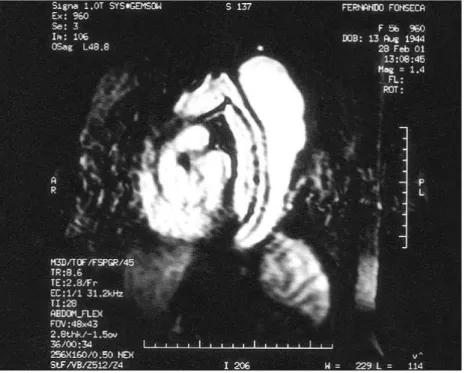
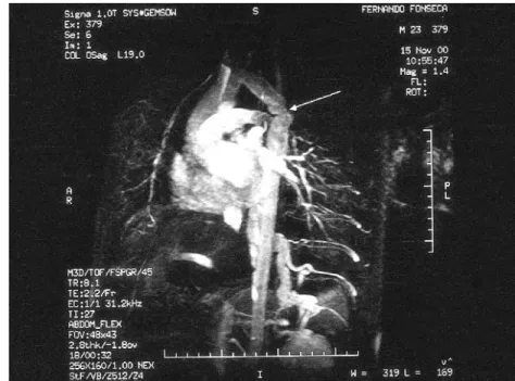
Fig. 1 A type B dissection with
a diminutive true lumen and a patent false lumen as indicated by equal signal intensity due to entry of contrast material into both lumens. The patency of the false lumen is a future predictor of expansion and complications, which occurred in this patient.
do no gadolinio, disponível comercialmente, tendo as imagens sido obtidas usando uma se-quência de time-of-flight (TOF) numa máquina de 1 Tesla, seguida de reconstrução tridimensio-nal. Descrevemos, ainda, as vantagens e des-vantagens potenciais da técnica.
MÉTODOS
Todas as imagens foram obtidas numa uni-dade de RM-GE 1,0 T Sigma, com um gra-diente de amplitude de 23 mT/m e um tempo de subida do gradiente de 460 microsegundos,
841
METHODS
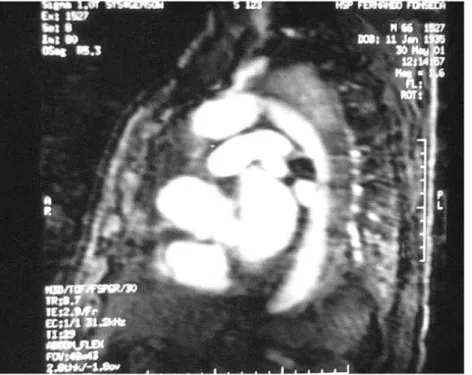
All imaging was performed on a GE 1.0 T Signa MR unit with a gradient amplitude of 23 mT/m and a gradient rise time of 460 micro-seconds with a standard body coil applied to the thorax. Seventeen patients with various aortic diseases underwent thirty-one exams over the last year. Nine patients had aortic aneurysms, three had coarctations of the aorta, five had dissections and five were postopera-tive follow-up scans. A localizing scan was fol-lowed by a Fast Spoiled Gradient Echo Scan Fig. 2 A type A dissection with
a filling defect close to the aortic root (arrow) due to absence of contrast entry into this area.
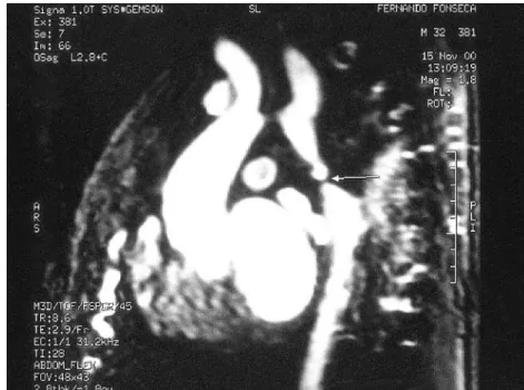
Fig. 3 Spoiled-gradient echo
ima-ges confirm the presence of orga-nized thrombus preventing entry of contrast in this area during the angiography scan in the prece-ding picture. Information ac-quired from this picture and the preceding one are complement-ary.
com uma cinta padronizada aplicada ao tórax. Dezassete doentes com várias doenças da aorta foram submetidos a 31 exames ao longo do úl-timo ano. Nove doentes tinham aneurisma da aorta, três tinham coartação da aorta, cinco ti-nham dissecções da aorta e em cinco casos tratou-se de exames de seguimento. Uma ima-gem localizada era seguida por uma imaima-gem de
Gradient Echo fast spoiled e uma sequência Time Of Flight (TOF). Para obter uma imagem
angiográfica segundo o longo eixo da aorta, o
imaging slab era colocado num corte axial
pre-viamente adquirido durante a imagem de loca-lização com o slab incluindo tanto a aorta as-cendente como a desas-cendente. Os parâmetros da sequência TOF-spoiled gradient echo (a se-quência do spoiled gradient echo usa um gra-diente para destruir a magnetização residual antes da excitação da sequência seguinte e é equivalente à sequência FLASH descrita em outros equipamentos) são as seguintes. Utili-zou-se uma largura de banda de 31,2 KHZ (a largura de banda variável é uma característica dos equipamentos GE com variações entre 15KHZ e 125KHZ, sendo os valores de TE e TR inversamente proporcionais aos valores da amplitude da banda) um TR (Tempo de Repe-tição) de 8,7 mseg e um TE (Tempo para Echo) de 2,9 mseg com um tempo de preparação de pré-imagem de 29 mseg (também variável em função da amplitude da banda). Cada corte ti-nha uma espessura de 2,8 mm e o número de segmentos dependia das dimensões da aorta. A
842
and a Time of Flight (TOF) sequence. For an angiography scan in the long axis of the aorta, the imaging slab was placed on a previously acquired axial slice during the localizing scan with the slab including both the ascending and the descending aorta. The parameters of the TOF-Spoiled Gradient Echo sequence (the spoiled gradient echo sequence uses a spoiler gradient to destroy residual magnetization prior to excitation of the next sequence and is equi-valent to the FLASH sequence described on other machines) are as follows. With a band-width of 31.2 kHz (the variable bandband-width is a feature of GE scanners, with ranges between 15 kHz and 125 kHz, the TE and TR values being inversely proportional to the value of the bandwidth), a TR (time to repetition) of 8.7 msec and a TE (time to echo) of 2.9 msec with a prescan preparation time of 29 msec (also variable based on bandwidth) was used. Indivi-dual slices were 2.8 mm thick and the number of slices depended on the dimensions of the aorta. The scan duration ranged between 17 and 26 seconds and all patients were advised to hold their breath during the scan period, af-ter taking a few deep inhalations prior to the scan. A field of view of 48x42 was used with a 256x160 matrix and one acquisition (NEX – 1). After imaging acquisition, reconstruction in a three-dimensional format was achieved with-in 6-8 mwith-inutes. No patient refused the exam
Fig. 4 A type B dissection
(sin-gle slice) with a thrombosed false lumen showing no entry of con-trast. Absence of patency is a good prognostic indicator and this patient has not had expan-sion of the aorta subsequently.
duração das imagens variou entre 17 e 26 se-gundos e todos os doentes foram informados de que deviam parar de respirar durante o período de registo das imagens, após tomarem algumas inspirações profundas antes de iniciar o re-gisto. Utilizou-se um campo de imagem de 48x42 com uma matriz de 256x160 e uma aquisição (NEX-1). Após a aquisição das ima-gens conseguia-se a reconstrução tridimensio-nal em 6-8 minutos. Nenhum doente recusou o exame devido a claustrofobia. Embora exista comercializada uma sequência coordenada de injecções – aquisição chamada SMARTPREP,
843
due to claustrophobia. Although a commer-cially available injection-acquisition coordinat-ing sequence called SMARTPREP is available on our scanner, we did not use the sequence during these studies. The gadolinium-based contrast agent, a concentration of 469 mg/ml or 0.5 mol/l (Magnevist®, Schering), was injected
as one bolus of 40 ml and imaging sequences were initiated seven seconds after completion of the bolus in patients with a heart rate over 100/sec and ten seconds for those below 100/sec. These parameters were decided upon by trial-and- error during the initial exams.
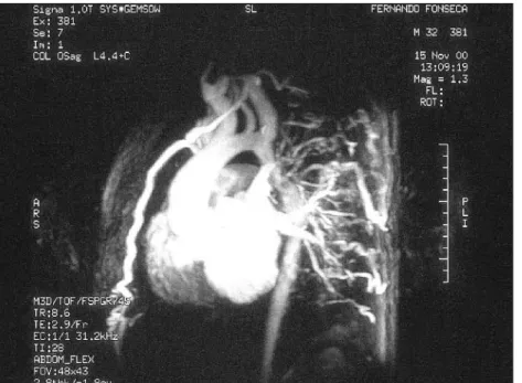
Fig. 5 The reconstructed image
showing the subclavian artery prior to the origin of the tear, de-monstrating this to be a type B dissection.
Fig. 6 The narrowest portion of a
coarctation as seen on a single slice. This diameter can be used for future reference. Notably no information on collateral activity is seen in this image.
não a usámos para estes estudos. O agente de contraste baseado no gadolinio, numa concen-tração de 469 mg/ml ou 0,5 mol/l (Magnevist®,
Schering) foi injectado sob a forma de bolus de 40 ml e, sete segundos mais tarde, iniciou-se a sequência de imagens nos doentes com fre-quência cardíaca superior a 100 bat/min. Nos doentes com frequência abaixo de 100 bat/min o intervalo de tempo foi 10 segundos. Estes parâmetros foram definidos por tentativa du-rante os exames iniciais.
RESULTADOS
Todas as imagens mostraram opacificação da aorta pelo contraste, após injecção do mesmo na altura da aquisição. Todos os doen-tes mantiveram a respiração suspensa durante o período de tempo necessário para a aquisição dos cortes. A qualidade das imagens foi ade-quada em todos os casos, incluindo aqueles em que a decisão cirúrgica foi baseada nos exames efectuados.
Nos três estudos em coartação da aorta, pu-deram visualizar-se as colaterais nas imagens de reconstrução 3D, e a porção mais estreita foi mais bem visualizada nos cortes simples. Compreensivelmente a circulação colateral re-duziu-se após a reparação operatória e embora não tivesse sido estudada previamente utili-zando este método, no doente com re-coartação da aorta, visualizou-se o aumento da circula-ção colateral. Embora a circulacircula-ção colateral te-nha sido visualizada ocasionalmente com as
844
RESULTS
All scans had opacification of contrast with-in the aorta after with-injection of contrast material at the time of acquisition of the scan (Figs.
1-8). All patients were also able to hold their
breath for the time period required for the ac-quisition of the slices. The quality of the im-ages was adequate in all cases, including those in which surgical decisions were based on the studies performed.
In the three studies with coarctation of the aorta, collaterals could be visualized in the re-constructed 3D scan and the narrowest portion was best seen in the single slices (Figs. 6-8). Collateral activity understandably was dimin-ished after operative repair and although not studied previously using this method, in the patient with re-coarctation of the aorta, enhanced collateral circulation was visualized (Fig. 8). Although collateral circulation could be visu-alized occasionally with gradient echo sequen-ces, the quality of the images was inferior to those obtained after contrast injection.
In those patients with aortic dissection, the flap could be distinguished as a dark linear image and the origin and the extent was easily delineated (Figs 1-2). The intimal tear was de-tected in four out of the five cases. Recons-tructed images in the axial plane provided images similar to those obtained by CT or fast spin echo, where the dimensions of the aorta, the true and the false lumen was measured. The patency of the false lumen could be easily discerned in those cases where contrast enhan-cement of both lumens could be demonstrated (Fig 1).
Fig. 7 The three-dimensional
re-construction shows extensive col-laterals, which presented consi-derable difficulties during operative repair. The internal mammary arteries are also clearly visualized close to the sternum. The follow-up scan after repair demonstrated absence of these collaterals.
845
sequências de gradient echo, a qualidade das imagens foi inferior à que se obteve após a in-jecção de contraste.
Nos pacientes com dissecção da aorta, o
flap pôde distinguir-se como uma imagem
linear escura, e a origem e extensão foram fa-cilmente delineados. A rotura da intima foi de-tectada em quatro dos cinco casos. A recons-trução de imagens no plano axial facultou imagens semelhantes às obtidas por CT ou spin
echo rápido, tendo sido utilizadas para a
medi-ção do verdadeiro e falso lume da aorta. A pa-tência do falso lume pôde ser facilmente iden-tificado nos casos em que se demonstraram, por contraste optimizado, ambos os lumens.
Nas imagens de aneurisma da aorta e ecta-sia anulo-aórtica, obtiveram-se dimensões das estruturas e imagens semelhantes às obtidas por angiografia convencional. As imagens re-construídas demonstraram claramente a orien-tação da junção sinotubular e do annulus. DISCUSSÃO
Embora os cortes isolados e a reconstrução tridimensional tenham fornecido excelentes imagens, consideramos que este método não pode nem deve substituir o gradient echo em sequências convencionais (com ou sem gra-dientes), devendo, no entanto, ser considerado como um método complementar. A sobreposi-ção de imagens dos dois métodos pode facultar informações complementares conforme de-monstrado no caso da dissecção do tipo A com
In the imaging of aneurysms of the aorta and annuloaortic ectasia, dimensions of the structures and images similar to those from conventional angiography were obtained. Re-constructed images clearly demonstrated the orientation of the sinotubular junction and the annulus.
DISCUSSION
Although the individual slices and the three-dimensional reconstructions provided ex-cellent images, we believe that the method cannot and should not replace conventional gradient echo (with or without spoiler grad-ients) sequences but should be considered complementary to them. Superimposing images of the two methods can provide supplementary information, as demonstrated in the case of type A dissection with a thrombosed false lu-men near the aortic valve but with patency more distally (Figs. 2-3). Patency of the false lumen is an independent predictor of mortality and the need for reoperation(3). Although
pa-tency of the false lumen can be discerned from the gradient echo images, sometimes it is diffi-cult due to motion artifact and distinguishing slow flow from intraluminal thrombus may not always be possible. Using contrast, it is vir-tually unmistakable as the false lumen will be perfused by contrast and have a signal inten-sity equivalent to that of the true lumen.
In those patients with coarctation of the aorta, we found that the individual slices
defin-Fig. 8 A coarctation operated on
twenty-two years prior to this exam showing remnant collaterals and mild recoarctation. A radio-femoral pulse delay and pressure difference was also present.
846
um falso lume trombosado perto da valvula aórtica mas com patência na parte distal. A patência do falso lume é um predictor inde-pendente da mortalidade e da necessidade de re-operação(3). Embora a patência do falso
lume possa ser perceptível a partir das ima-gens de gradient echo por vezes isso é difícil devido aos artefactos do movimento e a distin-ção entre o fluxo lento e o trombo intraluminal nem sempre é possível. Usando contraste, é virtualmente impossível a confusão uma vez que o falso lume é perfundido pelo contraste e tem uma intensidade do sinal equivalente à do verdadeiro lumen.
Nos doentes com coartação da aorta verifi-cámos que os corte isolados definem o local do estreitamento melhor do que as imagens de «gradiente ou spin echo» e as imagens recons-truídas facultam informação acerca da circula-ção colateral e sua extensão, que, num doente motivou consideráveis dificuldades durante a cirurgia. A observação clínica associada a um estudo de RM, quando possível, constitui a forma mais apropriada de seguir estes doentes no pós-operatório(4, 5).
A sequência de imagens que descrevemos aqui é rápida e proporciona imagens de exce-lente qualidade. A RM das doenças da aorta facilitou mudar para porpociona enormemente a tomada de decisão numa patologia muito complexa e a rapidez desta sequência deve permitir que seja usada para imagem das vá-rias doenças que compreendem os síndromes aórticos agudos (dissecção, hematoma intramu-ral e úlceras penetrantes). Embora possa ser difícil examinar doentes instáveis sob o ponto de vista hemodinâmico, monitorizados com equipamentos que impedem a sua entrada num aparelho de RM, muitos doentes com suspeita de síndrome aórtico agudo podem ser estuda-dos rapidamente usando esta técnica, em três ou quatro minutos. Outra vantagem da modali-dade de imagem que nós descrevemos é a sua capacidade de reconstrução de imagem em planos diferentes daquele em que foi adquirida (por ex. imagens axiais a partir de aquisições sagitais). Isto pode ajudar imenso nos hemato-mas intramurais e úlceras aórticas, uma vez que a imagem pode ser requerida frequente-mente para excluir a progressão para a dissec-ção, uma evolução previamente considerada ine-vitável, mas mais recentemente contestada(6, 7).
É também possível dispensar a fastidiosa aqui-sição de imagens de spin echo na referida si-tuação clínica uma vez que toda a informação
ed the site of the narrowing better than grad-ient or spin echo images and the reconstructed images provided information about collateral activity and extent, which in one patient pre-sented considerable difficulties during surgery (Figs. 6-7). A clinical exam coupled with a MR study, when possible, was deemed to be the most appropriate manner to follow these patients postoperatively(4, 5).
The imaging sequence we describe here is rapid and provides images of excellent quality. MR imaging of aortic diseases has greatly faci-litated decision-making in a very complex pa-thology and the rapidity of this sequence should allow it to be used for imaging of var-ious diseases that comprise the acute aortic syndromes (dissection, intramural hematoma and penetrating ulcers). Although it may be impossible to image hemodynamically unstable patients who are being monitored with equip-ment that precludes entry into a MR scanner, many patients with a suspected acute aortic syndrome can be imaged rapidly using this technique, within three to four minutes. Ano-ther advantage of the imaging modality we des-cribe is the ability to reconstruct images in planes other than the one that was acquired (e.g. axial images from sagittal acquisitions). This may help immensely in intramural hema-tomas and aortic ulcers, as imaging may be re-quired frequently to exclude progression to dissection, an outcome previously considered inevitable, but now challenged in concept(6, 7).
It is also possible to dispense with the cumber-some acquisition of spin echo images in such a clinical situation as all the information requir-ed can be garnerrequir-ed from the gradient echo and MR angiography sequences.
In conclusion, contrast-enhanced magnetic resonance angiography of aortic diseases is ra-pid, easily tolerated and provides indispens-able information complementary to that obtain-ed from conventional spin echo and gradient echo sequences, and should be performed in every patient undergoing an MR study of aortic pathology.
847
1. Erbel R, Alfonso F, Boileau C et al. Diagnosis and man-agement of aortic dissection. Recommendations of the Task Force on Aortic Dissection, European Society of Cardiology. Eur Heart J 2001;22:1642-81.
2. Arpasi PJ, Kostaki GB, Shetty AN, White RD, Simonetti OP. MR angiography of the thoracic aorta with an electrocar-diographically triggered breath-hold contrast-enhanced se-quence. Radiographics 2000;20:107-20.
3. Bernard Y, Zimmermann H, Chocron S et al. False lumen patency as a predictor of late outcome in aortic dissection. Am J Cardiol 2001;87:1378-82.
4. Therrien J, Thorne SA, Wright A, Kilner P, Somerville J. Repaired coarctation: A “cost-effective” approach to identify complications in adults. J Am Coll Cardiol 2000;35:997-1002. 5. Marx GR. “Repaired” coarctation in adults: Not a “simple” congenital heart defect. J Am Coll Cardiol 2000;35;1003-6. 6. Song J-K, Kim H-S, Kang D-H et al. Different clinical features of aortic intramural hematoma versus dissection in-volving the ascending aorta. J Am Coll Cardiol 2001;37: 1604-10.
7. Erbel R. Diseases of the thoracic aorta. Heart 2001;86: 227-34.
BIBLIOGAFIA / REFERENCES
Pedido de separatas para: Address for reprints: BOBAN THOMAS Serviço de Cardiologia Hospital Fernando Fonseca IC-19
2700 AMADORA
necessária pode ser recolhida a partir do
gra-dient echo e sequências angiograficas da RM.
Em conclusão, a angiografia por ressonân-cia magnética optimizada por contraste apli-cada a estudo das doenças da aorta, é rápida, facilmente tolerada e faculta informação com-plementar indispensável à obtida a partir dos exames convencionais de spin echo e gradient
echo devendo ser realizada em todos os
doen-tes submetidos a RM por patologia da aorta.