SUMMARY
Although several tendon sources are available for reconstructive surgical procedures, all have one or more shortcomings. The aim of this work was to evaluate if the extensor tendons of the hallux showed anatomical characteristics that could make them an additional source for tendon grafting procedures.
The authors performed a detailed morpho-metric analysis of the extensor tendons of the hallux in 26 lower limbs in order to evaluate the putative association of anatomical variants with hallux valgus, and to attempt to assess the feasibility of using part of the extensor apparatus of the hallux as a source of tendon for grafting procedures.
An accessory extensor hallucis longus ten-don was found in 92.3% of cases. The exten-sor hallucis brevis tendon length was 10.5 ± 0.6 cm; its width was 0.5 ± 0.1 cm, and its thickness varied between 1-2 mm, making it a potentially good candidate as a source of ten-don grafts. Several anatomical variations were observed, namely the fusion of the tendons of the extensor hallucis brevis and the accessory extensor hallucis longus muscles in the distal part of the foot.
This new therapeutic option, if implement-ed, would possibly increase the supply of
auto-genous donor tissue for reconstructive proce-dures, thereby enhancing the reconstructive surgeon’s armamentarium.
Key words: Extensor hallucis longus –
Exten-sor hallucis brevis – Variation – Tendon injuries – Tissue graft
INTRODUCTION
Tendon grafts are often needed in recon-structive surgery and in the realms of Ortho-pedics, Plastic Surgery, Maxillofacial Surgery, Burn Surgery, and even in Heart Surgery (Wehbe, 1994; Schenk et al., 2009; Terzis and Kyere, 2008). These tendon grafts can be used to reconstruct tendon or ligament defects, sta-bilize joints and maintain soft tissues in posi-tion (Breek et al., 1989). Recently, the use of the plantaris tendon has been proposed for atrioventricular valve repair (Shuhaiber and Shuhaiber, 2003).
Redundancy in the function of certain ten-dons has been known for decades (Brand, 1961), allowing several alternatives for tendon harvesting to become perfectly established. However, when a patient sustains extensive injuries, it is not uncommon for autologous tendons to be insufficient to reconstruct all
Morphometric analysis of the extensor
tendons of the hallux and potential
implications for tendon grafting
Diogo Casal
1,2, Diogo Pais
1, Maria Angélica-Almeida
2, Tiago Bilhim
1,
António Santos
1, João Goyri-O’Neill
11- Anatomy Department; Medical Sciences Faculty, New University of Lisbon, Portugal 2- Plastic and Reconstructive Surgery Department, São José Hospital, Lisbon, Portugal
Correspondence to:
Diogo Casal. Rua Luís Pastor de Macedo, N 32, 5D, 1750-159 Lisbon, Portugal. E-Submitted: February 22, 2010
the missing structures (Williamson and Richards, 2006). In addition, all tendon options currently in use for grafting proce-dures have one or more several limitations, namely: inconstancy; their removal results in a variable deficit in the donor region; and the surgical incisions required to perform their extirpation are located in body areas where healing is known to be suboptimal and thus results in conspicuous scars (Williamson and Richards, 2006; Tang, 2009). Therefore, any new alternative that might increase the supply of autologous tendons for reconstructive pro-cedures would be invaluable.
Supernumerary tendons in the hallucal extensor apparatus have been well document-ed for more than 125 years (Macalister, 1875; Gray, 1918; Gruber, 1875; Sarrafian and Topouzian, 1969). In 1976, Tate and Pachnik described an accessory tendon of the extensor hallucis longus in the majority of individuals (Tate and Pachnik, 1976). In the 1980s Kan-eff, Andreev and Stephanoff studied in detail the extensor tendons in the first ray of the foot, reporting several accessory tendons and over 20 different variations (Kaneff, 1986a, b; Kaneff and Andreev, 1983; Kaneff and Stephanoff, 1982). More recently, these find-ings have been reproduced by several authors (Denk et al., 2002; Bibbo et al., 2004; Hill and Gerges, 2008; Al-Saggaf, 2003; Bergman et al., 1988; Boyd et al., 2006; Aktekin et al., 2008).
Notwithstanding the reported high fre-quency of these accessory tendons, their clini-cal importance has been considered relatively minor, and their description is even omitted from many modern, comprehensive clinical anatomy textbooks (Hill and Gerges, 2008; Moore and Dalley, 2006). Moreover, the extensor tendons of the foot have not been used, as far as the authors know, as sources of tendon grafts (Chang, 2006; Tang, 2009).
Furthermore, certain authors have associat-ed certain variations in the extensor apparatus of the hallux to hallux valgus (Al-Saggaf, 2003), which is a common condition in which there is lateral deviation of the big toe, at the metatarso-phalangeal joint (Prosche et al., 2004). However, these findings have not been replicated by others and are still a matter of debate (Bibbo et al., 2004).
Thus in this work we studied the extensor tendons of the hallux from human cadavers in order to evaluate the potential of any of these tendons as a source of tendon grafts, and to
assess whether there might be any association between the morphometric features of these tendons and the presence of hallux valgus. MATERIALS ANDMETHODS
The study was performed on 26 lower extremities of freshly frozen adult human cadavers used for routine gross anatomical dis-sections at the Medical Sciences Faculty in Lis-bon, Portugal. Age at death was mostly between 60 and 85 (average 72.3) years. There were 7 men (53.8%) and 6 women (46.2%). They had had no prior surgical procedures in the leg or foot regions.
The dorsum of the foot and lower leg were carefully dissected, exposing the extensor ten-dons of the hallux from their origin to their insertion. Their origin, length, width, thick-ness and type of insertion were recorded, as well as the occurrence of hallux valgus. The mean width of each tendon was calculated based on the average of the widths measured at three points: tendon origin, middle portion of the tendon, and immediately before inser-tion, in the most distal place where it would be surgically possible to section the tendon for harvesting.
This research required no specific permis-sion from the ethics committee of our institu-tion, and conformed to the provisions of the Declaration of Helsinki (1995, revised 2000). Statistical analyses were performed using the PASWO 18.0 (IBM®) Statistical Analysis Software. The Chi-Square test was used to compare proportions, while Student’s t test and ANOVA were used for comparing means. A p value below 0.05 was considered statisti-cally significant. Mean values are represented by their numerical value ± standard deviation. RESULTS
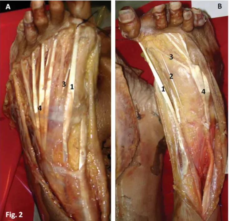
In all cases, the extensor apparatus of the hallux was composed of the extensor hallucis longus tendon (EHLp) and the extensor hallu-cis brevis tendon (EHB). An accessory exten-sor hallucis longus tendon (EHLa) was found in 92.3% of cases. Figure 1 portrays the usual composition of the extensor tendons of the hallux. The EHLa originated from the same muscular belly as the EHLp in all cases (Fig. 1). When present, the EHLa was placed medially to the EHLp (91.7%) since its ori-gin to its termination. Only in two feet, in Diogo Casal, Diogo Pais, Maria Angélica-Almeida, Tiago Bilhim, António Santos, João Goyri-O’Neill
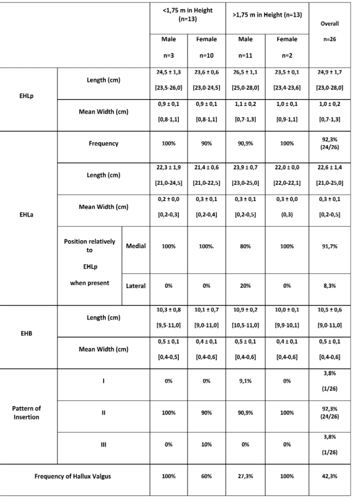
the same cadaver, was the EHLa absent (Fig. 2A). In both feet of one cadaver (8.3%) the EHLa was placed laterally to the EHLp and terminated in the medial portion of the EHB (Fig. 2B). In one foot, there were two separate EHLa, one of them with a normal diameter but the other much thinner, that inserted sep-arately at the base of the distal phalanx. These two separate EHLa were placed laterally to the EHLp.
Table 1 summarizes the results obtained globally, as well as those obtained after strati-fying for sex and height. The EHLa mean width (0.3 ± 0.1 cm) was significantly lower than that of the EHLp (1.0 ± 0.2 cm), corre-sponding to approximately one third. Howev-er, in one cadaver this EHLa had a mean width of 0.5 mm bilaterally; i.e., half of the mean width of the EHLp itself in that case. There were no statistically significant differences
between tendon length and width on the right and left sides.
The mean EHB length was 10.5 ± 0.6 cm, and its mean width was 0.5 ± 0.1 cm; i.e., half of the EHLp. The average thickness of the EHLp, EHLa, and of the EHB were remark-ably constant, being approximately 3-4 mm, 0.5-1 mm, and 1-2 mm, respectively.
The EHB terminated in the dorsal and medial aspect of the base of the proximal phalanx of the hallux in all cases. The pat-tern of insertion of the EHLp and the EHLa, by contrast, was variable. In most cases (92.3%), the tendons terminated separately: the EHLp at the base of the distal phalanx of the hallux and the EHLa in the medial aspect of the dorsum of the base of the prox-imal phalanx of the big toe. In the only cadaver in which there was no EHLa, the EHLp terminated in the usual fashion at the
Figure 1. Dorsal (A) and medial (B) views of the right foot showing the extensor tendons of the hallux. 1- Extensor hallucis longus tendon;
2- Accessory extensor hallucis longus tendon; 3- Extensor hallucis brevis tendon; 4- Extensor digitorum longus tendons; 5- Common mus-cle belly of the extensor hallucis longus musmus-cle giving off the extensor hallucis longus tendon and its accessory tendon.
base of the distal phalanx. In one case in which there were two EHLa, these two ten-dons and the EHLp terminated isolatedly at the base of the proximal and distal phalanx, respectively. Thus, according to the Al-Sagaff classification of the insertion of the extensors of the hallux, the type I pattern was found in 92.4% of cases, whereas pat-terns I and III were found in 3.8% each (Al-Saggaf, 2003).
Hallux valgus was more frequent in females (72.7%) than in males (27.3%), this difference being statistically significant (p = 0.02). No
other associations between the presence of hal-lux valgus and other parameters, namely type of insertion, were found.
DISCUSSION
The composition of the extensor tendons of the hallux in our series did not differ signifi-cantly from what has been described in the lit-erature, except for the prevalence of EHLa, which was 92.3% in our series; that is to say, much higher than that described originally by Diogo Casal, Diogo Pais, Maria Angélica-Almeida, Tiago Bilhim, António Santos, João Goyri-O’Neill
Figure 2. Dorsal view of the left (A) and right (B) feet of two different cadavers, showing anatomical variations in the extensor tendons of
the hallux. In Figure 2A there is no accessory extensor hallucis longus tendon. In Figure 2B the accessory extensor hallucis longus tendon is placed laterally to the main extensor hallucis longus tendon and fuses with the extensor hallucis brevis tendon. 1- Extensor hallucis longus tendon; 2- Accessory extensor hallucis longus tendon; 3- Extensor hallucis brevis tendon; 4- Extensor digitorum longus tendons.
Table 1. Morphometric features of the extensor tendons of the hallux in 26 feet. EHLp extensor hallux longus proprius tendon; EHLa
-extensor hallux longus accessorius tendon; EHB - -extensor hallux brevis tendon. Pattern of insertion of the -extensor apparatus is divided into three classes according Al-sagaff (2003). Values between [ ] represent the limits of variation of each variable.
Kaneff and Al-Sagaff, who reported values of 48.88% and 35%, respectively (Kaneff, 1986b; Al-Saggaf, 2003). However, our value is not significantly higher than that described recently by other authors, who described an EHLa in 70-87% of cases (Tate and Pachnik, 1976; Denk et al., 2002; Bibbo et al., 2003; Hill and Gerges, 2008). It is plausible that the differences found among the different authors may be due to population differences. Howev-er, given that our series of 26 specimens is rel-atively small (Table 2), we believe that further studies are warranted to test this hypothesis.
Al-Saggaf postulated that the presence of a supernumerary tendon of the extensor hallucis longus could be a predisposing factor for the development of hallux valgus (Al-Saggaf, 2003). However, this association was not replicated in subsequent investigations (Bibbo et al., 2004). Similarly, we also failed to iden-tify any statistically significant association between the presence of EHLa or any other morphometric feature and the presence of hal-lux valgus.
We found several anatomical variations regarding the EHLa. In 7.7% of cases this supernumerary tendon was absent. In addi-tion, although it was almost always located medially to the EHLp (91.7%) it was also placed lateral to it (8.3%). Interestingly, in one case the EHLa was placed laterally to the EHLp and terminated in the medial portion of the EHB, which in turn terminated in the usual fashion: in the dorsal and medial aspect of the base of the proximal phalanx of the hal-lux. This variation has been described for the
first time by Denk et al. (2002) and corre-sponds, as far as the authors know, to the sec-ond case reported in the literatur.
By performing a morphometric analysis of the extensor tendons of the hallux, we observed that the EHLa and the EHB had, on average, a width that was one third and one half of that of the EHLp, respectively. This observation was not mentioned in the litera-ture review we conducted, and may be of great interest for a better understanding of the functional aspects of the foot and their correlation with clinical findings. This knowledge could, for example, help explain why conservative treatment may suffice in a substantial number of cases of EHLp section or rupture, since the EHB and the EHLa will probably maintain the cut ends of the EHLp tendon close together, allowing tendon repair to occur spontaneously and thereby avoiding the need for surgery (Scaduto and Cracchiolo, 2000).
In addition, this study unequivocally sug-gests that the relatively large width and thick-ness of the EHB, as well as its significant length and constancy, would make EHB an excellent candidate as a source of tendon grafts. Moreover, this tendon fulfills all the other criteria currently accepted for donor ten-dons for tendon or ligament repair. In this sense, it is not situated too deeply, and hence would facilitate harvesting; no significant donor site loss would result from its harvest-ing, and its cross sectional diameter is not too large to hamper revascularization, while still being sufficient to provide enough autologus Diogo Casal, Diogo Pais, Maria Angélica-Almeida, Tiago Bilhim, António Santos, João Goyri-O’Neill
material for reconstruction (Williamson and Richards, 2006). Another potential advantage of using this tendon as a graft would be that the surgical incision necessary to harvest it would be placed in the dorsum of the forefoot, which corresponds to a place of the body where wounds usually heal inconspicuously and where scars are not easily visible (Park-house et al., 2006). This is a major advantage relative to other donor sites, such as the fore-arm and leg, where scars are frequently more noticeable (Parkhouse et al., 2006).
Functionally, there is strong evidence to suggest that harvesting this tendon would not result in any significant clinical deficit, since the tendon is routinely incorporated in the dorsalis pedis flap with no resulting impair-ment of the extension of the hallux (Furlow, 2009). Indeed, it is unanimously accepted that the main function of the EHB is only to aid the EHLp in extending the big toe at the metatarso-phalangeal joint (Moore and Dalley, 2006).
Additionally, the EHB compares favorably with the tendons commonly used in clinical practice in terms of certain anatomical charac-teristics. The palmaris longus tendon, for example, which is the most commonly used donor tendon in Hand Surgery (Williamson and Richards, 2006), is only slighter longer (10 to 12 cm), and has a similar width and thickness: 3-5 mm and 1-2 mm, respectively (Chui and Edgerton, 1990). Moreover, where-as the EHB is generally considered to be con-stant, the palmaris longus muscle is known to be absent in up to 12 to 25% of limbs (Williamson and Richards, 2006; Neumeister and Wilhelmi, 2006; Moore and Dalley, 2006). The plantaris tendon, which is also commonly used in reconstructive procedures, is also absent in 18% of limbs (Williamson and Richards, 2006).
In spite of all these potential advantages, the use of the EHB has been overlooked as a potential tendon donor site in Reconstructive Surgery (Chang, 2006; Williamson and Richards, 2006; Tang, 2009; Neumeister and Wilhelmi, 2006). We believe this is rather unfortunate, since the EHB would be particu-larly well suited for the repair of torn liga-ments of the hand, such as the collateral ligaments of the metacarpal-phalangeal joint of the thumb (Breek et al., 1989); the recon-struction of the A2 and A4 pulleys associated with the flexor tendons of the hand (Pederson et al., 2006) and, eventually, in some facial
reconstructive procedures, to contribute to soft tissue suspension (Terzis and Kyere, 2008). This proposal, if implemented, would increase the supply of autologous donor tissue for reconstructive procedures, thereby enhanc-ing the surgeon’s arsenal.
ACKNOWLEDGMENTS
The authors appreciate the proficiency of all Technical Staff members of the Depart-ment of Anatomy, in particular of Mr. Carlos Lopes and of Mr. Marco Costa.
REFERENCES
AKTEKIN M, UZMANSEL D, KURTOGLU Z, SANLI EC, KARA AB
(2008). Examination of the accessory tendons of extensor hal-lucis longus muscle in fetuses. Clin Anat, 21: 713-717. AL-SAGGAFS (2003). Variations in the insertion of the extensor
hallucis longus muscle. Folia Morphol, 62: 147-155.
BERGMANRA, AFIRIAK, MIYAUCHIR (1988). Illustrated encyclo-pedia of human anatomic variations.
BIBBOC, ANDERSONRB, DAVISWH (2003). Injury characteristics
and the clinical outcome of subtalar dislocations: a clinical and radiographic analysis of 25 cases. Foot Ankle Int, 24: 158-163. BIBBOC, ARANGIOG, PATELDV (2004). The accessory extensor
tendon of the first metatarsophalangeal joint. Foot Ankle Int, 25: 387-390.
BOYD N, BROCK H, MEIER A, MILLER R, MLADY G, FIROOZBAKHSH K (2006). Extensor hallucis capsularis:
fre-quency and identification on MRI. Foot Ankle Int, 27: 181-184.
BRANDPW (1961). Tendon grafting. J Bone Joint Surg, 43B:
444-453.
BREEKJC, TANAM, VANTHIEL TP, DAANTJECR (1989). Free tendon grafting to repair the metacarpophalangeal joint of the thumb. Surgical techniques and a review of 70 patients. J Bone
Joint Surg Br, 71: 383-387.
CHANGP (2006). Repair and grafting of tendon. In: MATHESSJ (ed.) Plastic Surgery. 2nd ed. Saunders, Philadelphia.
CHUIDTW, EDGERTONBW (1990). Repair and grafting of ten-don. In: MCCARTHYJG (ed.) Plastic Surgery. 1st ed. Saunders, Philadelphia.
DENKCC, OZNURA, SURUCUHS (2002). Double tendons at the
distal attachment of the extensor hallucis longus muscle. Surg
Radiol Anat, 24: 50-52.
FURLOWLT (2009). Dorsalis Pedis Flap. In: STRAUCHB, VASCONEZ
LO, HALL-FINDLAYEJ, LEEBT (eds.). Encyclopedia of Flaps. 3rd ed. Lippincott Williams and Wilkins, Philadelphia.
GRAY H (1918). Anatomy of the Human Body. Lea & Febiger, Philadelphia.
GRUBERW (1875). Über die Varietäten des Musculus extensor hal-lucis longus. Arch Anat Physiol Wissen Med, 565-589. HILLRV, GERGESL (2008). Unusual accessory tendon connecting
the hallucal extensors. Anat Sci Int, 83: 298-300.
KANEFFA (1986a). Die Aufrichtung des Menschen und die mor-phologische Evolution der Musculi extensores digitorum pedis unter dem Gesichtspunkt der evolutiven Myologie. Teil I.
KANEFFA (1986b). Die Aufrichtung des Menschen und die mor-phologische Evolution der Musculi extensores digitorum pedis unter dem Gesichtspunkt der evolutiven Myologie. Teil III.
Gegenbaurs Morphol Jahrb, 132: 681-722.
KANEFF A, ANDREEV D (1983). Über die organogenetische Entwicklung des M. extensor hallucis longus beim Menschen.
Anat Anz, 154: 237-244.
KANEFF A, STEPHANOFF A (1982). Vergleichend-anatomische Untersuchung des M. extensor hallucis longus beim Men-schen. Gegenbaurs Morphol Jahrb, 128: 690-701.
MACALISTERA (1875). Additional observations on muscular
anom-alies in human anatomy. Trans R Ir Acad Sci, 25: 1-130. MOORE KL, DALLEY AF (2006). Lower Limb. Clinically Oriented
Anatomy. 5th ed. Lippincott Williams and Wilkins, Philadelphia.
NEUMEISTERMW, WILHELMIBJ (2006). Flexor Tendon Repair. In: MCCARTHY JG, GALIANO RD, BOUTROS SG (eds). Current
Therapy in Plastic Surgery. 1st ed. Saunders, Philadelphia.
PARKHOUSEN, CUBISONTCS, HUMZAHMD (2006). Scar Revision.
In: MATHESSJ, HENTZVR (eds). Plastic Surgery. 2nd ed. Saun-ders, Philadelphia.
PEDERSONWC, STEVANOVICM, ZALAVRASCL, SHERMANR (2006). Reconstructive Surgery: Extensive Surgery to the Upper Limb. In: MATHESSJ (ed). Plastic Surgery. 2nd ed. Saunders, Philadelphia. PROSCHEH, FUHRMANNR, LINBW, FRÖBERR (2004). The
post-operative stability of the first metatarsal. Eur J Anat, 8: 55-59.
SARRAFIANSK, TOPOUZIANLK (1969). Anatomy and physiology of the extensor apparatus of the toes. J Bone Joint Surg Am, 51: 669-679.
SCADUTOAA, CRACCHIOLOA (2000). Lacerations and ruptures of
the flexor or extensor hallucis longus tendons. Foot Ankle Clin, 5: 725-736.
SCHENKS, MEIZERR, KRAMERR, AIGNERN, LANDSIEDLF, STEIN
-BOECKG (2009). Resection arthroplasty with and without
cap-sular interposition for treatment of severe hallux rigidus. Int
Orthop, 33: 145-150.
SHUHAIBERJH, SHUHAIBERHH (2003). Plantaris tendon graft for atrioventricular valve repair: a novel hypothetical technique.
Tex Heart Inst J, 30: 42-44.
TANGJB (2009). Flexor tendons. In: CHUNGKC, DISAJJ, GOSAIN
AK, KINNEYBM, RUBINJP (eds). Plastic Surgery: Indications
and Practice. 1st ed. Saunders, China.
TATER, PACHNIKRL (1976). The accessory tendon of extensor hal-lucis longus: its occurrence and function. J Am Podiatry Assoc, 66: 899-907.
TERZISJK, KYERESA (2008). Minitendon graft transfer for
sus-pension of the paralyzed lower eyelid: our experience. Plast
Reconstr Surg, 121: 1206-1216.
WEHBEMA (1994). Tendon graft anatomy and harvesting. Orthop
Rev, 23: 253-256.
WILLIAMSONDG, RICHARDSRS (2006). Flexor tendon injuries and reconstruction. In: MATHESSJ (ed). Plastic Surgery. Saunders, Philadelphia.