Biological Trace
Element Research
Editor-in-Chief
Gerhard N. Schrauzer
Editors
Peter Schramel
Yasushi Kodama
Alain Favier
Biological Trace
Element Research
Editor-in-Chief
Gerhard N. Schrauzer
Editors
Peter Schramel
Yasushi Kodama
Alain Favier
HumanaJournals.com
Sear
ch, Read and Download
Featured in this issue:
Possible Relationship
of Cadmium and Mercury
Levels with Hypertension
Age-Dependent Increase
of Magnesium in the Cerebral
Arteries
Systemic Markers of the Redox
Balance and Apolipoprotein E
Polymorphism
Systemic Markers of the Redox
Balance and Apolipoprotein E
Polymorphism in Atherosclerosis
The Relevance for an Integrated Study
P
AULAA. L
OPES,
1P
ATRÍCIAN
APOLEÃO,
1T
ERESAP
INHEIRO,
3F
ÁTIMAC
EIA,
1J
EAN-P
AULS
TEGHENS,
4M. L
EONERP
AVÃO,
5M. C
RISTINAS
ANTOS,
2 ANDA
NAM. V
IEGAS-C
RESPO*
,11
Centro de Biologia Ambiental and Departamento de
Biologia Animal, and
2Centro de Química e Bioquímica and
Departamento de Química e Bioquímica, Faculdade de Ciências,
Universidade de Lisboa, 1749-016 Lisboa, Portugal;
3
Laboratório de Feixe de Iões, Instituto Tecnológico e Nuclear (ITN),
Estrada Nacional 10, 2685-953 Sacavém, Portugal; and
4
Federation de Biochimie, Hôpital Edouard Herriot,
69437 03 Lyon, France and
5C.I.R.N., Universidade des Ac¸ores,
9500 Ponta Delgada, Portugal
Received June 21, 2005; Revised September 23, 2005; Accepted September 30, 2005
ABSTRACT
Prospective studies have demonstrated that an imbalance between oxidative damage and antioxidative protection can play a role in the development and progression of atherosclerosis. Also, genotypes with the apolipoprotein E ε4 allele have been associated with an increase risk for this pathology. Based on this knowledge, the aim of this study was to eval-uate indicators of the redox balance, trace elements, and apolipoprotein E allelic profile in subjects from the Lisbon population with clinically stable atherosclerosis, at risk for atherosclerotic events, and in healthy subjects for comparison. The activities of superoxide dismutase in erythrocytes and glutathione peroxidase in whole blood, plasma total thiols, and serum ceruloplasmin were kept unchanged among the three groups. Serum α-tocopherol was increased in atherosclerotic patients. Total malondialde-hyde in serum and protein carbonyls in plasma, which are indicators of
0163-4984/(Online)1559-0720/06/11201–0057 $30.00
lipid and protein oxidative damage, respectively, reached their highest values in risk subjects. The concentrations of potassium and calcium, in plasma and in blood cells, were slightly elevated in patients and might reflect an electrolytic imbalance. Regarding the apolipoprotein E polymorphism, atherosclerotic patients had an increased incidence of the high-risk geno-types for atherogenesis (ε3/ε4 and ε4/ε4). A multivariate model applied to the general population using most of the parameters clearly separated the three groups at study (i.e., the healthy group from the steady-state group of risk disease and from the atherosclerotic one). As shown by us, the use-fulness of biochemical and complementary genetic markers is warranted for a better knowledge on atherosclerosis molecular basis.
Index Entries:Atherosclerosis; antioxidant defenses; oxidative dam-age; trace elements; apolipoprotein E.
INTRODUCTION
Although mortality from coronary heart disease has declined recently, atherosclerosis and related vascular diseases are still the major causes of death in Western countries (1,2), including Portugal. Atherosclerosis is a chronic disease of large and medium-sized arteries, with hardening and loss of elasticity of the arterial walls and narrowing of the arterial lumen (1). High blood pressure, diabetes, smoking, and a diet rich in lipids and cho-lesterol clearly increase the likelihood of premature atherosclerosis, although other factors, such as age and the individual genetic makeup (as apolipoprotein E), might play an additional role (3–5). Apolipoprotein (apo) E gene expression affects the cholesterol metabolism (6,7) (more than any other gene identified so far), and the ε4 allele, in particular, has been associ-ated with coronary artery and heart diseases, stroke, cognitive dysfunction, and Alzheimer’s disease, as it promotes enhanced levels of both total cho-lesterol and low-density lipoprotein (LDL) chocho-lesterol in plasma (8,9).
The oxidation theory of atherosclerosis proposes that the formation of oxidized LDL in the subendothelial space of the artery wall represents a causative event for atherogenesis (5,10,11), through their enhanced uptake by unregulated macrophage scavenger receptor, and their immunogenic-ity, and capacity for inducing chemotactic, cytotoxic, and growth factors
(12). Antioxidants are thought to be potential antiatherogenic compounds (13). LDL is the primary carrier of cholesterol in the circulation, and its
components, unsaturated fatty acids and proteins, are sensitive to oxida-tion. Endothelial cells, smooth muscle cells, and macrophages can release free radicals and other oxidant species leading to LDL modification. How-ever, differences exist in the susceptibility of these lipoproteins to oxida-tive stress because small, dense LDL particles are more susceptible to oxidation than larger LDL subfractions and because LDL particles enriched with monounsaturated fatty acids are less prone to oxidation than those containing high levels of polyunsaturated fatty acids (14).
Although a number of randomized trials have failed to show any ben-efit from antioxidant treatment, there remains a preponderance of basic and clinical evidence in supporting the role of oxidative processes in atheroge-nesis (15–17). Fundamental research on redox status and oxidative damage mechanisms in atherosclerosis and associated pathologies (such as cardio-vascular disease and stroke) becomes an important goal and might help one to select more appropriate study designs in terms of preventive meas-ures in populations. The present study was developed under the scope of a project aiming to evaluate possible variations on redox balance markers, trace elements, and apo E polymorphism in subjects from the Lisbon pop-ulation, from the healthy to clinically stable atherosclerotic condition, including the hazardous situation of hyperlipidemia and/or hypertension. This article reports data on blood activities of superoxide dismutase (SOD) and glutathione peroxidase (GPx), plasma concentration of total thiols, and α-tocopherol levels in serum. The protein ceruloplasmin (Cp), which is con-sidered to have an antioxidant action and to be an inflammatory marker, was quantified in serum. Serum levels of total malondialdehyde (MDA) and plasma contents of protein carbonyls were evaluated as markers of oxidative modifications in lipids and proteins, respectively. The levels of essential elements—K (potassium), Ca (calcium), Fe (iron), Cu (copper), Zn (zinc), and Se (selenium)—playing a role in electrolytic mechanisms and oxidant/antioxidant pathways were determined in blood. The allelic pro-file of apo E was assessed for each subject and correlated with its clinical condition.
MATERIALS AND METHODS
Study Design
A total of 72 nonsmoker subjects from Lisbon, ranging in age from 40 to 78 yr, were included in the study and divided into 3 groups (Table 1). The pathological group was composed of ambulatory patients with a history of atherosclerotic disease: previous myocardial infarction, angina, and lower limb chronic arterial insufficiency, under therapy. Patients with acute or decompensated disorders (heart failure, respira-tory disorders, and other metabolic abnormalities) were excluded from the study. The subjects from the risk group were hypertensive and/or hyperlipidemic. The reference group was composed by healthy subjects whose lipid and blood pressure values were within normal ranges. All subjects gave informed consent and filled out a clinical report stating information relevant to the study, namely baseline characteristics and lifestyles (e.g., smoking habits). Subjects under dietary supplements or drugs with a known antioxidant capacity were excluded. The study was approved by the Human Ethics Committee of the National Health Insti-tute Dr. Ricardo Jorge and conformed to standard ethical guidelines for human research.
Blood Collection
The blood was collected by venipuncture after an overnight fast and aliquots were drawn into Z-Gel, lithium–heparin, and EDTA S-Monovette®
tubes. Serum and plasma were removed after centrifugation at 1500g for 10 min at 4°C. Lipid parameters and the enzyme activities were evaluated within 24 h. Samples for DNA extraction were stored at –70°C. Aliquots for elemental analysis were frozen at –20°C and those for total thiols, proteins, protein carbonyls, and Cp quantification were frozen at –80°C. For the determination of α-tocopherol levels and lipid peroxidation index, aliquots were kept under liquid nitrogen. Serum, plasma, and blood sam-ples from the three groups were analyzed in the same run.
Anthropometric Measurements
Body height and weight of the participants (without shoes and heavy clothing) were registered and body mass index (BMI = weight [in kg]/height2 [in m2]) was calculated as a measure of obesity. The World
Health Organization reports the following categories for BMI: obese (≥30 kg/m2), overweight (25.0–29.9 kg/m2), normal (18.5–24.9 kg/m2), and
underweight (<18.5 kg/m2) (18). Blood pressures were recorded to the
nearest mm Hg and hypertension was defined according to the guidelines provided by the National Institutes of Health (systolic blood pressure >140 mm Hg and/or diastolic blood pressure >90 mm Hg or under antihyper-tensive therapy) (19).
Lipid Profile Evaluation
High density lipoproteins (HDL) were separated by adding polyeth-ylene glycol to fresh samples in order to precipitate other lipoproteins (20). Total cholesterol, HDL cholesterol, and triglycerides concentrations in serum were estimated by using appropriate enzymatic kits (CHOD-PAP®,
HDL-C Plus®, and GPO-PAP®, respectively; Boehringer, Manheim,
Ger-many). LDL cholesterol concentration was estimated by the original Friedewald formula (21). Based on cholesterol and/or triglycerides levels in serum, subjects were divided into normolipidemics (with total choles-terol and triglycerides levels below 200 mg/dL and 150 mg/dL, respec-tively) and hyperlipidemics, who had one or both parameters above these reference values (22). At the laboratory, personnel were unaware of the sta-tus (patient, risk, or reference subject) of the specimens.
Determination of Enzyme Activities
Measurement of SOD activity in erythrocytes and GPx in whole blood were carried out with commercial kits (Ransod SD 125 and Ransel RS 506, respectively; Randox Laboratories, UK). For both enzyme activities, results were expressed per gram of hemoglobin, which was also determined by using a commercial kit (HG 980, Randox kit).
Determination of Total Thiols, α-Tocopherol,
and Ceruloplasmin
Total thiols in plasma were measured spectrophotometrically using the Ellman’s reagent (23). Serum α-tocopherol was evaluated by a reverse-phase high-performance liquid chromatography (HPLC) method, according to Julianto et al. (24). Cp in serum was determined by rate nephelometry using a commercial kit (CER Beckman-Array®System(s), Germany).
Determination of Total MDA and Protein Carbonyls
Malondialdehyde was measured in serum by liquid chromatogra-phy–mass spectrometry (LC/MS) according to a method derived from an initial ultraviolet (UV) version (25) with an electrospray ionization (ESI) positive detection mode and an internal standard (dideuterated tetra methoxypropane) for correction of MDA protein binding (26). The car-bonyl contents in plasma were evaluated according to Levine et al. (27) and expressed as nanomoles per milligram of protein, being the protein content determined based on the method of Lowry et al. (28).
Elemental Analysis
Concentrations of Fe, Cu, Zn, and Se in plasma and blood cells were determined by the PIXE (particle-induced X-ray emission) multielemental technique. This technique also allowed one to evaluate the levels of K and Ca. The methodology applied is described elsewhere (29). The analytical procedure was checked using Gent second-generation freeze-dried human serum reference material (30), which was analyzed together in each sam-ple batch. Differences in the certified value were below 5% for Fe and Se and below 10% for Cu, Zn, K, and Ca.
Apo E Genotyping
DNA extraction was achieved through the method of Miller et al. (31). Apo E genotyping started with a polymerase chain reaction (PCR) to amplify a 267-basepair fragment from the exon 4 of the apo E gene. The amplification products were then submitted to digestion with the HhaI restriction endonuclease, as described elsewhere (32). Each apo E allele yields a distinct pattern after electrophoresis on polyacrylamide gel
(32,33). Samples were analyzed in a random fashion and the laboratory
technician carrying out the genotyping procedures was blinded for the cholesterol levels of the samples concerned.
Statistical Analysis
Statistical analysis was performed using STATISTICA 5.0 and SPSS 10.0 softwares. Data were expressed as mean ± standard deviation. The presence of outliers was verified by running box plots, and when the
parameters did not fit a Normal distribution, a logarithmic transformation was performed (34). The values were analyzed using the Student’s t-test for independent samples, with a significance level of p < 0.05. Correlation analyses using the Pearson’s coefficient were also applied for assessing lin-ear relationships between the evaluated parameters. A forward stepwise discriminant analysis, using blood pressure, BMI, lipids (total cholesterol, HDL cholesterol, and triglycerides), SOD and GPx activities, nonenzy-matic parameters (total thiols, α-tocopherol, Cp), oxidative damage parameters (total MDA and protein carbonyls), and apo E polymorphism as variables, were also applied to help in predicting the shared variability relative to each clinical group (35).
Apo E allele frequencies were estimated using the gene-counting method, and a χ2goodness-of-fit test was applied to evaluate the genetic
Hardy–Weinberg equilibrium (36). Apo E categories were created as fol-lows: E2 (ε2/ε3), E3 (ε3/ε3), and E4 (ε3/ε4 and ε4/ε4) to explore the allelic effect and to increase statistical power (37). The differences in apo E genotypes between the studied groups were observed using a χ2
asso-ciation test.
RESULTS
Groups Description and Clinical Characterization
Table 1 presents clinical and biochemical characteristics of the study groups. Genders were pooled because no major differences were found between them (data not shown). Furthermore, the majority of females were at the postmenopausal stage and this fact contributes to the similar probability of both genders for the occurrence of atherosclerotic events. Also, Pedersen et al. (38) have reported no evident differences between females and males based on the prevalence and extent of atherosclerosis.
Body mass index classified the mean value for the reference group as a marginally normal one (Table 1), with 69% normal and 31% overweight persons. In turn, the risk group was composed of 32.5% normal, 50% over-weight, and 17.5% obese subjects and the pathological group by 25% nor-mal, 62.5% overweight, and 12.5% obese patients. Patients and risk subjects had higher blood pressures, but no differences were observed either for systolic or diastolic blood pressures between these two groups (Table 1). Higher values were found for total and LDL cholesterol levels in the serum of risk subjects and of patients relative to the reference group (Table 1). Serum triglycerides levels were enhanced in risk subjects and marginally higher in patients compared to levels of the reference ones (Table 1). Total proteins in plasma were increased in patients compared to risk subjects and the same trend was verified in relation to the reference group, although not reaching statistical significance (p = 0.069) (Table 1). As seen in Table 1, medicine intake was more pronounced in the
patho-logical group, being the conjugation of the three therapies verified in 6% of the patients.
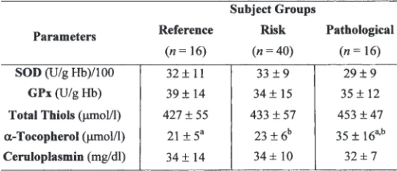
Markers of Antioxidant Defenses and Oxidative Damage
The activities of SOD in erythrocytes and GPx in whole blood and the plasma levels of total thiols kept unchanged across the three groups (Table 2). Serum α-tocopherol concentration was increased in atherosclerotic patients (Table 2). Ceruloplasmin levels in serum were similar for the three groups (Table 2). Figure 1 shows the variations of total MDA serum levels and plasma protein carbonyl contents among the three groups. Total MDA was similar between patients and references, but reached the highest values
Table 1
Baseline Characteristics for Each Group
Note: Data are expressed as mean ± standard deviation, except when
oth-erwise indicated. Values in each row sharing the same superscript are signif-icantly different (t-test, p < 0.05).
in risk subjects, this variation being statistically significant relative to the ref-erence group (Fig. 1A). In turn, the level of protein carbonyls was the lowest in the plasma of patients (Fig. 1B).
Elemental Analysis
Table 3 shows that the concentrations of K, either in plasma or in blood cells, were higher in patients in relation to risk subjects and in blood cells in relation to reference ones. Ca concentration in plasma was elevated in the pathological group (Table 3). Fe, Cu, and Zn levels remained unchanged across the three groups regardless of the biological
compart-Table 2
Activities of SOD in Erythrocytes and GPx in Whole Blood, Levels of Plasma Total Thiols, Serum α-Tocopherol,
and Cp for Each Group
Note: Data are expressed as mean ± standard deviation. Values in
each row sharing the same superscript are significantly different (t-test, p < 0.05).
Fig. 1. Markers of oxidative damage for each group: (A) total MDA in serum;
(B) protein carbonyls in plasma. Bars sharing the same superscript are
ment (Table 3). Risk subjects presented higher levels of Se in plasma rela-tive to reference ones (Table 3).
Distribution of apo E Genotypes
The distribution of apo E genotypes in this study is in Hardy–Wein-berg equilibrium (Table 4). The genotypes ε2/ε2 and ε2/ε4 were not found, although all three alleles were represented in our samples (Table 4). The
Table 3
Elemental Concentrations (mg/L) in Plasma and Blood Cell Fractions for Each Group
Note: Data are expressed as mean ± standard deviation. Values in each row sharing the same
superscript are significantly different (t-test, p < 0.05).
Table 4
Distribution of apo E Genotypes for Each Group
aχ2: Statistic between observed and expected values under
majority of subjects within each group were homozygous ε3/ε3 (Table 4). However, an increase in the occurrence of the risk genotypes for atheroge-nesis (ε3/ε4 and ε4/ε4) was observed from the reference group to patients, and an inverse trend was verified for the common ε3/ε3 genotype (Table 4). This last observation is reinforced by the allelic frequencies. Moreover, the proportion of the E3 genotypes category was proved to be different between the risk group and the others (reference/risk: χ2= 5.8, df = 1, p =
0.05; risk/pathological: χ2= 14.3, df = 1, p = 0.05). The same observation
could be applied for the E4 genotypes category between the reference and pathological groups (χ2= 4.5, df = 1, p = 0.05).
Multivariate Analysis
The variations of the parameters based on atherosclerosis disease sta-tus were assessed by discriminant analysis (Fig. 2). The first function (p = 0.000) to which the variables total cholesterol, systolic blood pressure, diastolic blood pressure, total MDA, HDL cholesterol and triglycerides were related to (discriminant loadings 0.562, 0.271, 0.255, 0.237, 0.184, and 0.180, respectively) explained 62% of the variations and separated the ref-erence group from the others. The second discriminant function (p = 0.000) was associated with α-tocopherol, protein carbonyls, total thiols, and apo E polymorphism (discriminant loadings –0.545, 0.281, –0.254, and –0.167, respectively) explained the remaining 38% of the variations and segre-gated the pathological group from the risk group (Fig. 2).
DISCUSSION
Oxidative processes and inflammation are key components of athero-sclerosis, from fatty streak formation to plaque rupture and thrombosis. Recent clinical studies have identified potentially clinically useful markers of oxidative stress and inflammation (17). In this study, we report on the usefulness of the relationship between those parameters and the major risk factors (including genetic markers) in humans from health to atheroscle-rosis disease.
Variations in Antioxidant Defenses and Oxidative Markers
Both SOD and GPx activities, either of patients or of subjects from the risk group, were not different from those of reference subjects. SOD is an important antioxidant enzyme and, contrary to our findings, some studies have reported decreased SOD activity in the erythrocytes of subjects suf-fering from ischemia (39). It is believed that low SOD activity contributes to higher levels of the superoxide radical, which can be implicated in the pathogenesis of atherosclerosis (40). The protective effect of GPx in partic-ular on lipoperoxidation is reinforced because this enzyme not only elimi-nates hydrogen peroxide but also converts some lipid hydroperoxides to
nontoxic alcohols, acting as a chain-breaking antioxidant (41). In studies on atherosclerotic obliterans patients, a decline in the antioxidative barrier of plasma because of reduced GPx activity has been reported (42) and experimental evidence exists for a weak glutathione-related enzymatic antioxidant shield in human atherosclerotic plaques, resulting from the absence of GPx activity in atherosclerotic tissue (43). In contrast, Wein-brenner et al. (44) suggested that oxidative stress might occur in patients with stable coronary heart disease, based on enhance activities of SOD in erythrocytes and GPx in whole blood, even in patients clinically stable and under medical treatment, like the patients of our study. Thus, contradic-tory findings exist for these enzyme activities in the atherosclerotic condi-tion. The comparison of data from different studies must be made carefully because, in general, the experimental conditions are not identical, including some of the major risk factors associated with this pathology, such as cigaret smoking (Lopes et al., unpublished data) and medicine intake. In fact, recent data suggested that statins, in addition to their lipid lowering activity, can reduce the production of reactive oxygen species and increase the resistance of LDL to oxidation (45).
Thiols are compounds containing sulfhydryl residues, and in biologi-cal systems, they are involved in protein synthesis and structure and play an important role in redox-sensitive signal transduction, immune regula-tion, and coordinating the antioxidant defense network (46). Disturbances in tissue thiol homeostasis have been linked to several human health dis-orders (46). In our study, the levels of total thiols in plasma were similar in patients and in reference subjects. However, patients with coronary artery
Fig. 2. Scatterplot of the canonical discriminant functions, indicating the centroids (cross) for each group (R, square: reference group; RK, diamond: risk group; P, circle: pathological group) (for details see the Materials and Methods section).
disease have been described as having decreased concentrations of plasma protein thiols (47).
Vitamin E is the major lipid-phase antioxidant protecting against oxidative lipid damage (5). It is thought to be effective in preventing ath-erosclerosis (12), because a local or generalized diminution of α-tocopherol concentration caused by dietary or oxidative factors can stimulate cell growth and lead to atherosclerosis progress (48). However, supplements of dietary vitamin E in clinical trials have not prevented consistently cardiac attacks in humans with established coronary heart disease, and vitamin E might become an oxidant agent (2,49,50). Such contradictory results have questioned the role of vitamin E as a protective agent in human athero-sclerosis (51). In the present study, the highest levels of serum α-tocopherol were found in atherosclerotic patients, but this finding disagrees with oth-ers (as discussed later in this subsection) and could be justified by a healthy diet recommended by physicians. In turn, the serum level of α-tocopherol for the risk group was similar to that of the reference one, which is not in accordance with data reported elsewhere on decreased serum α-tocopherol concentration in subjects with various coronary risk factors including male gender, higher age, smoking habits, altered serum lipid profile, and obesity (52). It is not clear whether the vitamin E decrease is a result of the ongoing atherosclerotic process by itself or contributes to its further aggravation (12). In several clinical trials, patients with coronary heart and artery diseases and after stroke presented unchanged α-toco-pherol status (12,47,53).
In the present study, a positive correlation between total MDA and pro-tein carbonyls (r = 0.33; p < 0.05) was found, supporting the view that dif-ferent biomolecules can be damaged by oxidants. No differences were found in MDA concentration between healthy and atherosclerotic subjects, which agrees with data published by Weinbrenner et al. (44) for males with coro-nary heart disease. However, previous studies have shown that subjects with ischemic heart disease, unstable angina, stable angina, or myocardial infarction have higher levels of MDA (11,54). One possible explanation for our results is the presence of both genders in the pathological group because estrogens could act as a confounding factor on the circulating levels of MDA, as stated by Fabbi et al. (4). Also, the apparent absence of lipid per-oxidation in these patients might be a result of the tight and successful reg-ulation of lipids contents by statins, mainly total cholesterol, whose mean value, although increased, did not reach a hazardous level (above 250 mg/dL) (4,45). Notwithstanding, higher values of total MDA were found in risk subjects with hyperlipidemic profile compared to reference ones, which could be eventually explained by the absence of statins intake. In addition, this result supports our own previous report in which the hyperlipidemia condition (without atherosclerotic events) led to higher lipoperoxidation levels, quantified through TBARS method (Lopes et al., unpublished data).
The evaluation of the carbonyl groups in plasma proteins is a respected marker of free-radical reaction intensity (27,55) in aging and in
various diseases (56–58). In our study, lower levels of protein carbonyls were found in the pathological group, which had a slightly higher protein concentration compared to the reference group. This result is not in accor-dance with previous findings that defend the accumulation of protein car-bonyls in vascular lesions as a common phenomenon in most, if not all, types of vascular damage (56). However, in our study, the higher plasma protein level in the pathological group could lead to a carbonyl/protein ratio lower than that observed in the other groups. In turn, higher car-bonyls and lower protein concentrations were found in the plasma of risk subjects, suggesting, for this group, signs of free-radical attack. These results are in fact corroborated by the negative correlation observed between total proteins and protein carbonyls (r = –0.74; p < 0.05).
Variations in Elemental Status
In this study, potassium and calcium were slightly elevated in patients, suggesting alterations in elemental homeostasis probably the result of changes in cellular ion transport related to the atherosclerotic process. Thus, the variations found for these elements might reflect an electrolytic imbal-ance. On the other hand, in vitro studies have reported an intracellular cal-cium rise due to oxidized LDL (59), which promotes the activation of T-cells
(60) and could establish an interesting relationship among the oxidative
stress, the inflammatory process, and the electrolytic balance.
Iron blood levels remained unchanged, but total proteins correlated pos-itively with this element in plasma (r = 0.33; p < 0.05). This association could be justified by the existence of several iron-containing proteins, such as fer-ritin, transferrin, haemoglobin, myoglobin, or the hemin moiety (61). Recent studies have demonstrated that iron deposition is prominent in human ath-erosclerotic lesions, and animal experiments have further revealed that the severity of atherosclerosis can be markedly influenced by iron overload or deficiency (62). In fact, iron has been inconsistently associated with cardio-vascular disease in spite of the plausible hypothesis as to how transition metal might accelerate the progression of atherosclerosis (63). Free iron is deleterious to cells because it catalyzes the generation of hydroxyl radicals from superoxide and hydrogen peroxide via the Fenton reaction (62).
In this study, unchanged blood levels of copper and iron in athero-sclerotic patients agrees with the maintenance of serum Cp concentration. Cp is a α2-globulin acting as a multicopper oxidase playing a role in iron
metabolism, through its ferroxidase activity (42,61,64). The physiologic importance of this protein might include antioxidant activity by promot-ing iron mobilization or scavengpromot-ing free radicals and, thus, preventpromot-ing metal-catalyzed free-radical tissue damage. On the other hand, the puta-tive role of Cp as a risk factor for atherosclerosis and cardiovascular dis-ease has been attributed to its pro-oxidant activity to modify LDL
(61,64,65). Previous studies on atherosclerosis obliterans showed that Cp
in this pathology (42,66). This correlation supports in part the positive associations found in our study between Cp and copper (in plasma and blood cells: r = 0.73, r = 0.45; p < 0.05, respectively), although the absolute Cp concentration has been measured instead of its activity.
Zinc levels, either in plasma or in blood cells, were similar for all the studied groups. In contrast, Isra and Majewski (66) have found zinc lev-els greatly enhanced in the serum of atherosclerosis obliterans patients. The meaning of this finding is not clear, as several studies propose zinc to be antiatherogenic, through either its inhibition of LDL oxidation or its role in inhibition of the oxidative-stress-responsive factors involved in destruction of vascular endothelial cell integrity (3,67). Furthermore, severe and moderate zinc deficiency has been reported to cause oxidative damage to proteins, lipids, and DNA in rat testes, which might be the result of iron accumulation or a reduction in zinc-dependent antioxidant processes (68).
Concentrations of selenium in plasma can provide useful indicators of human selenium intake and status (69). In this study, risk subjects presented higher levels of selenium in plasma than the reference ones, which might be related to the presence of approximately 80% of hyperlipidemic subjects in the risk group (70). This association could in part be the result of the bind-ing of selenium to lipoproteins, mainly LDLs, which are recognized as hav-ing significant amounts of selenium (71). Moreover, epidemiological studies are known to be controversial with respect to the association of this element and the occurrence of cardiovascular diseases (72), although a protective role has been attributed to selenium in reducing the risk for heart diseases and cancer (73,74).
Variations in apo E Polymorphism
In recent years, several polymorphisms playing a role in the develop-ment and progression of atherosclerosis have been identified in relation to blood coagulation, endothelial function, and lipid metabolism (75), as is the case of apo E. The serum cholesterol concentration is in part deter-mined by the common apo E polymorphism and the ε4 allele appears to be associated with elevated serum cholesterol concentration (6,76–78) and to contribute to an increased risk for coronary artery disease and atheroscle-rosis (37,79,80). In this study, the high-risk genotypes for atherogenesis (ε3/ε4 and ε4/ε4) were found in a higher percentage in patients, similarly to results of Stengard et al. (81,82) and to those of Pirim et al. (83). These hazardous genotypes were also found in risk subjects. These findings agree with the above-mentioned association between the ε4 allele and the incidence of atherosclerosis. Moreover, intervention trials in humans as well as experimentation in animal models have demonstrated a connec-tion between apo E and atherosclerosis. It is the case of experiments with apo E-null mutant mice that are hypercholesteremic animals, which rap-idly developed atherosclerotic lesions, either when kept on an atherogenic diet or on a normal chow (84).
FINAL REMARKS
The maintenance of some antioxidant defenses in atherosclerotic subjects observed in this study could be consequence of medical treat-ment. Nevertheless, a multivariate model applied to all the subjects clearly discriminated the three groups at study (i.e., the healthy group from the group in a steady state of risk disease and from the atheroscle-rotic one). These findings point out the need for further coordinated studies using complementary markers, both at biochemistry and genet-ics levels, in order to enlighten our knowledge about atherosclerosis molecular mechanisms.
ACKNOWLEDGMENTS
This work forms part of the project “Markers of the prooxidant/ antioxidant balance and characterization of the allelic profile of Apo E in inhabitants of Lisbon and Ponta Delgada”, supported by Fundação para a Ciência e a Tecnologia (POCTI/ESP/41008/2001) and CBA (Centro de Biologia Ambiental). Paula Alexandra Lopes is grateful for a PhD grant (PRAXIS XXI/BD/21444/99) and for financial support from Instituto de Investigação Científica Bento da Rocha Cabral. The authors are indebted to the National Health Institute Dr. Ricardo Jorge for the support in the blood collection and assessment of serum lipid profile.
REFERENCES
1. B. Frei, On the role of vitamin C and other antioxidants in atherogenesis and vascular
dysfunction, Proc. Soc. Exp. Biol. Med. 222, 196–204 (1999).
2. J. W. Heinecke, Clinical trials of vitamin E in coronary artery disease: is it time to
recon-sider the low density oxidation hypothesis? Curr. Atherosclerosis Rep. 5, 83–87 (2003).
3. B. Hennig, M. Toborek, and C. J. McClain, High-energy diets, fatty acids and
endothe-lial cell function: implications for atherosclerosis, J. Am. Coll. Nutr. 20(2), 97–105 (2001).
4. P. Fabbi, G. Ghigliotti, C. Brunelli, et al., Intense lipid peroxidation in premature
clini-cal coronary atherosclerosis is associated with metabolic abnormalities, J. Lab. Clin.
Med. 143, 99–105 (2004).
5. A. Munteanu, J. -M. Zingg, and A. Azzi, Anti-atherosclerotic effects of vitamin E: myth
or reality?, J. Cell Mol. Med. 8(1), 59–76 (2004).
6. J. E. Eichner, S. T. Dunn, G. Perveen, D. M. Thompson, K. E. Stewart, and B. C. Stroehla,
Apolipoprotein E polymorphism and cardiovascular disease: a huge review, Am. J.
Epi-demiol. 155(6), 487–495 (2002).
7. S. Bhakdi, An hypothesis for the immunopathogenesis of atherosclerosis, Clin. Nephrol.
60, S49–S52 (2003).
8. R. W. Mahley, T. L. Innerarity, S. C. Rall, Jr., and K. H. Weisgraber, Plasma lipoproteins:
apolipoprotein structure and function, J. Lipid Res. 25, 1277–1294 (1984).
9. R. W. Mahley, Apolipoprotein E: cholesterol transport protein with expanding role in
10. L. P. L. Van de Vijver, A. F. M. Kardinnal, D. E. Grobbee, H. M. G. Prince, and G. Van
Poppel, Lipoprotein oxidation, antioxidants and cardiovascular risk: epidemiologic evidence, Prostaglandins Leukotrienes Essential Fatty Acids 57, 479–487 (1997).
11. I. Durak, M. Kaçmaz, M. Y. B. Çimen, Ü. Büyükkoçak, and H. S. Öztürk, Blood
oxi-dant/antioxidant status of atherosclerotic patients, Int. J. Cardiol. 77, 293–297 (2001).
12. M. Feki, M. Souissi, E. Mokhtar, et al., Vitamin E and coronary heart disease in
Tunisians, Clin. Chem. 46(9), 1401–1405 (2000).
13. S. R. Thomas and R. Stocker, Molecular action of vitamin E in lipoprotein oxidation:
implications for atherosclerosis, Free Radical Biol. Med. 28(12), 1795–1805 (2000).
14. S. Ylä-Herttuala, LDL oxidation and atherogenesis, in Natural Antioxidants and Food Quality in Atherosclerosis and Cancer Prevention, J. T. Kumpulainen and J. T. Salonen, eds.,
The Royal Society of Chemistry, Cambridge, pp. 7–9 (1996).
15. B. P. Yu, Cellular defenses against damage from reactive oxygen species, Physiol. Rev.
74, 139–162 (1994).
16. A. J. C. Slooter, M. L. Bots, L. M. Havekes, et al., Apolipoprotein E and carotid artery
atherosclerosis: the Rotterdam study, Stroke 32, 1947–1952 (2001).
17. M. H. Shishehbor and S. L. Hazen, Inflammatory and oxidative markers in
atheroscle-rosis: relationship to outcome, Curr. Atherosclerosis Rep. 6, 243–250 (2004).
18. Expert Panel on the Identification, Evaluation, and Treatment of Overweight in Adults.
Clinical guidelines on the identification, evaluation and treatment of overweight and obesity in adults: executive summary, Am. J. Clin. Nutr. 68, 899–917 (1998).
19. A.-C. Santos and H. Barros, Prevalence and determinants of obesity in an urban
sam-ple of Portuguese adults, Public Health 117, 430–437 (2003).
20. F. C. Ballantyne, R. S. Clarck, H. S. Simpson, and D. Ballantyne, HDL and LDL
sub-fractions in myocardial infarction in control subjects, Metabolism 31, 433–437 (1982).
21. W. T. Friedewald, R. I. Levy, and D. S. Fredrickson, Estimation of the concentration of
low-density lipoprotein cholesterol in plasma, without use of the preparative ultracen-trifuge, Clin. Chem. 18(6), 499–502 (1972).
22. Expert Panel on Detection, Evaluation, and Treatment of High Blood Cholesterol in
Cho-lesterol in Adults. Executive summary of the third report of the National ChoCho-lesterol education program (NCEP) expert panel on detection, evaluation and treatment of high blood cholesterol in adults (adult treatment panel III), JAMA 285, 2486–2497 (2001).
23. J. Sedlak and R. H. Lindsay, Estimation of total, protein-bound, and nonprotein
sulfhydryl groups in tissue with Ellman’s reagent, Anal. Biochem. 25, 192–205 (1968).
24. T. Julianto, K. H. Yuen, and M. Noor, Simple HPLC method for determination of
α-toco-pherol in human plasma, J. Chromatogr. B. 732(1), 227–231 (1999).
25. J. P. Steghens, A. L. Van Kappel, I. Denis, and C. Collombel, Diaminonaphtalene, a new
highly specific reagent for HPLC-UV measurement of total and free malondialdehyde in human plasma or serum, Free Radical Biol. Med. 31(2), 242–249 (2001).
26. G. Peiro, J. Alary, J. P. Cravedi, E. Rathahao, J. P. Steghens, and F. Guéraud,
Dihydroxy-nonene mercapturic acid, a urinary metabolite of 4-hydroxynonenal, as a biomarker of lipid peroxidation, Biofactors 24(1–4), 89–96 (2005).
27. R. D. Levine, D. Garland, C. N. Oliver, et al., Determination of carbonyl content in
oxidatively modified proteins, Methods Enzymol. 186, 464–478 (1990).
28. O. H. Lowry, N. J. Rosebrough, A. L. Farr, and A. R. Randall, Protein measurement with
the Folin phenol reagent, J. Biol. Chem. 193, 265–275 (1951).
29. M. A. Barreiros, T. Pinheiro, M. F. Araújo, M. M. Costa, M. Palha, and R. C. Silva,
Qual-ity assurance of X-ray spectrometry for chemical analysis, Spectrochim. Acta B 56(11), 2095–2106 (2001).
30. J. Versieck, L. Van Ballenberghe, A. De Kesel, et al., Certification of a second-generation
biological reference material (freeze-dried human serum) for trace element determina-tions, Anal. Chim. Acta 204, 63–75 (1998).
31. S. A. Miller, D. D. Dykes, and H. F. Polesky, A simple salting out procedure for
extract-ing DNA from human nucleated cells, Nucleic Acids Res. 16, 1215 (1988).
32. J. E. Hixson and D. T. Vernier, Restriction isotyping of human apolipoprotein E by gene
amplification and cleavage with Hha I, J. Lipid Res. 31, 545–548 (1990).
33. P. Richard, G. Thomas, M. P. Zulueta, et al., Common and rare genotypes of human
apolipoprotein E determined by specific restriction profiles of polymerase chain reac-tion-amplified DNA, Clin. Chem. 40(1), 24–29 (1994).
34. J. H. Zar, Biostatistical Analysis, 3rd ed., Prentice-Hall International, London (1996). 35. B. G. Tabachnick and L. S. Fidell, Using Multivariate Statistics, Harper Collins College,
New York (1996).
36. S. W. Guo and E. A. Thompson, Performing the exact test of Hardy–Weinberg
propor-tion for multiple alleles, Biometrics 48, 361–372 (1992).
37. L. Braeckman, D. De Bacquer, M. Rosseneu, and G. De Backer, Apolipoprotein E
poly-morphism in middle-aged Belgian men: phenotype distribution and relation to serum lipids and lipoproteins, Atherosclerosis 120, 67–73 (1996).
38. H. S. Pedersen, G. Mulvad, W. P. Newman, and D. A. Boudreau, Atherosclerosis in
coronary arteries and aorta among Greenlanders: an autopsy study, Atherosclerosis 170, 93–103 (2003).
39. D. Yucel, S. Aydogdu, S. Cehreli, et al., Increased oxidative stress in dilated
cardiomy-opathic heart failure, Clin. Chem. 44, 148–154 (1998).
40. T. Fukai, R. J. Folz, U. Landmesser, and D. G. Harrison, Extracellular superoxide
dis-mutase and cardiovascular disease, Cardiovasc. Res. 55, 239–249 (2002).
41. J. M. C. Gutteridge, Lipid peroxidation and antioxidants as biomarkers of tissue
dam-age, Clin. Chem. 41(12 Pt. 2), 1819–1828 (1995).
42. M. Piorunska-Stolzmann, M. Iskra, and W. Majewski, The activity of cholesterol
esterase and ceruloplasmin are inversely related in the serum of men with atheroscle-rosis obliterans, Med. Sci. Monit. 7(5), 940–945 (2001).
43. D. Lapenna, S. de Gioia, G. Ciofani, et al., Glutathione-related antioxidant defenses in
human atherosclerotic plaques, Circulation 97, 1930–1934 (1998).
44. T. Weinbrenner, M. Cladellas, M. I. Covas, et al., and the SOLOS Study Investigators,
High oxidative stress in patients with stable coronary heart disease, Atherosclerosis 168, 99–106 (2003).
45. R. S. Rosenson, Statins in atherosclerosis: lipid-lowering agents with antioxidant
capa-bilities, Atherosclerosis 173, 1–12 (2004).
46. C. K. Sen and L. Packer, Thiol homeostasis and supplements in physical exercise, Am. J. Clin. Nutr. 72, 653S–669S (2000).
47. P. Tosukhowong, S. Sangwatanaroj, S. Jatuporn, et al., The correlation between markers
of oxidative stress and risk factors of coronary artery disease in Thai patients, Clin.
Hemorheol. Microcirc. 29(3–4), 321–329 (2003).
48. N. K. Özer and A. Azzi, Effect of vitamin E on the development of atherosclerosis, Tox-icology 148, 179–185 (2000).
49. R. Brigelius-Flohé, F. J. Kelly, J. T. Salonen, J. Neuzil, J.-M. Zingg, and A. Azzi, The
Euro-pean perspective on vitamin E: current knowledge and future research, Am. J. Clin.
Nutr. 76, 703–716 (2002).
50. C. Schneider, Chemistry and biology of vitamin E, Mol. Nutr. Food Res. 49, 7–30 (2005). 51. P. A. Lopes, M. C. Santos, L. Vicente, and A. M. Viegas-Crespo, Effect of cigarette
smok-ing on serum α-tocopherol and the lipid profile in healthy humans, Clin. Chim. Acta
348(1–2), 49–55 (2004).
52. K. Miwa, S. Okinaga, and M. Fujita, Low serum α-tocopherol concentrations in subjects
with various coronary risk factors, Circ. J. 68, 542–546 (2004).
53. C. Sánchez-Moreno, J. F. Dashe, T. Scott, D. Thaler, M. F. Folstein, and A. Martin,
Decreased levels of plasma vitamin C and increased concentrations of inflammatory and oxidative markers after stroke, Stroke 35, 163–168 (2004).
54. L. Tamer, N. Sucu, G. Polat, et al., Decreased serum total antioxidant status and
ery-throcyte-reduced glutathione levels are associated with increased serum malondialde-hyde in atherosclerotic patients, Arch. Med. Res. 33(3), 257–260 (2002).
55. J. Renke, S. Popadiuk, M. Korzon, B. Bugajczyk, and M. Wozniak, Protein carbonyl
groups’ content as a useful clinical marker of antioxidant barrier impairment in plasma of children with juvenile chronic arthritis, Free Radical Biol. Med. 29(2), 101–104 (2000).
56. T. Miyata, R. Inagi, K. Asahi, et al., Generation of protein carbonyls by glycoxidation
and lipoxidation reactions with autoxidation products of ascorbic acid and polyunsat-urated fatty acids, FEBS Lett. 437, 24–28 (1998).
57. P. Mecocci, M. C. Polidori, L. Troiano, et al., Plasma antioxidants and longevity: a study
on healthy centenarians, Free Radical Biol. Med. 28(8), 1243–1248 (2000).
58. Ü. Mutlu-Türkoglu, E. Ilhan, S. Öztezcan, A. Kuru, G. Aykaç-Toker, and M. Uysal,
Age-related increases in plasma malondialdehyde and protein carbonyl levels and lympho-cyte DNA damage in elderly subjects, Clin. Biochem. 36, 397–400 (2003).
59. C. R. W. Kuhlmann, M. Schäfer, F. Li, et al., Modulation of endothelial Ca2+-activated
K+ channels by oxidized LDL and its contribution to endothelial production,
Cardio-vasc. Res. 60, 626–634 (2003).
60. C. Mazière, P. Morlière, Z. Massy, et al., Oxidized low-density lipoprotein elicits an
intracellular calcium rise and increases the binding activity of the transcription factor NFAT, Free Radical Biol. Med. 38, 472–480 (2005).
61. P. L. Fox, B. Mazumder, E. Ehrenwald, and C. K. Mukhopadhyay, Ceruloplasmin and
cardiovascular disease, Free Radical Biol. Med. 28(12), 1735–1744 (2000).
62. L. -Y. Chau, Iron and atherosclerosis, Proc. Natl. Sci. Counc. ROC (B) 24(4), 151–155
(2000).
63. M. C. Corti, M. Gaziano, and C. H. Hennekens, Iron status and risk of cardiovascular
disease, Ann. Epidemiol. 7(1), 62–68 (1997).
64. C. Beaumont, Mécanismes moléculaires de l’homéostasie du fer, Med. Sci. (Paris) 20(1),
68–72 (2004).
65. K. Klipstein-Grobusch, D. E. Grobbee, J. F. Koster, et al., Serum caeruloplasmin as a
coronary risk factor in the elderly: the Rotterdam Study, Br. J. Nutr. 81, 139–144 (1999).
66. M. Iskra and W. Majewski, Copper and zinc concentrations and the activities of
ceru-loplasmin and superoxide dismutase in atherosclerosis obliterans, Biol. Trace Element
Res. 73(1), 55–65 (2000).
67. B. Hennig, P. Meerarani, M. Toborek, and C. J. McClain, Antioxidant-like properties of
zinc in activated endothelial cells, J. Am. Coll. Nutr. 18(2), 152–158 (1999).
68. P. Evans and B. Halliwell, Micronutrients: oxidant/antioxidant status, Br. J. Nutr.
85(Suppl. 2), 67–74 (2001).
69. G. H. Lyons, G. J. Judson, J. C. R. Stangoulis, L. T. Palmer, J. A. Jones, and R. D.
Gra-ham, Trends in selenium status of South Australians, Med. J. Aust. 180, 383–386 (2004).
70. P. A. Lopes, M. C. Santos, L. Vicente, et al., Trace element status (Se, Cu, Zn) in healthy
Portuguese subjects of Lisbon population: a reference study, Biol. Trace Element Res. 101, 1–17 (2004).
71. V. Ducros, F. Laporte, N. Belin, A. David, and A. Favier, Selenium determination in
human plasma lipoprotein fractions by mass spectrometry analysis, J. Inorg. Biochem.
81(1–2), 105–109 (2000).
72. J. T. Salonen, G. Alfthan, J. Pikkarainen, J. K. Huttunen, and P. Puska, Association
between cardiovascular death and myocardial infarction and serum selenium in a matched-pair longitudinal study, Lancet 52, 175–179 (1982).
73. J. Nève and Y. Palmieri, First symposium on human health related aspects of selenium
research in Europe, J. Trace Elements Med. Biol. 14, 116–121 (2000).
74. M. R. Kafai and V. Ganji, Sex, age, geographical location, smoking, and alcohol
and Nutrition Examination Survey, 1988–1994, J. Trace Elements Med. Biol. 17(1), 13–18 (2003).
75. M. G. Andreassi, Coronary atherosclerosis and somatic mutations: an overview of the
contributive factors for oxidative DNA damage, Mutat. Res. 543, 67–86 (2003).
76. D. A. Boudreau, W. D. Scheer, G. T. Malcom, G. Mulvad, H. S. Pederson, and E. Jul,
Apolipoprotein E and atherosclerosis in Greenland Inuit, Atherosclerosis 145, 207–219 (1999).
77. S. R. Srinivasan, C. Ehnholm, A. Elkasabany, and G. S. Berenson, Apolipoprotein E
polymorphism modulates the association between obesity and dyslipidemias during young adulthood: the Bogalusa Heart Study, Metabolism 50(6), 696–702 (2001).
78. G. M. Novaro, R. Sachar, G. L. Pearce, D. L. Sprecher, and B. P. Griffin, Association
between apolipoprotein E alleles and calcific vascular heart disease, Circulation 108, 1804–1808 (2003).
79. C. Ehnholm, M. Lukka, T. Kuusi, E. Nikkilä, and G. Utermann, Apolipoprotein E
poly-morphism in the Finnish population: gene frequencies and relation to lipoprotein con-centrations, J. Lipid Res. 27, 227–235 (1986).
80. T. B. Shea, E. Rogers, D. Ashline, D. Ortiz, and M.-S. Sheu, Apolipoprotein E deficiency
promotes increased oxidative stress and compensatory increases in antioxidants in brain tissue, Free Radical Biol. Med. 33(8), 1115–1120 (2002).
81. J. H. Stengard, K. E. Zerba, J. Pekkanen, C. Ehnholm, A. Nissinen, and C. F. Sing,
Apolipoprotein E polymorphism predicts death from coronary heart disease in a lon-gitudinal study of elderly Finnish men, Circulation 91, 265–269 (1995).
82. J. H. Stengard, J. Pekkanen, C. Ehnholm, A. Nissinen, and C. F. Sing, Genotypes with
the apolipoprotein ε4 allele are predictors of coronary heart disease mortality in a lon-gitudinal study of elderly Finnish men, Hum. Genet. 97, 677–684 (1996).
83. I. Pirim, F. Polat, E. Akarsu, Y. N. Sahin, and E. Bozkurt, Apolipoprotein-E genotyping
in patients with coronary heart disease, Turk. J. Med. Sci. 31, 229–233 (2001).
84. M. L. Sentman, T. Brännström, S. Westerlund, et al., Extracellular superoxide dismutase
deficiency and atherosclerosis in mice, Arteriosclerosis Thromb. Vasc. Biol. 21, 1477–1482 (2001).