2018
UNIVERSIDADE DE LISBOA
FACULDADE DE CIÊNCIAS
Using a Systems Approach to Identify the Mechanism of Action of
Correctors
Doutoramento em Biologia
Especialidade de Biologia de Sistemas
Nikhil T Awatade
Tese orientada por:
Professora Margarida D. Amaral e Dr. Rainer Pepperkok
2018
UNIVERSIDADE DE LISBOA
FACULDADE DE CIÊNCIAS
Using a Systems Approach to Identify the Mechanism of Action of
Correctors
Doutoramento em Biologia
Especialidade de Biologia de Sistemas
Nikhil T Awatade
Tese orientada por:
Professora Margarida D. Amaral e Dr. Rainer Pepperkok
Júri:Presidente:
● Doutor Rui Manuel dos Santos Malhó, Professor Catedrático Faculdade de Ciências da Universidade de Lisboa
Vogais:
● Doutor Karl Kunzelmann, Professor
Faculty of Biology and Pre-Clinical Medicine da University of Regensburg, Alemanha; ● Doutora Ana Colette Pereira de Castro Osório Maurício, Professora Associada com Agregação Instituto de Ciências Biomédicas Abel Salazar (ICBAS) da Universidade do Porto;
● Doutora Margarida Sofia Pereira Duarte Amaral, Professora Catedrática Faculdade de Ciências da Universidade de Lisboa (orientadora); ● Doutora Maria Margarida Perestrello Ramos, Professora Auxiliar Faculdade de Ciências da Universidade de Lisboa.
Documento especialmente elaborado para a obtenção do grau de Doutor
I Would Like to Dedicate This Thesis To My PARENTS....
"Nothing is as important as passion. No matter what you want to do with your life, be passionate." -Jon Bon Jovi
i
SummaryCystic Fibrosis (CF), the most common life-shortening genetic disorder among Caucasians, is caused by mutations in the gene encoding the Cystic Fibrosis Transmembrane Conductance Regulator (CFTR) protein, an ion channel expressed at the apical membrane of epithelial cells.
High-throughput screens (HTS) identified several novel molecules potentially targeting the underlying CFTR defect but only for some patients: potentiator VX-770 (Ivacaftor/Kalydeco), for subjects bearing G551D and other gating mutations, the combination corrector/potentiator VX-809 (Lumacaftor)/VX-770 (Orkambi) for F508del-homozygous patients and another similar combination VX-661 (Tezacaftor)/VX-770 is under approval.
The main objective of this PhD work was to study new compounds that correct the basic CF defect, by rescuing CFTR protein traffic and function, focusing both on individual responses of CF patients with different CFTR mutations to these new drugs, and their mechanism of action.
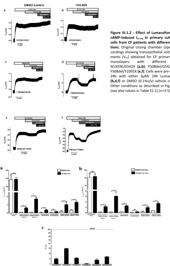
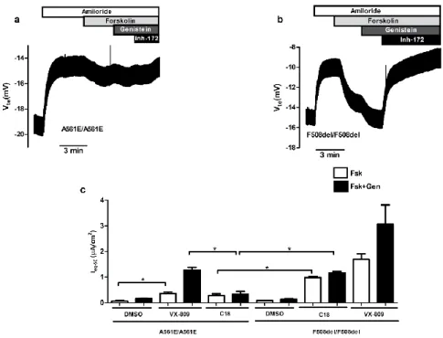
Chapter 1 focusses on the measurement of functional responses on human bronchial epithelial cells (HBE’s) derived from CF lung explants bearing different CFTR mutations to VX-809 namely: A561E, N1303K, G542X and Y1092X. Our data showed a positive response of A561E/A561E to VX-809 and F508del/Y1092X but not F508del/G542X.
In Chapter 2, we evaluated the efficacy of CFTR modulators (correctors/potentiators) in physiologically relevant tissues, namely rectal biopsies, intestinal organoids, (HBE’s) and human nasal epithelial cells (HNE’s), from CF patients with rare CFTR mutations. Data obtained here showed that neither R560S nor H1079P-could not be rescued by any of the CFTR modulators, but 3849+10kbC>T and R334W and c.120del23-CFTR were rescued by VX-770 alone or with VX-809.
In Chapter 3 we evaluated the efficacy of two novel CFTR correctors (B9, E12) in primary HBE cells, and three novel compounds E-act mimics (C2, C5, and C7) as enhancers of alternative Cl- channel TMEM16A
in human intestinal organoids.
In Chapter 4 (final) we assessed the effect of CFTR modulators and their possible additivity with F508del-CFTR genetic revertants 4RK, R1070W, and G550E to understand the mechanism of action of small molecule correctors and another variant diacidic ER exit code DD/AA in CFBE mCherry cells expressing these varinats by Ussing chamber analysis with or without CFTR modulators. Our data show that C18 and VX-661 and low temperature (But not VX-809) rescued DD/AA to the cell surface and genetic revertants restore the channel function without any CFTR modulator.
Altogether, results from this work bring new insights into how the CFTR genotype may influence CFTR function and response to CFTR modulators and how each patient should be assessed individually for the responsiveness to the CFTR modulators towards personalized therapeutics.
ii
ResumoA Fibrose Quística (FQ) é a doença autossómica recessiva letal mais comum na população Caucasiana, afetando cerca de 1 em 2500-6000 nados vivos, e com uma frequência de portadores de 1 em cada 25 indivíduos. Esta doença é causada por mutações no gene CFTR (do inglês Cystic Fibrosis
Transmembrane Conductance Regulator), localizado no braço longo cromossoma 7. A proteína CFTR é
expressa na membrana apical das células epiteliais, onde funciona como um canal de cloreto (Cl-),
regulando o transporte de sal e de água. A CFTR também é descrita como uma proteína reguladora de outros canais iónicos. Até à data já foram descritas mais de 2000 mutações no gene CFTR, para a mioria das quais porém, as consequências funcionais continuam por esclarecer. A mutação F508del (representando a deleção do aminoácido fenilalanina na posição 508 da proteína) é responsável por 85% dos cromossomas FQ a nível mundial, sendo assim a mutação mais comum nestes doentes. A FQ é uma doença caracterizada por múltiplas manifestações em diferentes órgãos, tais como vias respiratórias superiores e inferiores, pâncreas, ductos biliares, trato gastrointestinal, ductos deferentes, glândulas sudoríparas, algumas células do sistema imunológico e outros tecidos, sendo, contudo, a doença pulmonar a principal causa de morbilidade e mortalidade. Os doentes com FQ podem apresentar quadros clínicos muito diferentes, mas têm em comum a presença dum muco espesso, que impede que o transporte/limpeza mucociliar seja eficiente, levando à retenção de bactérias (principalmente Pseudomonas aeruginosa) que originam infeções respiratórias recorrentes e inflamação crónica, e à perda progressiva da função pulmonar. Além dos sintomas respiratórios, outros sintomas clássicos de FQ incluem uma elevada concentração de electrólitos no suor (parâmetro utilizado no principal teste de diagnóstico), insuficiência pancreática exócrina, cirrose hepática, obstrução intestinal e infertilidade masculina. A esperança média de vida para indivíduos recém-nascidos com FQ em 2010 com a mutação mais comum, foi estimada em 37 anos.
O desenvolvimento de metodologias de alto rendimento (“High-throughput screens”, HTS) transformou de forma significativa o panorama terapêutico, passando duma visão terapêutica uniformizada - "one-size-fits-all" - para uma visão de medicina personalizada, utilizando terapias específicas para cada mutação. Através destas metodologias de HTS foi possível identificar diversas novas moléculas com potencial de correção dos defeitos da CFTR ao nível do DNA, do RNA, ou da proteína. Entre elas, o potenciador, VX-770 (Ivacaftor/Kalydeco), direcionado a indivíduos com a mutação G551D ou outras mutações que afetem a regulação da abertura ("gating") do canal CFTR; a combinação corretor/potenciador VX-809 (Lumacaftor)/VX-770 (conhecida como Orkambi), para indivíduos homozigóticos para a mutação F508del; e mais recentemente (ainda em aprovação) a combinação VX-661 (Tezacaftor)/VX-770 para estes mesmos indivíduos e outros com a mutação F508del e uma segunda outras mutação com atividade residual. Contudo, a combinação VX-809/VX-770 apenas demonstrou ter um efeito moderado na melhoria da doença pulmonar. O mecanismo de ação destes compostos ainda não é totalmente conhecido e, apesar dos avanços na terapêutica da FQ, nem todas as mutações da CFTR são passiveis de correção farmacológica. Por isso, torna-se pertinente avaliar as respostas a novos moduladores da CFTR e aos existentes por parte de outras mutações em modelos ex vivo, como tecidos/células primárias dos pacientes (por exemplo células epiteliais nasais humanas, células brônquicas humanas) ou como biópsias retais, permitindo assim não só avaliar a eficácia do modelador para um determinado genótipo da CFTR, como também permitir prever o benefício clínico de forma individualizada para cada paciente. A ativação, através de fármacos, de outros canais de Cloreto (como por exemplo o TMEM16A) continua a ser uma estratégia atrativa para compensar a ausência de atividade da CFTR para compensar a ausência de canais CFTR. A ativação
iii
funcional do TMEM16A poderá ajudar em particular os pacientes com FQ cujas mutações são insensíveis a terapias moduladoras da CFTR.
O principal objetivo deste trabalho de doutoramento consistiu no estudo de novos compostos que corrigem o defeito básico desta doença, resgatando a proteína CFTR (Cystic Fibrosis Transmembrane Conductance Regulator) que se encontra mutada na doença genética Fibrose Quística (FQ), focando-se quer nas respostas individuais de pacientes com FQ e diferentes mutações CFTR a estes novos fármacos, quer no seu mecanismo de ação.
O primeiro objetivo (Capítulo 1) desta tese focou-se na quantificação da atividade da CFTR em culturas primárias de células epiteliais brônquicas (Human bronchial epithelial cells - HBE’s), derivadas de pulmões de pacientes com FQ. Com este intuito, foi avaliado o efeito do VX-809 em mutações já descritas como tendo defeitos semelhantes à F508del (classe II) - A561E e N1303K - e mutações de classe I, como as G542X e Y1092X. Para tal, utilizámos monocamadas de células HBEs cultivadas em filtros porosos e posteriormente analisadas em micro câmaras de Ussing com perfusão continua. Os resultados mostraram que a mutação A561E em homozigotia responde positivamente ao tratamento com VX-809, com um aumento de 7 vezes comparativamente a células controlo incubadas com DMSO, representando cerca de 6% de recuperação quando comparadas com células sujeitos controlo (indivíduos sem FQ). O VX-809 também mostrou ter um impacto positivo no genótipo F508del/Y1092X, observando-se uma resposta de 7% comparativamente a células controlo, e quase o dobro do observado para o genótipo F508del/G542X (cerca de 4% vs células controlo). Deste estudo também podemos observar que as células que apresentam somente uma cópia da mutação F508del têm uma resposta menor ao tratamento com VX-809. Adicionalmente, observámos diferenças significativas nas respostas ao VX-809 entre células de diferentes pacientes homozigóticos para a mutação F508del (por exemplo, Dador 1 – cerca de 5%, e Dador 2 – 15% de recuperação da atividade da CFTR vs células normais). Estes estudos demonstram a importância de análises de modelos ex vivo para uma terapêutica mais personalizada na FQ e reforça o tópico principal deste estudo: que cada paciente deveria ter uma avaliação individual da capacidade de resposta aos diferentes moduladores da CFTR. No Capítulo 2, o principal objetivo foi avaliar a eficácia dos moduladores da CFTR (Corretores/Potenciadores) em tecidos fisiologicamente relevantes. Para isso utilizámos diversos modelos celulares, como biópsias rectais (realizando o mesmo protocolo usado no diagnóstico de FQ), organoides intestinais derivados dessas bióipsias, e culturas primárias de células brônquicas e de células nasais. Estes estudos focaram-se principalmente em indivíduos com mutações no gene da CFTR extremamente raras (chamadas mutações órfãs). As mutações raras estudadas neste trabalho foram: R560S, H1079P, 3849+10kbC>T, R334W e c.120del23.
Resumidamente, para caracterizar e avaliar a eficácia dos moduladores da CFTR em células com o genótipo R560S, usámos material de pacientes - organoides intestinais – assim como linhas celulares CFBE expressando esta mutação de forma estável. Os resultados mostram que não houve correção da mutação R560S com nenhum dos moduladores testados, nem nas células CFBE nem nos organoides intestinais, ao contrário do que foi observado para a mutação F508del. Estudos funcionais e bioquímicos da CFTR com a mutação H1079P revelaram que esta proteína mutante não apresenta função como canal de cloreto. O uso dos moduladores CFTR e agentes “read through” (para ultrapassar codões stop prematuros) não aumentaram a função da CFTR em materiais de pacientes com o genótipo H1079P/W1282X e a respetiva medição da atividade basal quer por camara de Ussing quer em organoides intestinais mostrou ausência de atividade da proteína CFTR. A caracterização funcional
iv
de organoides intestinais com o genótipo 3849+10kbC>T mostrou que esta mutação apresenta função residual da CFTR. O tratamento com VX-809 não teve qualquer efeito na atividade residual da mutação 3849+10kbC>T mas esta foi significativamente melhorada pelo potenciador, clinicamente aprovado, VX-770, sozinho ou combinado com o VX-809. A análise da atividade basal da CFTR em três tecidos diferentes (biópsia rectal, organoides do intestino e células do epitélio nasal) de pacientes com a mutação R334W demonstrou atividade residual de CFTR com esta mutação. Além disso, a combinação do corretor VX-809 e dos potenciadores Genisteina ou VX-770 (com ou sem VX-809) teve um efeito positivo na atividade da CFTR tanto nos organoides intestinais como nas células respiratórias. No entanto verificou-se alguma variabilidade na resposta dos 3 diferentes pacientes com esta mutação. A avaliação funcional da atividade da CFTR em linhas celulares com a mutação c.120del23 mostrou uma resposta positiva ao tratamente apenas com potenciador ou qualquer combinação de corretor/potenciador face à resposta basal (células tratadas com DMSO).
Com este objetivo, conseguimos estabelecer metodologias para a análise da CFTR em culturas de células humanas do epitélio nasal e organoides intestinais, duas ferramentas extremamente importantes para analisar a atividade da CFTR de forma personalizada para cada paciente.
Estes estudos evidenciam a importância de avaliar os efeitos dos moduladores da CFTR em diferentes modelos fisiológicos, para uma melhor caracterização da resposta de cada modelador antes da sua administração a pacientes com FQ.
O terceiro objetivo (Capítulo 3) deste trabalho focou-se na avaliação da eficácia de dois novos corretores da CFTR (B9 e E12) em céulas derivadas de pulmões explantados de pacientes com FQ. Adicionalmente, identificámos três novos compostos - análogos ao E-act, um estimulador do canal alternativo de cloreto TMEM16A (C2, C5 e C7) que parecem ter um efeito positivo na função deste canal. Com efeito, os resultados obtidos mostram que o composto E12, em conjunto com o VX-809, tem um efeito aditivo significativo. Estudos em organoides intestinais com os análogos do E-act levaram à identificação de três estimuladores da função do canal TMEM16A. Mostrámos que o composto C2 leva a um aumento significativo da resposta ao ATP em organoides intestinais e, consequentemente, na função do canal TMEM16A. Os resultados obtidos demonstraram assi que estas células são uma boa ferramenta para a descoberta de novos corretores da CFTR, assim como estimuladores do canal TMEM16A.
No quarto e último objetivo (Capítulo 4) deste trabalho pretendia-se avaliar o efeito dos moduladores da CFTR e a possível aditividade com os revertentes genéticos da F508del 4RK, R1070W, G550E – e da variante DA/AA em linhas celulares CFBE que os expressam estavelmente. Os resultados obtidos mostraram que o defeito de tráfego da variante DA/AA-CFTR foi corrigido pelos corretores C18 e VX-661, bem como pela incubação das células a baixa temperatura. Porém, o VX-809 teve um efeito moderado nesta variante. Demonstrou-se ainda que os revertentes F508del-4RK, F508del-R1070W e F508del-G550E restabeleciam alguma função do canal mesmo na ausência dos moduladores. Os compostos aqui estudados demonstram que existe variabilidade no efeito aditivo com os revertentes genéticos, fornecendo assim pistas para um possível mecanismo de ação, exercido através da ligação a motivos estruturais específicos.
Em resumo, os resultados obtidos durante o presente trabalho de doutoramento trouxeram novos conhecimentos sobre a influência do genótipo CFTR na função deste canal e na resposta a moduladores, e como sobre a importância, quer de sistemas celulares heterólogos, quer de materiais
v
derivados dos pacientes (e fisiologicamente mais relevantes) para a análise das respostas funcionais desta proteína com diferentes mutações, em abordagens de medicina personalizada.
vi
AcknowledgementsForemost, I would like to express my sincere gratitude to my Supervisor Professor Margarida D Amaral for the continuous support of my PhD study and related research, for her patience, motivation, and immense knowledge. Besides this, I want to thank Professor Margarida Ramos for introducing me to the world of Physiology and partly supervising my progress and giving valuable inputs during PhD. I want to thank present and past members of the Amaral lab for making my five years stay in Portugal memorable. Thanks to Marisa Sousa for sharing all the knowledge about Ussing chamber. Thanks to Simao Luz, Veronica Felício, Goncalo Prista, Susana Igreja, Sara Canato, Sara Afonso, Joao Fernandes, Joao Amorim, Ana Marta Romao, Inna Uliyakina, Ana Cachaco and Hugo Botelho who made my initial stay in Lisbon (Junior Fellowship period) very pleasant and comfortable. Thanks to Joao Santos, Ines Pankonien, Sofia Ramalho, Iris Lameiro, Iris Silva, Joana Lérias, Margarida Quaresma, Madalena Pinto, Catarina Baptista, Luis Sousa, Filipa Simoes, Arsénia Massinga, Karina Mendes and Miqueias Lopes. Thanks to my Lab managers Marta Palma, Jose Murias (for sharing all the useful information about Photography) and Sofia Correia for providing all the necessary lab reagents whenever required. Thanks to Lab secretary Renata Vincent, Filipa Coutinho, Carolina Varela and Andreia Reis for handling all administrative and beaurocracy related issues during my stay at FCUL. Thanks to Luis Marques for IGC trips for Organoids swelling assays. My sincere thanks to Professor Jeffery Beekman for providing me an opportunity to spend six weeks in his lab in Utrecht and kindly sharing all the technical aspects related to Organoids technique.
Thanks to PI’s from the lab, Professor Luka Clarke (for CF subject sample collection and Thesis proofreading), Professor Paulo Matos, Professor Anabela Ramalho and Professor Carlos Farinha for all the scientific discussions. Thanks to Doctors and Nurses from Santa Maria and D Estefania hospital for providing all the patient related samples. Big thanks to CF Subjects for contributing to CF research by providing necessary material. Thanks to our collaborators from Valencia and Prague for sending very valuable CF/non-CF lung explants. Thanks to my colleagues from BioSys PhD program. Thanks to IGC microscope unit for providing all the facilities for Organoids imaging. Thanks to C8 building security staff for letting me work during weekends. Thanks to FCT for my PhD Fellowship. Finally, thanks to my Flat partners Mohammad, Andre, and Joao for sharing an apartment with me for 5 years.
Last but not the least, I want to thank my Family; my parents and to my brother Nandan and Sister Neelima, Monali and my Dog Lili for supporting me throughout my PhD.
vii
According to the provisions of article 31 of the Regulation of Postgraduate Studies of the University of Lisboa, Dispatch no. 2950/2025, published in the Diário da República - 2nd Series - nº 57 - March 23, 2015, results were used in this dissertation were included in the following articles:
1. Awatade NT, Ramalho S, Felício V, Silva IAL, Botelho HM, De Poel E, Vonk A, Beekman JM, Farinha CM, Amaral MD (2018) R560S: a class II CFTR mutation that is not rescued by current modulators.
Manuscript submitted to Journal of Cystic Fibrosis.
2. Awatade NT, Uliyakina I, Farinha CM, Clarke LA, Mendes K, Solé A, Pastor J, Ramos MM, Amaral MD (2015) Measurements of Functional Responses in Human Primary Lung Cells as a Basis for Personalised Therapy for Cystic Fibrosis. E-Biomedicine 2: 147-153. [PMID: 26137539]
In compliance with the provisions of the aforementioned regulation, the author clarifies that the ex-periments that led the elaboration of the results presented here, as well as the interpretation and discussion thereof were his responsibility, except when stated otherwise. The results obtained by other authors were included with their authorization to facilitate the understanding of the works and are indicated in the respective figures and methodologies.
In addition to the above, there were additional articles published in international journals containing results obtained during the present PhD:
1. Liu J, Bihler H, Farinha CM, Awatade NT, Romão AM, Mercadante D, Cheng Y, Musisi I, Jantarajit W, Wang Y, Cai Z, Amaral MD, Mense M, Sheppard DN (2018) Partial rescue of F508del-CFTR chan-nel gating with modest improvement of protein processing, but not stability by a dual-acting small molecule. Br J Pharmacol. [DOI: 10.1111/bph.14141] [PMID: 29318594].
2. Lérias JR*, Pinto MC*, Botelho HM, Awatade NT, Quaresma M, Silva IAL, Wanitchakool P, Schreiber R, Pepperkok P, Kunzelmann K, Amaral MD (2017) A Novel Microscopy-Based Assay Identifies Ex-tended Synaptotagmin-1 (ESYT1) as a Regulator of Anoctamin 1 Traffic. BBA- Mol Cell Res 1865: 421-431. (*1st co-authorship) [PMID: 29154949]
3. Pereira JFS, Awatade NT, Loureiro CA, Matos P, Amaral MD, Jordan P (2016) The third dimension: new developments in cell culture models for colorectal research. Cell Mol Life Sci 73: 3971–3989 [PMID: 27147463]
4. Srivastava JK*, Awatade NT*, Bhat HR, Kmit A, Mendes K, Ramos M, Amaral MD, Singh UP (2015) Pharmacological evaluation of Hybrid thiazolidin-4-one-1,3,5-triazines for NF-κB, biofilm and CFTR activity. RSC Adv 5: 88710. (*1st co-authorship) [DOI: 10.1039/c5ra09250g].
5. Botelho HM, Uliyakina I, Awatade NT, Proença MC, Tischer C, Sirianant L, Kunzelmann K, Pepper-kok P, Amaral MD (2015) Protein Traffic Disorders: an Effective High-Throughput Fluorescence Mi-croscopy Pipeline for Drug Discovery. Sci Rep 5: 9038. [PMID: 25762484].
viii
Table of Contents
Summary ... i
Resumo ... ii
Acknowledgements ... vi
List of Abbreviations ...xi
I. GENERAL INTRODUCTION ... 1
1 Cystic Fibrosis Overview ... 2
1.1 A Brief History ... 2
1.2 Pathophysiology of Cystic Fibrosis ... 2
2 The CFTR gene, Mutations, and Protein ... 3
2.1 Functional Classes of CFTR mutations ... 4
2.2 CFTR protein – Biosynthesis, Trafficking, Degradation and Endoplasmic Reticulum
Quality Control ... 5
2.3 The Cl
-Channel Function of CFTR ... 7
2.4 Regulation and Activation of the CFTR Cl
-channel ... 7
2.5 Recent Advances in CFTR structure ... 8
3 Electrolyte transport in CF... 9
3.1 Sweat glands ... 10
3.2 Airways ... 11
3.3 Intestinal tract ... 11
3.3 CFTR as Regulator of Epithelial Ion Trasnport ... 12
3.4 CFTR as regulator of other ion channels ... 13
4 CFTR Function Measurements to Establish a Diagnosis of CF ... 14
4.1 Sweat Test ... 14
4.2 Nasal Potential Difference ... 15
4.3 Voltage/Current Measurements in Rectal Biopsies ... 16
4.4 Evaporimetry Test ... 17
5 Novel Biomarkers Based on CFTR Function Measurements ... 18
5.1 Human Bronchial Epithelial Cells (HBE’s) ... 18
5.2 Human Nasal Epithelial cells (HNE’s) ... 19
5.3 Intestinal Organoids ... 19
6 Cystic Fibrosis Therapeutic Approaches ... 21
6.1 CFTR modulators – Personalised Medicine ... 21
6.2 Repair of CFTR protein synthesis ... 21
6.3 Repair of CFTR protein folding and trafficking ... 22
ix
6.5 Repair of Defective CFTR Splicing ... 24
6.6 Alternative non-CFTR Cl
-secretory pathways ... 24
7. Objectives of Present PhD Work ... 27
II. MATERIALS AND METHODS ... 28
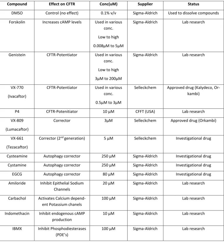
2.1 Chemicals, Compounds, and Statistical Analysis ... 29
... 30
2.2 Primary cell Culture ... 31
2.3 Stably Expressing cell lines ... 33
2.4 Functional analysis ... 33
2.4.2 Ussing chamber ... 34
2.5 The Forskolin Induced swelling assay in Primary Intestinal Organoids ... 35
2.6 Western Blot ... 36
2.7 Rectal biopsy immunostaining ... 36
2.8 mRNA and extraction from native cells, cDNA synthesis and transcript analyses ... 36
III. RESULTS AND DISCUSSION ... 37
Chapter 1. Measurement of Functional Responses on Human Primary Lung Cells as a
Basis for Personalized Therapy for Cystic Fibrosis ... 38
1.1 Abstract ... 38
1.2. Introduction ... 39
1.3 Results ... 40
1.4. Discussion ... 43
Chapter 2. Correlations among different CFTR biomarkers in patient-derived materials
... 46
2.1 R560S is a class II mutation that is not rescued by current modulators ... 46
2.1.1. Abstract ... 46
2.1.2. Introduction ... 47
2.1.3. Results ... 48
2.1.4 Discussion ... 52
2.2. Functional Assessment of Rare CFTR H1079P Mutation ... 54
2.3. Functional Assessment of Correctors and Potentiators on Organoids with the
3849+10kbC>T/F508del CFTR Genotype ... 63
2.4 CFTR Modulators Enhance Function R334W-CFTR both in Intestinal Organoids and
Conditionally Reprogrammed Human Nasal Epithelial Cells... 69
2.5 Assessment of the Efficacy of Correctors and Potentiators on Rare CFTR 120del23
(Class VI) Mutation ... 78
Chapter 3. Assessment of Novel Compounds Rescuing F508del-CFTR and Enhancing
TMEM16A Function in Human Epithelial Cells/ Tissues ... 84
x
Chapter 4. Additivity of CFTR modulators with Genetic Revertants of F508del-CFTR ... 90
IV. CONCLUSIONS AND FUTURE PERSPECTIVES ... 99
IV.1 Patient-derived models used to study CFTR modulators ... 100
IV.2. Personalized therapies: Repurposing approved drugs for rare CFTR mutations . 102
IV.3 Assessment of the effect of Compounds as potential therapeutic agent for CF .. 104
IV.4 Additivity of CFTR modulators with Genetic revertants of F508del-CFTR ... 104
IV.5 Future Perspectives ... 104
V. REFERENCES ... 105
xi
List of Abbreviations
aa Amino acid
ABC ATP-binding cassette
ACTV Amphotericin, ceftazidime, tobramycin, and vancomycin (antibiotics cocktail) ALI Air liquid interface (culture)
ASL Airway surface liquid
ATP Adenosine 5 Trisphosphate
AUC Area under the curve
BEGM Bronchial epithelial growth medium
BHK Baby hamster kidney cell line
Ca2+ Calcium ion
CaCC Calcium-activated Cl- channel
cAMP Cyclic Adenosine 5’ Monophosphate
CBAVD Congenital bilateral absence of the vas deferens
CCH Carbachol
CF Cystic fibrosis
CFBE41o Cystic fibrosis human bronchial epithelial cell line CFTR Cystic fibrosis transmembrane conductance regulator
Cl- Cl- ion
COP Coat protein I/II
CRC Conditionally reprogrammed cells
CRISPR Clustered regularly interspaced short palindromic repeats
DD/AA Di-acidic code
DIOS Distal intestinal obstruction syndrome
DMSO Dimethyl sulfoxide
DMEM Dulbecco’s modified eagle medium
DMEM-F12 1:1 mixture of DMEM with F12 Ham’s medium
DTT Dithiothreitol
EDTA Ethylenediamine tetraaceticacid
ENaC Epithelium sodium channel
ER Endoplasmic reticulum
ERQC Endoplasmic reticulum quality control
FDA Food and drug administration
FEV1 Forced expiratory volume in first second
FIS Forskolin induced swelling assay
Fsk Forskolin
FVC Forced vital capacity
Gen Genistein
H+ Hydrogen ion
HBE Human bronchial epithelial cells
HCO3- Bicarbonate ion
Hdj Human DnaJ homologue
HEPES N-(2-hydroxyethyl)-piperazine-N-(2-etanesulphonic acid)
HNE Human nasal epithelial cells
Hsp Heat shock protein
HTE Human tracheal epithelial cells
HTS High throughput screening
IBMX 3-isobutyl-1-methylxanthine
Ieq-sc Equivalent short-circuit current
K+ Potassium ion
xii
MI Meconium ileus
MoA Mechanism of action
MSD Membrane spanning domain
Na+ Sodium ion
NBCS New born calf serum
NBD Nucleotide binding domain
NCM Noggin conditioning medium
NHEJ Non-homologous end joining
NMD Non-sense mediated decay
NPD Nasal potential difference
Orkambi Lumacaftor and Ivacaftor combination ORCC Outwardly rectifying Cl- channel
PCL Periciliary layer
PCR Polymerase chain reaction
PBS Phosphate buffer saline
PDE Phosphodiesterases
PGE Prostaglandins
PK Protein kinase
PI Pancreatic Insufficiency
PS Pancreatic Sufficiency
PTC Premature termination codon
QPIT Quantitative pilocarpine iontophoresis
RCM Rspondin conditioning medium
RD Regulatory domain
ROCK Rho A kinase
ROMK Renal outer medullary K+ channel
RT Room Temperature
Rte Transepithelial resistance
SDS Sodium Dodecyl Sulfate
SLC26 Solute carrier 26 family
UTP Uridine 5’Triphosphate
Vte Transepithelial voltage
% v/v Percentage expressed in volume/volume
WCM Wnt-3a conditioning medium
1
2
1 Cystic Fibrosis Overview
1.1 A Brief History
“Woe to the child which when kissed on the forehead tastes salty. He is bewitched and soon must die”.
[This text from Northern European folklore is the first reference to the disease known today as Cystic Fibrosis] Cystic Fibrosis (CF) is the most common life-shortening genetic disease among Caucasians with an incidence of 1 in 2500-6000 live births and affects approximately 80,000 people worldwide (Bobadilla et al, 2002, Farrel, 2008, Rodrigues et al, 2009). In 1936, Fanconi et al were probably the first to refer to the disease as “Cystic fibrosis with bronchiectasis” (Fanconi et al, 1936). In 1938, Dr Dorothy Andersen (US) gave the first clear pathophysiological description of CF: she noted that most destruction occurred in the pancreas and so called it “Cystic Fibrosis of Pancreas” (Andersen DH, 1938). In 1946, Andersen and Hodges presented the first conclusive evidence that CF was a genetic disorder, by studying the pattern of disease inheritance in families. They concluded that this disease resulted from an autosomal recessive mutation whereby two copies of the mutant gene were needed to cause the disease (Anderson & Hodges, 1946). A heat wave in New York in 1949, resulted in an increase of the treatment for dehydration of children with CF compared to other children (Kessler & Andersen, 1951). Surprised by this event Paul di Saint Agnese noticed that children with CF lost an excessive amount of salt in sweat (Di Sant’Agnese et al, 1953). This significant observation had clinical benefits and resulted in the establishment of a sweat chloride (Cl-) test by Gibson & Cooke (1959), the first
method of diagnosis, which is still used today (for more details refer 4.1).
In the 1980’s more knowledge was gained about the molecular basis of the disease by Quinton PM, who used sweat ducts to identify altered Cl- transport (Quinton, 1983). About the same time, Knowles
and Boucher identified increased sodium reabsorption (Knowles et al, 1983). In 1989, the CF gene was discovered by the joint efforts of three research groups - Lap-Chee Tsui’s in Toronto, Francis Collins’ in Michigan and Robert Williamson’s in London - and its identity was verified using cells derived from sweat ducts (Kerem et al, 1989, Riordan et al, 1989, Rommens et al, 1989). This gene encodes a cAMP-regulated Cl- channel, the Cystic Fibrosis Transmembrane Regulator (CFTR). Finally, the cause of the CF
was known to be linked to mutations in this protein. The first mutation identified was F508del-CFTR, corresponding to a three-base pair deletion that leads to an absence of phenylalanine at position 508 of the CFTR protein. In 1990, Drumm et al performed patch-clamp analysis of whole-cell clones and showed that the anion efflux responses were due to cAMP stimulation of Cl- conductance (Drumm et
al, 1990).
1.2 Pathophysiology of Cystic Fibrosis
CF is caused by mutations in the CFTR gene, which encodes the CFTR protein, a protein that plays a major role in Cl- transport and HCO
-3 conductance, as well as in regulating other ion channels and
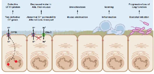
transporters. CFTR is a transmembrane protein expressed at the apical surface of epithelial cells and in glands that produce mucus, sweat, saliva, tears, and digestive enzymes. CF causing mutations prevent the channel from proper functioning, leading to abnormal ion transport a process that leads to dehydration of the airway surface liquid (ASL) (Figure I.1.1). As a consequence, CF airways produce abnormally thick, sticky mucus, which impairs mucociliary clearance (MCC) and obstructs the smaller airways, causing disseminated bronchiectasis - a characteristic feature of CF. The thick mucus and reduced MCC also traps bacteria (eg. Pseudomonas aeruginosa) in the airways originating recurrent respiratory infections and chronic inflammation, which leads to progressive loss of lung function.
3
Besides these respiratory symptoms, classical forms of CF also include a high Cl- concentration in the
sweat (used for CF diagnosis), exocrine pancreatic insufficiency (PI) - present in 85% of subjects with CF, hepatic cirrhosis, intestinal obstruction (includes distal intestinal obstruction syndrome at a later age and meconium ileus that occurs in 10-17% of subjects within the first days of life) and male infertility due to azoospermia attributed to congenital absence of vas deferens, although at variable presentations (Welsh & Smith, 1995; Zielesnki & Tsui 1995; Rowe et al 2001; Bell et al 2013).
Figure – I.1.1 A cascade of pathophysiology in CF lung disease. The mechanism of CF dysfunction starts with
the primary CFTR gene defect and ultimate leads to severe lung deficiency. CFTR, cystic fibrosis transmembrane conductance regulator; ASL, airway surface liquid; ENaC, epithelial Na+ channel. [Adapted
from Amaral MD, 2015]
2 The CFTR gene, Mutations, and Protein
The CFTR gene (or ABCC7) is a large gene (~190 kb) located on the long arm of chromosome 7, band 31-32 (7q31-q32). The CFTR gene comprises of 27 coding exons that after splicing result in a mRNA of about 6.5kb which is translated into a protein with 1480aa residues with a molecular weight of ~170kDa. The protein encoded by the CFTR gene is a Cl- channel located in the apical membrane of
epithelial cells (Riordan et al, 1989). Structure of the CFTR gene, mRNA and protein are shown in figure I.1.2.
Figure I.1.2 - From CFTR gene
to protein. Scheme
representing the CFTR gene, mRNA and protein. N –
N-terminus; TM –
transmembrane domain; NBD – nucleotide-binding domain; R – regulatory domain; C – C terminus [Image from MD Amaral, included here with permission].
4
2.1 Functional Classes of CFTR mutationsAlmost 2,000 CFTR variants have been reported so far in the cystic fibrosis mutation database (www.genet.sickkids.on.ca), which can be grouped into seven classes (De Boeck & Amaral, 2016). These classes can be also called ‘theratypes’ since they are defined by the respective CFTR modulator strategy according to the respective molecular/cellular defect (figure I.1.3). The rationale is that mu-tations within the same class can be treated by the same therapeutic strategy. Regarding severity, Class I-III, VI and VII are associated with severe CF phenotype that consists of a major lung, pancreatic and liver disease, while class IV and V are associated with milder CF and also a higher survival rate.
Figure I.1.3 - The classification system for CFTR mutations. Class I mutations disrupt protein synthesis; class
II impair CFTR protein traffic to the cell surface; class III affect gating of the channel; class IV cause reduced conductance; class V lead to reduced levels of CFTR protein, class VI affect the stability of the protein at the cell surface; class VII are responsible for a total absence of mRNA and are considered at present ‘unrescuable’ by small molecules targeting CFTR. [Figure adapted from De Boeck & Amaral, 2016]
Class I mutations are responsible for altering the production of protein and this class mainly includes
the nonsense mutations and those that produce a premature termination codon (PTC) in the nucleotide sequence (mainly due to frameshift or splicing). These PTC produce truncated CFTR proteins that do not reach the cell membrane. 10% of the worldwide CF population is affected by this class of mutations (CF genetic analysis consortium). Examples of this class of mutations are G542X (Described in Chapter 1) and R553X and patients with these mutations have minimal CFTR function.
Class II mutations are those that result in defective CFTR trafficking/processing and are thus unable to
reach the cell surface. These mutations lead to ubiquitination and degradation in the endoplasmic reticulum and Golgi body. Small amounts of CFTR do reach the cell membrane but they mainly display abnormal ion transport. One example is the F508del mutation, which accounts for 70% of the CF cases worldwide. This mutation is responsible for both defective trafficking and gating and the patients bearing this mutation have minimal CFTR function.
Class III mutations do not affect CFTR traffic to the apical membrane but these proteins have defects
in gating/regulation, which means there is no significant movement of Cl- ions through the CFTR
channel. G551D and S1251N are examples of this class. Patients with these mutations have minimal CFTR function.
Class IV mutations also do not affect CFTR traffic to the apical membrane but these proteins have
defective conductance properties which reduce mobility of the Cl- ions through the CFTR channel.
5
and are located within the membrane spanning domains. Patients with these mutations have some residual CFTR activity.
Class V mutations produce a remarkably reduced amount of CFTR protein however it preserves the
normal function at the apical plasma membrane. These mutations are caused by alternative splicing defects that result in improper processing of mRNA. For example, 3849+10kbC>T (Described in Chapter 2.3) and 2789+5G>A are two class V mutations and patients with these mutations have residual CFTR activity.
Class VI mutations create increased protein instability and shorter residence time at the cell surface,
mainly caused by C-terminal truncation. In this class of mutations biosynthesis, processing and macroscopic Cl- channel function of truncated CFTRs are essentially normal, however the degradation
rate of the mature, complex-glycosylated form is 5- to 6-fold faster than the wild type CFTR (Haardt et al., 1999). Rescued F508del and c120del23 (Described in Chapter 2.5) belong to this class.
Class VII mutations were proposed (Boeck & Amaral, 2016) and this class arose from the division of
the traditional class I mutations into a new class I (with stop-codon mutations) and a new class VII (with no mRNA transcription). Mutations from this class are called unrescuable because they cannot be rescued by pharmacological agents. N1303K (described in Chapter 1) and R560S (described in Chapter 2.1) mutations are two examples and these mutations remained unresponsive to available CFTR pharmacological agents.
Although this classification is helpful to rationalize therapies, for a high number of CFTR mutations (especially for rare ones) it is still unknown what are the associated defects and the respective mutation classes. Variation in severity of disease within patients strongly influenced by factors such as polymorphisms in the CFTR gene, modifier genes, environmental factors and nutritional status may also worsen the severity of different CF mutations. This explains why it is difficult to predict the clinical outcome of an individual patient based only on the CFTR genotype. The most common Class II mutation - F508del-CFTR - alters the CFTR mRNA structure and reduces the translational efficiency. However even knowing that F508del is a typical class II mutation, other detailed studies also show that it belongs to at least two additional classes - (Lazrak, et al 2013). Moreover, it is often found that patients with the same CFTR genotype (e.g., F508del/F508del) have significantly different clinical responses to CFTR modulating drugs (Wainwright et al, 2015; Awatade et al, 2015).
2.2 CFTR protein – Biosynthesis, Trafficking, Degradation and Endoplasmic Reticulum Quality Con-trol
CFTR protein has to undergo a number of cellular processes and quality checks before it reaches the apical membrane. Like other integral membrane glycoproteins, CFTR assembly begins with the for-mation in the endoplasmic reticulum (ER) where it is core glycosylated (Cheng et al, 1990).
6
Figure I.1.4 - Western blot showing the band B and band C. [Adapted from
O’Riordan et al, 2000]
This immature form of CFTR specifically refers to the Band B, has a molecular mass of about 135-140 kDa on SDS-PAGE (figure I.1.4) and it represents the less complex core-glycosylated form that has not reached the Golgi. After passing through the ER quality control (ERQC), the final step of CFTR pro-cessing occurs in the Golgi with a glycosylation step that involves the conversion of mannose-enriched side chain to a mature complex oligosaccharide attached at the aspargine residues in the fourth extra-cellular loop within MSD2. In Western blot analysis this fully glycosylated form of CFTR is known as band C and has a molecular mass of about 170-180 kDa see figure I.1.4 (Cheng et al, 1990).
The most frequent mutant protein - F508del-CFTR - fails to acquire a native conformation, since it is almost completely retained at the ER, and targeted to degradation via the ubiquitin-proteasome path-way. Stringent quality control mechanisms in the ER can discriminate normally folded from abnormally folded proteins and ensure that only correctly folded proteins exit the ER and undergo Golgi matura-tion (Farinha and Amaral, 2005).
Figure I.1.5 - Fate of CFTR molecules synthesized on ER-associated ribosomes. [Adapted from Riordan, 1999]
CFTR is cotranslationally incorporated into the ER membrane. Core-glycosylated chains are attached to the protein, to which calnexin binds. In addition, cytosolic chaperons such as Hsc70, Hdj-2 and CHIP bind and ubiquitination may occur. Hsc70 and its co-chaperone Hdj2 play an important role in the early steps of wt-CFTR biogenesis and facilitate its folding process. Conformational maturation from the ER is accompanied by dissociation of calnexin cytosolic chaperones. Fully loaded CFTR is protected from degradation, but misfolded proteins that do not acquire native conformation are substrates of
7
ubiquitinating enzymes and are degraded by the proteasome. The export competent population trav-els from the ER to the Golgi apparatus, where complex oligosaccharides chains are completed. From the trans-Golgi network mature CFTR is then delivered to the apical membrane by clathrin-coated ves-icles. Endocytic recycling of this protein and degradation of some internalized protein by lysosomal proteases accounts for the turnover of CFTR from the cell membrane.
2.3 The Cl- Channel Function of CFTR
CFTR plays an important role in epithelial Cl- transport, both as a Cl- channel and as a regulator of other
channels and transporters, including the epithelial sodium channel, potassium channels, ATP release mechanisms, anion exchangers, sodium bicarbonate transporters and aquaporin water channels. It is mainly located in the apical membrane of epithelial cells of tissues such as kidney, pancreas, intestine, heart, vas deferens, sweat duct, and lung. CFTR is a member of the ATP-binding cassette (ABC) protein superfamily, a group of transporters that uses the energy of ATP hydrolysis to drive the transport of substrates into (importers) or out of (exporters) the cell (Hwang and Kirk, 2014). After identification of the CFTR gene, several functional studies confirmed CFTR as the affected gene in CF and its protein product as an epithelial cAMP-regulated Cl- channel. Transfection of the CFTR wild-type into cultured
CF airway and digestive epithelial cells regained the affected Cl- transport (Anderson et al, 1991,
Kartner et al, 1991, Bijman et al, 1993, Bear et al, 1992), confirming its role.
Furthermore, compelling evidence by Cheng et al in 1991 through the expression of wt-CFTR in artificial lipid bilayers, resulted in Cl- channels with the characteristic properties of CFTR associated
conductances (Cheng et al, 1991) which include:
a) CFTR has low unitary conductance (6-10pS) (Sheppard & Welsh, 1999; Dawson et al, 1999); b) CFTR shows a linear current-voltage relationship and does not display voltage dependent activation or inactivation (Sheppard & Welsh, 1999);
c) Ion selectivity is measured by “relative permeability” and “relative conductance”. CFTR shows different permeability and conductance to different ions. A CFTR permeability sequence is SCN> NO3> Br> Cl> I> acetate and the corresponding conductance sequence is Cl> NO3> Br>
acetate >I >SCN (Sheppard & Welsh, 1999; Dawson et al, 1999);
d) CFTR is an ion channel and not a pump. Also, its pore must remain open to both sides of the membrane to allow anion diffusion down an electrochemical potential gradient (Hwang & Kirk, 2014)
e) CFTR activity is regulated by cAMP dependent phosphorylation and by intracellular nucleotides (Sheppard & Welsh, 1999).
2.4 Regulation and Activation of the CFTR Cl- channel
CFTR is a member of the ATP-binding cassette (ABC) transporter superfamily, which bind ATP and use the energy to facilitate the transport of substrates across cellular membranes (Hyde et al, 1990, Hig-gins, 1992). CFTR is a large integral membrane protein and it is a cAMP-regulated Cl- channel (Sitton,
8
CFTR protein structure is composed of five functional domains: Two hydrophobic membrane spanning domains (MSD1 and MSD2) each consisting of six transmembrane (TM) helixes (figure I.1.6) form the anion-conducting pore (Sheppard and Welsh, 1999). Two hydrophobic membrane associated domains form the Nucleotide binding domains (NBD 1 and NBD 2) and Regulatory (R) domain.
Figure I.1.6 - Model of the proposed structural do-mains of CFTR. MSD: membrane spanning dodo-mains,
NBD: nucleotide binding domains, R: regulatory do-main, PKA: cAMP dependent protein kinase A. [Adapted from Sheppard and Welsh, 1999]
R domain is a unique feature of CFTR among all ABC transporters and contains many charged residues and multiple consensus phosphorylation sites. The regulatory R domain contains Protein Kinase A (PKA) and Protein Kinase C (PKC) phosphorylation sites and phosphorylation by PKA results in nucleo-tide binding and hydrolysis at the NBD 1 and NBD 2, which in turn causes opening of the channel al-lowing Cl- to move out of the cell (McCarty et al, 2000). Both the amino (N) and carboxyl (C) terminal
tails of CFTR channel are located in the cytoplasm and play a pivotal role in mediating interactions with a variety of binding proteins. These physical interactions are tightly regulated and are important in the compartmentalized regulation of CFTR function. Apart from PKA, there are other kinases reported to be involved in CFTR mediated Cl- secretion such as PKC, src kinase, AMP-dependent protein kinase,
Casein kinase 2 (Seibert et al, 1999; Treharne et al, 2009; Kongsuphol et al, 2009), SYK-spleen tyrosine kinase (Luz et al, 2011) and Serine/threonine kinase WNK4 kinase (Mendes et al, 2011). Apart from cAMP regulation of CFTR Cl- channel, both the β
2-adrenergic and the Adenosine 2b receptor have an
important role in regulating CFTR mediated Cl- secretion in human airways. These two different
signal-ling pathways mainly act through G-protein coupled receptors that release Gs, stimulate AC and raise cAMP which in turns activates CFTR (Hentchel-Franks et al, 2004, Stanton and Guggino, 2006).
2.5 Recent Advances in CFTR structure
Recently Zhang & Chen, 2016 reported the structure of the zebrafish CFTR protein in the dephosphor-ylated state (See figure I.1.7 A) and in the absence of ATP, by cryogenic electron microscopy. Zebrafish CFTR shares about 55% overall sequence identity to human CFTR. The interesting feature of this model was a novel interfacial motif, which was called “lasso motif” because of its shape (shown as a green ribbon in figure I.1.7 A). The lasso motif is located in the vicinity of the R domain (Zhang & Chen, 2016). The main structural features include funnel shaped ion conduction pathway which consists of a large vestibule, positively charged residues in the entire length of the funnel and closure of the channel in the extracellular surface by a single gate.
9
Figure I.1.7 – Zebrafish and human CFTR protein structures. A represents zebrafish dephosphorylated CFTR
structure (PDB ID - 5UAK, adapted from [Zhang & Chen, 2016]); B represents zebrafish phosphorylated CFTR structure (PDB ID - 5W81, adapted from [Zhang et al, 2017]); C represents human dephosphorylated CFTR structure (PDB ID - 5UAK, adapted from [Liu et al, 2017]). MSD 1 - light blue, MSD 2 - Dark blue, NBD 1 - Red, NBD 2 – Orange, Lasso motif - Green, ATP – Sticks, R domain – Yellow, Mg2+ - Green spheres.
Noticeably, authors from this study were able to map 46 positions which are mainly affected by CF causing mutations. In another study by the same authors the structure of zebrafish CFTR in its phos-phorylated/ATP-bound conformation was published (See figure I.1.7 B), and it exhibited many of the features anticipated for the open channel state, such as disengagement of the R domain from the molecular center, dimerization of the NBD’s and a lateral opening for a Cl- flow to the cytosol (Zhang
et al, 2017).
Liu et al 2017 finally illustrated a cryogenic electron microscopy structure for human CFTR (See figure I.1.7 C) in the dephosphorylated/ATP-free form. This structure bears a close resemblance to the zebrafish ortholog. The human CFTR structure reveals an important piece of evidence - the helix be-longing to the R domain docked inside the intracellular vestibule precluding channel opening (see yel-low ribbon in Figure I.1.7 C) (Liu et al, 2017).
In summary, these recently published structures provide a novel molecular understanding and struc-tural knowledge to understand human CF and facilitate innovative therapeutic interventions.
3 Electrolyte transport in CF
Conclusive evidence of increased salt loss in sweat (Di Sant’Agnese et al, 1953; Gibson and Cooke, 1959) and the anomalies in exocrine secretions of CF subjects (Dearborn, 1976) led to comprehensive research of the bioelectrical properties of different epithelial tissues, such as the sweat glands, the respiratory tract and the gastrointestinal tract.
10
3.1 Sweat glandsThe sweat gland primarily consists of a secretory coil and a reabsorptive duct as shown in figure I.1.8. Sweat production in the secretory coil of non-CF individuals is induced by, at least, two different cell components, namely Adrenergic and Cholinergic (Reddy et al, 1992a; Reddy et al 1992b).
Figure I.1.8 - A diagram of the two components and two steps required in the exocrine gland secretion. In
general, as shown here for the sweat gland, the secretory coil (or acinus in other glands) first secretes a primary fluid that is isotonic with the interstitial fluid and is usually stimulated mainly by cholinergic agonists that stimulate normal sweating in CF. Alternatively, adrenergic agonists can stimulate normal glands to secrete primary fluid, but generally at lower maximum rates. The beta-adrenergic stimulation characteristically fails in CF patients due to absence or malfunction of its target, the CFTR Cl- channel in the
apical membrane of secretory cells. As the isotonic fluid is secreted, it is expressed through the remaining tubule (reabsorptive duct) to exit the gland. [Adapted from Quinton, 2007]
Adrenergic stimulation leads to an intracellular increase in cAMP signaling thereby activating CFTR. Cholinergic stimulation increases the intracellular Ca2+ dependent Cl- secretion. In this way, adrenergic
and cholinergic agonists lead to Cl- secretion in the secretory coil, which drives the electrolyte and fluid
secretion to produce sweat that is isotonic with plasma. In CF subjects, the beta-adrenergic stimulation characteristically fails due to the defective CFTR. The cholinergic pathway is not disturbed in CF subjects, which justify the normal Cl- secretion, and production of isotonic sweat in the secretory coil
(Sato et al, 1991).
In the reabsorptive duct, the electrolyte and fluid transport is directed by the basolateral Na+-K+
-ATPase, by pumping Na+ out of the cell (Bijman & Quinton, 1984), thus creating a low intracellular Na+
concentration (Figure I.1.8). This triggers the passive Na+ movement into the cell across the apical
membrane. In non-CF individuals, electroneutrality is maintained by Na+ absorption accompanied by
Cl- diffusion, and since the sweat duct is impermeable to water, sweat becomes hypotonic at the end
of the sweat duct. In CF subjects, an absence of CFTR leads to impaired diffusion of Cl- together with
Na+ absorption, thereby preventing NaCl reabsorption that results in increased Cl- and to a lesser
extent increased Na+ concentration in the sweat of CF subjects. These abnormalities in electrolyte
transport in CF subjects monitored/diagnosed by Sweat Cl- test or by an Evaporimetry procedure (refer
11
3.2 AirwaysCFTR plays an important role either as an absorptive or as a secretory pathway for Cl- /HCO
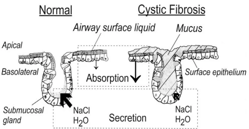
3- ions in
both respiratory and intestinal epithelia. The apical surface of airway epithelium is lined with a thin fluid coating called the Airway Surface Liquid (ASL). This is a two-layer system composed of a pericilliary layer where the cilia beats and typically this layer is composed of 1% mucins, 1% salt and 98% water and varies from 7-70µM in height (Sims and Horne, 2013), with 7µM being the height of the cilia (Harvey et al, 2011) and more superficial gel layer that constitutes an efficient barrier against microorganisms. The mucus is produced by the submucosal glands and goblet cells, while its hydration is controlled by transepithelial transport.
Figure I.1.9 - Model of the airway epithelium consisting of an absorptive surface (expressing epithelial Na+
channels ENaC and (CFTR)) and secretory submucosal glands. [Adapted from Kunzelmann & Mall, 2001]
The regulation of ASL in the normal airway epithelia is ensured by the ENaC, CFTR, and CaCC for the maintenance of PCL volume. Proper hydration/volume of the normal airway surface is maintained by active transport processes that control the mass of salt (NaCl) on airway surfaces, with water following by osmosis (Matsui et al, 2000). The adequate balance between absorption and secretion ensures the net transport of ions across the epithelium. CFTR has shown to have an inhibitory effect on the ENaC channel (Rubenstein et al, 2011) and the defective CFTR in CF subjects is thought to cause ENaC hyperactivation leading to enhanced Na+ absorption followed by increased water absorption out of
the ASL and into the cell by osmosis leads to hyperabsorption in CF epithelium. These results in a reduction in ASL layer, submucosal glands are no longer cleared from mucus, and MCC is largely impaired in CF airways as shown in figure I.1.9 (Kunzelmann and Mall, 2011).
3.3 Intestinal tract
The mechanism of fluid absorption and secretion in the colonic epithelium functions as in the airway epithelium. Epithelium transport in the colon is represented by a net absorption of NaCl, short chain fatty acids (SCFA) and water, allowing very little water and salt content in faeces. Moreover, colonic cells also secrete mucus, bicarbonate, and KCl. The function of the small intestine is especially to secrete fluid, while in the colon mainly fluid absorption takes place (Kunzelmann and Mall, 2002) as shown in the figure I.1.10. Absorption within the colonic epithelium can be electrogenic which occurs via ENaC or is electroneutral via parallel Na+/H+ and Cl-/HCO
12
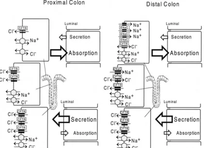
Figure I.1.10 - Models for electrolyte transport in proximal and distal colonic epithelium and expression of different ion transporters along the crypt axis. Electroneutral NaCl absorption (parallel Na+/H+ and Cl-/HCO3
-exchange) dominates in the surface epithelium and is also present in the crypts. Electrogenic Na+ absorption
via the epithelial Na+ channel (ENaC) takes place in the surface epithelium and upper crypts of the distal colon.
The cystic fibrosis transmembrane conductance regulator (CFTR) is expressed throughout the colonic epithelium and dominates in the crypts. [Adapted from Kunzelmann and Mall, 2002]
As shown in figure I.1.10 defective cAMP-mediated Cl- secretion occurs in different regions of the colon
(proximal) and rectum (distal). In Ussing chamber studies, it has been shown that, stimulation of colonic Cl- secretion by increasing both intracellular cAMP and Ca2+. Non-CF subjects upon cAMP and
cholinergic activation display negative voltage deflection whereas tissues from CF subjects show only a K+ secretory response which is depicted as positive voltage deflection in open circuit studies. This
defective colonic transport in CF subjects plays an important role in CF diagnosis (Refer Fig I.1.14).
3.3 CFTR as Regulator of Epithelial Ion Trasnport
Besides its function as Cl- channel, CFTR has also been shown to regulate several other channels and
transporters, thus being a general regulator of ion transport in epithelia. CFTR is found to be expressed in luminal membranes of both secretory and absorptive epithelia, playing a predominant role in both cAMP- and Ca2+-activated secretion of electrolytes (see figure I.1.11).
13
Figure I.1.11 - Cell models of the mechanism of electrolyte secretion and electrolyte absorption in the airway and intestinal epithelia. (a) In secretory cells, Cl- is taken up from the basolateral (blood) side by the Na+-K+-2Cl- (NKCC1) cotransporter. K+
is recycled via basolateral K+ channels, and Na+ is pumped out of the cell by the Na+-K+-ATPase. Cl- leaves the cell via luminal
(apical) CFTR Cl- channels, and Na+ is secreted via the paracellular shunt following the electrical driving force generated by
the lumen negative transepithelial voltage. K+ is also secreted to the luminal side via luminal K+ channels. Depending on the
tissue, intracellular cAMP is increased and secretion is activated by adenosine (airways) or prostaglandin E2 (PGE2, intestine).
(b) In absorptive epithelial cells, Na+ is taken up by ENaC. Cl- is transported via the basolateral shunt and probably via CFTR
Cl- channels. Na+ is pumped out of the cell by the basolateral Na+-K+-ATPase, whereas Cl- and K+ leave the cell via Cl- and K+
channels, respectively. In cells that coexpress CFTR and ENaC, CFTR stimulation and/or expression leads to inhibition of ENaC. Thus, cAMP-mediated stimulation may shift the epithelium from absorption towards secretion of NaCl. [Image reproduced from Kunzelmann and Mall, 2001].
In secretory epithelia, Cl- is absorbed on the basolateral side by the Na+-K+-2Cl- (NKCC1) co-transporter,
accumulating Cl- inside the cell in preparation for secretion through CFTR when it receives the
appro-priate stimulus at the apical membrane (figure I.1.11a). Apart from luminal Cl- secretion, CFTR also
regulates reabsorption of electrolytes by controlling the activity of the amiloride sensitive epithelial Na+ channel (ENaC; figure I.1.11b). When activated via the PKA-dependent pathway, it is believed that
CFTR inhibits ENaC, thus reducing Na+ absorption (Kunzelmann et al., 2001). In CF epithelia, both the
secretion and absorption of electrolytes are found to be impaired.
3.4 CFTR as regulator of other ion channels
In addition to acting as an anion channel, CFTR may act as a regulator of other ionic channels present in epithelia:
1) Stutts et al in 1995 have shown that stimulation of CFTR by cAMP agonists inhibits the amiloride-sensitive epithelial Na+ channels (ENaC) and increased ENaC activity in CF respiratory epithelia
(Stutts et al, 1995).
2) In addition to ENaC, CFTR has also been correlated to the regulation of other Cl- channels such as
the ORCC as presented by Schwiebert in 1999 and Jovov B in 1995 (Jovov et al, 1995; Schwiebert et al, 1999).
3) CFTR is shown to enhance K+ channels function (ROMK-renal outer medullary K+ channels) and
enhancement of the sulphonylurea sensitivity of the ROMK2 (McNicholas et al, 1996).
4) CFTR also plays an important role in the regulation of Calcium-activated Cl- channels (CaCC) (Wei
et al, 2001; Tarran et al, 2002); also regulate TRPV4, which provide the Ca2+ signals for regulatory
volume decrease (RVD) in airway epithelium (Arniges et al, 2004).
5) Apart from these activities, CFTR and the Cl- bicarbonate exchangers (SLC26A3) and (SLC26A6) are
mutually enhanced by a physical association between the R domain of CFTR and the STAS domain of the SLC26 transporters, an effect mediated by PKA dependent phosphorylation of the R domain of CFTR (Ko et al, 2004).
6) Moreover, the presence of CFTR was also detected in intracellular vesicles where it may play a role in intracellular and intravesicular pH regulation (Lukacs et al, 1992; Barasch et al, 1991), CFTR is also shown to control exocytosis/endocytosis processes (Jouret et al, 2007; Raggi et al, 2011). 7) CFTR also provides an “anchoring platform” at the cell membrane where specialized
microdomains anchored to CFTR include PDZ-domain proteins, kinases, transport proteins, myosin motors, Rab GTPases and SNARES (Guggino & Stanton, 2006).
14
4 CFTR Function Measurements to Establish a Diagnosis of CF
Traditionally, the diagnosis of CF is based on clinical symptoms suggestive of the disease and/or a positive family history. Such symptoms include mostly those affecting the airways and the gastrointestinal tract, but also those affecting other systems (see above).
Nevertheless, to confirm a diagnosis of CF, it is necessary to obtain evidence of CFTR dysfunction through the identification of two CFTR gene mutations previously assigned as CF disease causing, two tests showing a high Cl- concentration in sweat (>60 mEq/L), distinctive transepithelial nasal potential
difference measurements and/or assessment of CFTR (dys) function in native colonic epithelia ex vivo (Ratjen & Doring 2003; Rosenstein & Cutting 1998; Farrell et al 2008). For individuals with symptoms suggestive of CF but intermediate sweat Cl- values (30 - 60 mEq/L), the need for additional proof of
CFTR function (through NPD measurements or CFTR functional assessment in rectal biopsies) is particularly important.
4.1 Sweat Test
The term sweat testing generally refers to the quantitative or qualitative analysis of sweat to determine electrolyte concentration, conductivity, or osmolality for the confirmation of a CF diagnosis (Mishra et al, 2005). The sweat test is the most widely used to diagnose CF. It is based on Darling and co-workers (1953) observation that there is a high concentration of Na+ and Cl- in the sweat of patients
with CF. Measurement of sweat was first implemented by Gibson and Cooke in 1959 known as GC technique, also called the quantitative pilocarpine iontophoresis (QPIT), and is considered to be the most accurate method to diagnose CF (Gibson and Cooke, 1959).
Figure I.1.12 - Sweat Cl- concentrations related to CF diagnosis. [Adapted from Davies, 2012]
In a sweat test, localized sweating is produced by iontophoresis of the cholinergic drug, pilocarpine nitrate into the selected area of the skin, where it increases the intracellular calcium concentration and stimulate sweat production by opening the calcium-activated Cl- channels (Mishra et al,2005).
Stimulated sweat is then absorbed by a special filter paper and analyzed for the content of Cl-. CF is
diagnosed when the Cl- concentration in sweat is greater than 60mmol/L (Farrell and Koscik, 1996) as
shown in figure (Fig.I.1.12). The values between 40mmol/L and 60mmol/L are borderline and require follow-up, careful observation, repeat sweat testing and extended CFTR mutation analysis. The normal sweat Cl- values are 10-35mmol/L (Shwachman et al, 1981).
![Figure I.1.5 - Fate of CFTR molecules synthesized on ER-associated ribosomes. [Adapted from Riordan, 1999]](https://thumb-eu.123doks.com/thumbv2/123dok_br/15656880.1059767/21.892.213.628.656.973/figure-fate-molecules-synthesized-associated-ribosomes-adapted-riordan.webp)
![Figure I.1.7 – Zebrafish and human CFTR protein structures. A represents zebrafish dephosphorylated CFTR structure (PDB ID - 5UAK, adapted from [Zhang & Chen, 2016]); B represents zebrafish phosphorylated CFTR structure (PDB ID - 5W81, adapted from [](https://thumb-eu.123doks.com/thumbv2/123dok_br/15656880.1059767/24.892.167.732.57.440/zebrafish-structures-represents-zebrafish-dephosphorylated-structure-represents-phosphorylated.webp)