F
ACULDADE DEM
EDICINA DEL
ISBOA
What is the role of the cell
adhesion protein N-cadherin
in the molecular and
morphological asymmetries
of the Hensen's node
Raquel Valente Mendes
Supervisor | Leonor Saúde, PhD
Doutoramento em Ciências Biomédicas
Especialidade | Ciências Morfológicas
Todas as afirmações efectuadas no presente documento são da exclusiva responsabilidade do seu autor, não cabendo qualquer responsabilidade à Faculdade de Medicina de Lisboa pelos conteúdos nele apresentados.
A impressão desta dissertação foi aprovada pelo
Conselho Cientifico da Faculdade de Medicina de Lisboa
em reunião de (22 de Julho de 2014).
AGRADECIMENTOS i
ABSTRACT iii
RESUMO vi
ABBREVIATIONS ix
INDEX OF FIGURES xi
INDEX OF TABLES xiv
INDEX OF MOVIE LEGENDS xv
C
HAPTERΙ
G
ENERALI
NTRODUCTIONI.1
L
EFT-‐R
IGHTA
SYMMETRY INV
ERTEBRATES2
I.2
S
TEPS INL
EFT-‐R
IGHT ASYMMETRY ESTABLISHMENT5
I.3
B
REAKING THE INITIAL SYMMETRY7
I.3.1 The conceptual F-‐molecule model 7
I.3.2 Different mechanisms to break symmetry 8
I.3.2.1 Motile cilia in the node 8
I.3.2.1.1 Directional Nodal Flow 10
I.3.2.1.2 Two Models for generating Left-‐Right asymmetric gene
expression by Nodal Flow 11
I.3.2.2 Other players in Left-‐Right patterning prior to the
formation of cilia 13
I.3.2.2.1 Gap junction communication (GJC) 16
Ι.3.2.2.2 Ion Flux 17
Ι.3.2.2.3 Motor Proteins/Cytoskeleton 19
Ι.3.2.2.4 Serotonin (5HT) 20
Ι.3.2.3 Putting it all together -‐ Intracellular Model 22
Ι.3.2.4 Planar cell polarity (PCP) 25
Ι.4
C
HICKEN EMBRYO AS A MODEL SYSTEM27
Ι.4.1 Early development of the chick 28
Ι.4.1.1 Cleavage 28
Ι.4.1.2 Gastrulation 30
I.4.2 Hensen's node, a Left-‐Right inducer 37
I.4.2.1 Left side gene expression in the node 40
I.4.2.2 Right side gene expression in the node 42
I.4.3 Left-‐Right asymmetry, downstream of the Node 43
I.4.3.1 Nodal in the left Lateral Plate Mesoderm and asymmetric
gene expression 45
I.4.3.2 Right Lateral Plate Mesoderm and asymmetric gene
expression 46
I.4.4 Stabilization of side-‐specific gene expression –
− Midline Barrier Model 47
I.5
L
ATE STEPS:
A
SYMMETRIC ORGAN MORPHOGENESIS51
I.6
E
VOLUTIONARY CONSERVATION55
I.7
C
ADHERINS57
I.7.1 Cadherins are involved in multiple Biological Processes 60
I.7.2 Structural and functional organization of the cytoplasmic
domain 61
I.7.3 Interactions with the Actin Cytoskeleton 64
I.8
C
ADHERIN EXPRESSION AND FUNCTION IN DEVELOPMENT65
I.8.1 Cadherins and morphogenetic movements 65
I.8.1.1 Convergent and Extension movements 65
I.8.1.2 Border Cell Migration 66
I.8.2 Classical cadherins in cell sorting 67
I.9
N-‐
CADHERIN70
I.9.1 N-‐cadherin in development 71
I.9.1.1 N-‐cadherin and Left-‐Right asymmetries 74
I.9.1.2 N-‐cadherin and collective cell migration 75
I.9.2 N-‐cadherin and cancer 77
I.10
A
IMS AND SCOPES OF THIS THESIS80
COLI BACTERIA
II.2
A
NTI-‐
SENSERNA
PROBE SYNTHESIS88
II.3
O
LIGONUCLEOTIDES90
II.4
P
OLYMERASEC
HAINR
EACTION(PCR)
90
II.5
DNA
CONSTRUCTS91
II.6
E
MBRYOLOGICAL METHODS93
II.7
D
RUG TREATMENT93
II.8
B
EAD IMPLANTATION93
II.9
I
N VITRO CHICK EMBRYO ELECTROPORATION INN
EW CULTURE95
II.10
C
ELL LABELLING IN THEH
ENSEN'
S NODE WITH THE LIPOPHILIC DYESD
II
ANDD
IO
95
II.11
W
HOLE-‐
MOUNT IN SITU HYBRIDIZATION96
II.12
T
ISSUE EMBEDDING AND PREPARATION OF CRYOSTAT SECTIONS96
II.13
I
MUNOFLUORESCENCE98
II.14
F
LUORESCENCE IMAGING98
II.15
L
IVE IMAGING98
II.16
S
TATISTICAL ANALYSIS99
C
HAPTERΙΙΙ
N-
CADHERIN LOCKS LEFT-
RIGHT ASYMMETRYBY ENDING THE LEFTWARD MOVEMENT OF
H
ENSEN'
S NODE CELLS
A
BSTRACT102
I
NTRODUCTION103
III.1
R
ESULTS104
III.1.1 N-‐cadherin asymmetric expression in Hensen's node is generated by the leftward cell movements downstream of the H+/K+-‐ATPase pump
105
III.1.3 Blocking N-‐cadherin activity perturbs Left-‐Right
asymmetry establishment 110
III.1.4 Inhibition of Fgf signalling on the left side of the node, rescues normal gene expression in the left Lateral Plate Mesoderm
114 III.1.5 Overexpression of N-‐cadherin on the right side of the
node does not affect Left-‐Right gene expression in the node and Lateral Plate Mesoderm
116
III.2
D
ISCUSSION118
S
UPPLEMENTARY DATASI.I MNCD2 recognizes chicken N-‐cadherin and weakly blocks
cell-‐cell adhesion 124
SI.2 N-‐cadherin asymmetric activity halts the leftward cell
movements in Hensen's node 128
SI.3 Blocking N-‐cadherin activity perturbs Left-‐Right
asymmetry establishment 129
C
HAPTERΙV
N-
CADHERIN PRESERVES THEA
SYMMETRICMORPHOLOGY OF THE NODE AND ITS
LEFT
-
RIGHT CELL IDENTITY
A
BSTRACT136
I
NTRODUCTION137
IV.1
R
ESULTS139
IV.1.1 N-‐cadherin preserves the asymmetric morphology of the
Hensen's node 139
IV.1.2 Asymmetric number of cells between both sides of Hensen's node underlies its morphological asymmetry 140 IV.1.3 N-‐cadherin prevents cell mixing in the Hensen's 145
IV.2
D
ISCUSSION149
IV.2.1 Asymmetric morphology of the Hensen's node is
C
HAPTERV
N
OTCH TARGET GENE−
CH
ES6-1,
A NOVELPLAYER IN LEFT
-
RIGHTA
SYMMETRY
A
BSTRACT156
I
NTRODUCTION157
V.1
R
ESULTS164
V.1.1 cHes6-‐1 asymmetric expression is downstream of the
H+/K+ATPase pump 164
V.1.2 cHes6-‐1 asymmetric expression requires Nodal signalling 165 V.1.3 cHes6-‐1 asymmetric expression seems to be conserved
between chicken and mouse but not in quail embryos 167
V.2
D
ISCUSSION 170V.2.1 Asymmetric expression of cHes6-‐1 is H+/K+-‐ATPase pump
dependent 170
V.2.2 Nodal signalling activates cHes6-‐1 on the left mesoderm
lateral to the primitive streak 171
V.2.3 cHes6-‐1 asymmetric expression seems to be conserved between chicken and mouse but not in quail embryos 172 V.2.4 A fate map of the cHes6-‐1-‐expressing region is needed 173
C
HAPTERVI
C
ONCLUSIONS ANDF
UTURED
IRECTIONS
C
ONCLUSIONS178
F
UTUREW
ORK182
VI.1 What directs cell movements towards the left side of the
node during stage HH4? 186
VI.2 What is inhibiting fgf8 expression in the rostral region of
the node? 185
VI.3 Find out what causes the asymmetric morphology of the
node 185
VI.4 Investigate the functional relevance of asymmetric levels
VI.6 Fate mapping cHes6-‐1-‐expressing region 188
Gostaria de agradecer a todas as pessoas que, directa ou indirectamente me permitiram chegar até aqui, que aumentaram os meus conhecimentos e me aperfeiçoaram.
Em primeiro lugar gostaria de agradecer à Leonor, a minha orientadora, por me ter guiado durante todo este percurso, por todo o apoio, ensinamentos e porque sempre acreditou e depositou confiança no meu trabalho. MUITO OBRIGADA LEONOR!
A todas as pessoas que fizeram e fazem parte do nosso grupo (SD, UDEV e L. Saúde lab.), que permitiram que o ambiente fosse sempre descontraído, animado e estimulante.
Obrigada Rita Fior pelos teus conselhos, as tuas ideias, a tua energia e a tua boa disposição (mesmo quando havia ABBA a acompanhar...). Obrigada Susaninha, por seres uma Amiga, vou ter saudades tuas! Obrigada às minhas colegas de doutoramento, Sofia Azevedo, Rita Pinto e Sara Fernandes. Sara, obrigada pela companhia da noite e pelos lanchinhos partilhados.
Ana Ribeiro, obrigada por seres tão simples e boa colega de trabalho. Obrigada Ana Margarida, por me teres ajudado na etapa final do meu trabalho. Obrigada às duas pelos cafézinhos depois de almoço e pelas conversas que ajudaram tanto a descontrair.
Raquel Lourenço, obrigada por me teres ensinado e partilhado os teus conhecimentos de galinha. Obrigada também à Susana Lopes.
Muito obrigada Gabriel, por me teres ensinado tudo o que sei em BioImaging e por toda a ajuda que me prestaste desde o início do meu estágio até agora.
Flipe V. Boas, obrigada pelas discussões sobre o Hes.
Agradeço também ao Joaquin, por me ter ensinado a pôr as "beads", pela troca de ideias e por ser tão prestável.
Agradeço também ao meu comité de tese, Ana Tavares, Domingos Henrique e António Jacinto, pelas reuniões e discussões construtivas.
Obrigada à equipa do BioImaging, José Rino, António Temudo e Ana Nascimento, pela ajuda sempre presente e por serem tão profissionais.
Obrigada às minhas amigas, Joana Tato e Andreia Pinto pelos lanches, almoços de Natal, amigos secretos e conversas que nos ajudaram a esquecer os momentos difíceis.
Finalmente, um OBRIGADA aos meus pais e à minha mana, por todo o encorajamento e por se terem sempre preocupado.
E ao Pedro, OBRIGADA! Por seres o meu companheiro em tudo, no meu trabalho e na minha vida. Por teres estado sempre lá! Por me teres SEMPRE incentivado, apoiado, ajudado e por me teres acompanhado nesta luta (de noite e de dia).
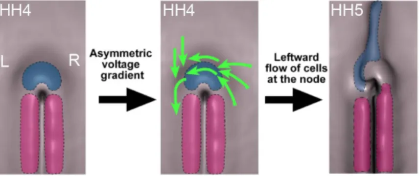
In contrast to the symmetric appearance from the outside, the vertebrate body is asymmetric on the inside, if we consider the distribution of the internal organs. Problems in the correct establishment of the internal organs may give rise to several human developmental disorders. Therefore, it is imperative to understand how left-‐right (LR) asymmetry is initiated and moreover, how it is maintained. One of the symmetry breaking mechanisms in vertebrates is the leftward fluid flow generated by cilia in the embryonic node. However, the chicken embryo is an exception, since the establishment of asymmetry occurs much earlier than the appearance of cilia. Indeed, the asymmetric expression of shh and fgf8 is established by a leftward cell movement around the Hensen's node at stage HH4. It was also uncovered that the activity of the H+/K+ATPase pump is upstream of this leftward cell movement. In the chicken embryo, one of the earliest molecules asymmetrically expressed in the node is the cell adhesion N-‐cadherin. Both, mRNA and protein show a preferential localization on its right side. It has been shown that inhibition of N-‐ cadherin function at early stages leads to a LR mispositioning of the chicken heart, suggesting that N-‐cadherin plays a role in LR patterning. The main objective of this thesis was to uncover what is the role of N-‐ cadherin in the establishment/maintenance of the molecular and morphological asymmetries of Hensen's node. I show that asymmetric N-‐ cadherin is under the control of both, the H+/K+ATPase activity and the
leftward cell movements. I also show that when N-‐cadherin's function is blocked, the node cells on the right side instead of stopping the leftward
asymmetric expression of fgf8 and nodal in the node, becoming symmetric and severely reduced, respectively. Consequently, an incorrect expression of cer1 and snai1 is translated to the left lateral plate mesoderm (LPM). On the basis of these results, the information that is given by the LPM for the heart formation is distorted, resulting in a range of abnormal heart looping phenotypes. Conversely, the inhibition of Fgf signalling on the left side of the node rescued the normal expression of cer1 and snai1 in the LPM and the correct looping of the heart. This work shows that N-‐cadherin's asymmetric localization in the chicken's node is crucial to terminate the leftward cell movements, and therefore, it is an essential step in the establishment of LR asymmetry.
I also evaluated the role of N-‐cadherin in the maintenance of the node's morphological asymmetry. Blocking N-‐cadherin's function compromised the asymmetric morphology of the node, not through cell shape changes but by changing the difference in the number of cells between both sides of the node. In addition, I propose that different levels of N-‐cadherin between the right and the left side of the node are important to avoid cell mixing in the node region. As a consequence, cells on the left and on the right side of the node are kept unmixed so they can preserve their molecular identity.
Finally, I also demonstrate that in the chicken embryo the novel asymmetrically expressed gene cHes6-‐1 is downstream of the ion exchanger H+/K+-‐ATPase and the Nodal signalling pathway. Unexpectedly,
the asymmetric localization of cHes6-‐1 in the mesoderm lateral to the primitive streak is not shared with the quail. Instead, this asymmetric
functional relevance of this asymmetric expression in the mesoderm lateral to the primitive streak, and moreover to which asymmetric structure(s) will this particular territory give rise to, is still an unanswered question.
K
EYWORDS:
left-‐right asymmetry, Hensen's node, leftward cell movement, N-‐cadherin, cell-‐sorting, Notch signalling, Hes6-‐1, cell fate.Embora externamente os vertebrados se apresentem como organismos simétricos, o seu interior é assimétrico, tendo em conta a distribuição dos órgãos internos. O incorrecto estabelecimento da assimetria dos órgãos internos pode originar vários distúrbios no desenvolvimento humano. Como tal, é necessário não só compreender como é que a assimetria esquerda-‐direita (ED) é estabelecida, mas também como é mantida. Um dos mecanismos de quebra de simetria conhecido nos vertebrados é feito por cílios que geram um fluído para o lado esquerdo no nó embrionário. No entanto, o embrião de galinha é uma excepção, uma vez que o estabelecimento da assimetria ocorre muito antes do aparecimento de cílios. Na galinha, a expressão assimétrica de shh e fgf8 é estabelecida por um movimento de células que ocorre para o lado esquerdo no nó de Hensen durante o estadio HH4. Sabe-‐se ainda, que a actividade da bomba H+/K+-‐ATPase está a juzante destes movimentos celulares. No embrião de
galinha, uma das primeiras moléculas expressas assimetricamente no nó é a molécula de adesão celular N-‐caderina. Tanto o seu mRNA como proteína, estão localizados preferencialmente no lado direito do nó. Demonstrou-‐se que a inibição da N-‐caderina no início do desenvolvimento da galinha promove a incorrecta localização do coração, sugerindo que a N-‐caderina desempenha um papel na padronização da assimetria ED. O principal objectivo desta tese foi descobrir qual o papel da N-‐caderina na criação/manutenção das assimetrias moleculares e morfológicas do nó de Hensen. É aqui demonstrado, que a localização assimétrica da N-‐caderina não só está sob o controlo da actividade da bomba protónica -‐ H+/K+-‐
células do lado direito do nó em vez de cessarem a sua migração para o lado esquerdo no final do estadio HH4, continuavam a executar este tipo de movimento, até ao final do estadio HH6. Este deslocamento contínuo das células para o lado esquerdo do nó, altera a expressão normal do fgf8 e do nodal no nó, tornando-‐se simétrica e reduzida, respectivamente. Consequentemente, a expressão do cer1 e do snai1 é incorrectamente transferida para o lado esquerdo da mesoderme da placa lateral (MPL). Como efeito dos resultados anteriores, a informação dada pela MPL para a formação do coração é incorrecta, resultando na formação de corações com diversos fenótipos anormais. No entanto, se inibirmos a sinalização Fgf do lado esquerdo do nó, conseguimos não só recuperar a expressão normal do cer1 e snai1 na MPL, como a correcta posição do coração. Este trabalho, mostra que a localização assimétrica da N-‐caderina no nó de Hensen é crucial para parar os movimentos das células do nó para o lado esquerdo, tratando-‐se assim, de um ponto crítico para o estabelecimento da assimetria ED.
No sentido de promover um entendimento mais abrangente acerca da função da N-‐caderina no nó, fui ainda avaliar qual o seu papel na conservação da assimetria morfológica no nó de Hensen. Verificou-‐se, que quando a função da N-‐caderina é bloqueada, a morfologia assimétrica do nó é comprometida, não porque as suas células mudam de forma, mas porque é alterada a diferença no número de células entre os dois lados do nó. Observámos ainda, que a acumulação N-‐caderina do lado direito do nó poderá estar envolvida no processo de segregação celular entre os seus dois lados. Ou seja, a N-‐caderina poderá estar a garantir que as células do
Por fim, demonstro ainda que o gene cHes6-‐1 está expresso assimetricamente no embrião da galinha, é dependente da via de sinalização da bomba H+/K+-‐ATPase e da via de sinalização Nodal.
Surpreendentemente, a sua localização assimétrica na mesoderme lateral à linha primitiva não está conservada entre a galinha e a codorniz. No entanto, esta assimetria é partilhada entre a galinha e o mHes6 no ratinho. No geral, estes resultados revelam um possível novo candidato na cascata da assimetria ED. No entanto, porque é que este gene está assimetricamente localizado na mesoderme lateral à linha primitiva ou qual(ais) a(s) estrutura(s) assimétrica(s) a que poderá dar origem, é ainda uma questão por responder.
P
ALAVRAS-‐C
HAVE:
assimetria esquerda-‐direita, nó de Hensen, movimento celular para o lado esquerdo, N-‐caderina, segregação celular, sinalização Notch, Hes6-‐1, destino celular.cAct-‐RIIa chicken Activin type IIa receptor AJ Adherens Junction
AP Anterior Posterior ASE side-‐specific enhancer
bHLH basic Helix Loop Helix
BMP Bone Morphogenetic Protein BBR Boeringer Blocking Reagent BSA Bovine Serum Albumin CAMs Cell Adhesion Molecules
C. elegans Caenorhabditis elegans
cDNA complementary DNA
CSL mammalian CBF-‐1, Drosophila Supressor of Hairless and C. elegans Lag-‐1
DIG Digoxigenin
DiI 1,1’-‐dioctadecyl-‐3,3,3’-‐tetramethylindocarbocyamine perchlorate
DiO 3,3’-‐dioctadecyloxacarbocyanine perchlorate
DV Dorso Ventral
DNA Deoxyribonucleic Acid
EMT Epithelial-‐to-‐Mesenchymal Transition FGF Fibroblast Growth Factor
GFP Green Fluorescent Protein GJC Gap Junction Comunication GRP Gastrocoel Roof Plate her Zebrafish hes-‐related genes
hes Hairy Enhancer of Split genes HH Hamburguer and Hamilton stage
H+/K+/ATPase Hydrogen-‐potassium adenosine triphosphatase
H+-‐V-‐ATPase Vacuolar H(+)-‐ATPase subunit A Iv Inversus viscerum
LPM Lateral Plate Mesoderm LR Left-‐Right
Lrd Left-‐right dynein MAM Mastermind Min minutes
mRNA messenger RNA
NICD Notch Intra-‐Cellular Domain NLS Nuclear Localization Sequence NVP Nodal Vesicular Particle OD Optical Density
o.n overnight
PCP Planar Cell Polarity PS Primitive Streak
PSM Pre-‐Somitic Mesoderm RA Retinoic Acid
Raldh2 Retinaldehyde dehydrogenase 2 RNA Ribonucleic Acid
sec seconds
SELI Self-‐Enhancement and Lateral Inhibition SHH Sonic hedgehog
YFP Yellow Fluorescent Protein ZO-‐1 Zonula Ocludens
C
HAPTERΙ
Figure 1 Human laterality disorders 3
Figure 2 Left–right axis determination in a vertebrate
embryo 5
Figure 3 The F-‐molecule model. 6
Figure 4 Leftward flow is generated by the posterior tilt of
nodal cilia 8
Figure 5 Establishment of LR asymmetry in the mouse
embryo 12
Figure 6 Conserved mechanisms of LR patterning among
phyla over developmental stages 21
Figure 7 A model of the LR pathway based on cytoplasmic
motor protein movement 23
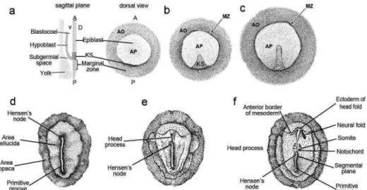
Figure 8 Early gastrulation stages in chick development 28 Figure 9 Schematic representation of a transversal section
from the blastoderm of a chicken embryo at stage HH4.
32
Figure 10 Mesodermal cell fates during vertebrate
gastrulation 36
Figure 11 Leftward cell movements generates asymmetry in
the chicken Hensen's node 38
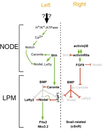
Figure 12 Schematic representation of the genes involved in
chick left-‐right asymmetry pathway 46
Figure 13 The classic cadherin–catenin protein complex 63
Figure 14 Cadherin in morphogenetic movements. 65
Figure 15 The role of cadherin in cell sorting and positioning 69
promoted by leftward cell movements downstream of the H+/K+-‐ATPase pump
Figure 2 Transient leftward cell movements in Hensen's node are promoted by asymmetric N-‐cadherin activity
108 Figure 3 Loss of N-‐cadherin activity affects asymmetric
gene expression in Hensen's node and Lateral Plate Mesoderm
113 Figure 4 N-‐cadherin controls cer1 and snai1 expression in
the Lateral Plate Mesoderm by controlling Fgf signalling
115 Figure 5 N-‐cadherin overexpression on the right side of
the node does not affect asymmetric gene 117 Figure 6 Proposed model for N-‐cadherin function in the
establishment of Left-‐Right asymmetry in the chick embryo
120
Figure S1 MNCD2 recognizes chicken N-‐cadherin and
weakly blocks cell-‐cell adhesion. 126
Figure S2 Transient leftward cell movements in Hensen's node are promoted by asymmetric N-‐cadherin activity.
129 Figure S3 Loss of N-‐cadherin activity affects asymmetric
gene expression in Hensen's node and Lateral Plate Mesoderm
130
C
HAPTERΙV
Figure 1 Blocking N-‐cadherin activity impacts on Hensen's
node asymmetric morphology 140
Figure 2 Blocking N-‐cadherin activity does not affect cell
shape in the Hensen's node 144
Figure 3 Cell mixing in the Hensen's node at different
Figure 1 The H+/K+ATPase pump activity modulates the
expression of cHes6-‐1 165
Figure 2 Nodal induces cHes6-‐1 expression 167
Figure 3 hes6 expression pattern in different organisms 169
C
HAPTERΙ
Table 1 Some of the asymmetrically expressed genes inthe chicken embryo 50
C
HAPTERΙΙ
Table 1 Appropriate restriction enzyme and RNA
polymerase for each probe 90
Table 2 Oligonucleotides 91
C
HAPTERΙΙΙ
Table 1 Number of cells that crossed the midline at different stages of development in control versus Anti-‐N-‐cad treated embryos
109
Movie 1 Time-‐lapse movie of a chicken embryo electroporated on the right side with Kaede-‐NLS photoconvertible fluorescent protein and treated with IgG2a control antibody.
121
Movie 2 Time-‐lapse movie of a chicken embryo
electroporated on the right side with Kaede-‐NLS photoconvertible fluorescent protein and treated with anti-‐N-‐cad antibody.
121
Movie 3 Time-‐lapse movie of a chicken embryo
electroporated on the right side with full-‐length N-‐ cadherinYFP
121 Movie S1 Time-‐lapse movie of a chicken embryo
electroporated with Kaede-‐NLS photo-‐convertible fluorescent protein on the right side.
132 Movie S2 Time-‐lapse movie of a chicken embryo
electroporated with Kaede-‐NLS photo-‐convertible fluorescent protein on the right side and treated with the N-‐cadherin blocking MNCD2 antibody.
132
INTRODUCTION
INTRODUCTION
In this Chapter, I will start by presenting a general overview on left-‐right (LR) asymmetry establishment, which is the central topic of this thesis. I will begin by describing its components and its regulators in different model systems, and then I will focus in one specific model -‐ the chicken embryo. I will give a brief summary of the initial steps of chicken embryo development, and I will describe how its left-‐right (LR) asymmetry is initiated, stabilized, propagated and lastly, translated into the asymmetric positioning of the heart. Finally, I will briefly review the function of a major family of cell adhesion proteins, the cadherins, during development and, in particular, the role of in N-‐cadherin role during embryonic development, LR asymmetries, and its relation to abnormal cell migration.
I.1 Left-Right Asymmetry in Vertebrates
The vertebrate body plan exhibits outwardly a bilateral symmetry along the mediolateral axis. Internally, however, the distribution of the organs such as the heart, stomach, and intestines has both an asymmetric structure and asymmetric position. The same general pattern of LR asymmetry is conserved in vertebrates and the LR patterning is remarkably similar (reviewed in (Fujinaga 1997)), suggesting that the asymmetric structure and arrangement of organs is required for their normal function.
The orientation of the LR axis is not by chance, with a predisposition of nearly 100% for a precise handedness (situs solitus) (Fig. 1a), which implies
the existence of genetic mechanisms that tightly regulate patterning along the LR axis. The failure to properly pattern the LR axis results in distinct classes of laterality defects: situs inversus corresponds to a situation where the position of the internal organs is completely reverted as a mirror-‐ image (Fig. 1b), left or right isomerism are situations where bilateral symmetry is not broken and two left or right sides will form (Figs. 1c, d) (Fliegauf, Benzing et al. 2007).
Figure 1| Human laterality disorders. (a) Schematic illustration of normal left–right body
asymmetry (situs solitus). (b-‐d) Three laterality defects that affect the lungs, heart, liver, stomach and spleen. (b) Situs inversus totalis is a complete mirror-‐image reversal of organ asymmetry. (c) Left isomerism: two left sides are formed. (d) Right isomerism: two right sides are formed. R-‐Right; L-‐Left. Adapted from (Fliegauf, Benzing et al. 2007).
Over the last 20 years, there was an effort to understand LR axis patterning and to identify the molecular and cellular mechanisms that regulate the asymmetric development. These regulatory mechanisms have been revealed through the identification of asymmetrically expressed genes and the characterization of targeted mutations that lead to LR problems. Several discoveries have helped to build a model on how LR asymmetry is initiated, stabilized, propagated, and translated into asymmetric organogenesis during development of vertebrate embryos.
I.2 Steps in Left-Right asymmetry establishment
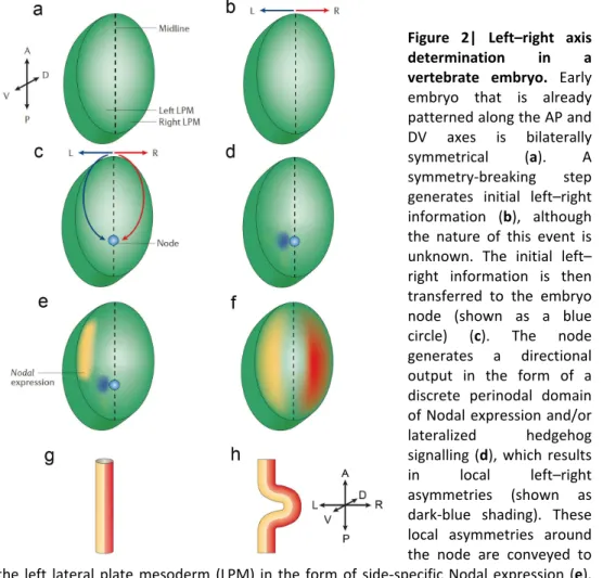
In general terms, the establishment of LR asymmetry can be divided into four steps: 1) initial breaking of LR symmetry in or near the organizer (node) (Figs. 2a-‐d); 2) transfer of LR signals from the node to the lateral plate mesoderm (LPM) (Fig. 2e); 3) LR asymmetric expression of signalling molecules to the left side in the lateral plate and (Fig. 2f) and 4) LR morphogenesis of the visceral organs induced by these signalling molecules (Fig. 2h). Studies on different vertebrates, such as zebrafish, frog, chick and mouse have contributed for this view of the establishment of LR asymmetry.
Figure 2| Left–right axis determination in a vertebrate embryo. Early
embryo that is already patterned along the AP and DV axes is bilaterally symmetrical (a). A symmetry-‐breaking step generates initial left–right information (b), although the nature of this event is unknown. The initial left– right information is then transferred to the embryo node (shown as a blue circle) (c). The node generates a directional output in the form of a discrete perinodal domain of Nodal expression and/or lateralized hedgehog signalling (d), which results in local left–right asymmetries (shown as dark-‐blue shading). These local asymmetries around the node are conveyed to the left lateral plate mesoderm (LPM) in the form of side-‐specific Nodal expression (e). Broad domains of expression of left-‐ and right-‐side specific genes (yellow and red, respectively) are then established (f), transferring laterality information to the organ primordia (a structure that represents a single primordium is shown in (g), which, in turn, execute LR asymmetrical morphogenetic programs (illustrated as the directional looping of the organ primordium in (h). Adapted from (Raya and Izpisua Belmonte 2006).
However, a unifying mechanism that that breaks symmetry has not yet been found and different strategies seem to be used by different vertebrates.
I.3 Breaking the initial symmetry
I.3.1 The conceptual F-molecule model
The vertebrate body plan is progressively organized according to the establishment of three body axes during embryonic morphogenesis: antero-‐posterior (AP), dorso-‐ventral (DV) and finally left-‐right (LR). Since the LR axis is the last to be determined during development, it could be assumed that using the pre-‐existing positional cues the LR polarity could be generated.
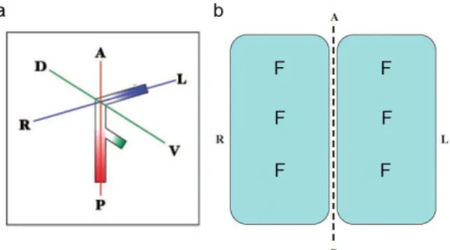
In the early 1990s, Wolpert and colleagues postulated a model called the F-‐molecule model (Brown and Wolpert 1990). This hypothetical molecule of F-‐shape would have three arms. When two arms are aligned with the AP and DV axes, the third arm would be automatically aligned along the future LR axis. If the third arm has intrinsic polarity, like the plus and minus ends of microtubules, cells on the left and right sides of the embryo would be polarized asymmetrically with regard to the LR orientation (Fig. 3). This model turns out to be correct in principle: as later studies revealed, the cilium of the node cells and its cytoskeletal structure could fill the role of the F-‐molecule (see below).
Figure 3| The F-‐molecule model. (a) Hypothetical F-‐molecule with three arms. The upper
arm (the one aligned with the LR axis) is polarized. (b) Two cells facing the midline (dotted line) contain F molecules. Two cells are asymmetric with regard to the midline. A-‐ anterior; P-‐posterior, L-‐left; R-‐right; D-‐dorsal; V-‐ventral. Adapted from (Hamada 2008).
I.3.2 Different mechanisms to break symmetry
The fact that the first asymmetrically expressed genes are found at the LR organizer (node in mice, Hensen's node in the chick, the gastrocoel roof plate in Xenopus, and Kupffer’s vesicle (KV) in zebrafish) (Levin, 2005) drew attention to this structure as the place where the initial LR decisions might be taken. Evidence from various experimental approaches and model organisms demonstrates that the embryo node has a crucial role for LR asymmetrical patterning in all vertebrates (Capdevila, Vogan et al. 2000; Mercola and Levin 2001; Palmer 2004). Nevertheless, compelling evidence from several model organisms support the possibility that LR symmetry breaking events might occur prior to gastrulation and therefore prior to node formation.
I.3.2.1
Motile cilia in the node
The role of cilia in the establishment of LR asymmetries has been best studied in the mouse embryo. An elegant series of experiments done in the mouse node provided the first experimental evidence for the role of cilia in LR axis determination (Nonaka, Tanaka et al. 1998; Okada, Nonaka et al. 1999; Takeda, Yonekawa et al. 1999). Interestingly, this model is completely consistent with a previously observed correlation between
situs abnormalities and ciliary dysfunction in humans, the so called
Kartagener's syndrome (Afzelius 1976). In these affected individuals a reversion of the localization of the organs is associated with immotile sperm and defective cilia in their airways (Afzelius 1976). Thus, this association indicated that motile cilia might control LR asymmetry.
This conclusion was later supported by the analysis of the inversus
which have randomized LR patterning, were shown to have a mutation in the left-‐right dynein (lrd) (Supp et al. 1997), an important component of the ciliary motion motor. And in fact, it was then shown that human patients with Kartagener's syndrome present mutations in this motor protein.
Equally important, was the discovery that cells in the murine ventral node are monociliated. These cilia are composed of 9 + 0 microtubules arrangement, rather than the 9 + 2 conventional motile cilia arrangement in normal ciliated cells. Therefore, based on their structure, these monocilia were thought to lack motility (Bellomo, Lander et al. 1996). However, it was later discovered that the nodal monocilia 9 + 0 have an accelerated movement and that this generates a leftward extracellular fluid flow (nodal flow) around the node (Hirokawa, Tanaka et al. 2006). This leftward nodal flow is able to determine laterality by a yet unknown mechanism (Fig. 4b) (Nonaka, Tanaka et al. 1998; Nonaka, Yoshiba et al. 2005).
Figure 4| Leftward flow is generated by the posterior tilt of nodal cilia. (a) Scanning
electron micrographs of wild-‐type mouse nodes in ventral view. (b) Generation of leftward flow by tilted cilia rotation in the node. Arrows-‐monocilia. Upper, anterior (A); lower, posterior (P); left of the figure, right (R); right of the figure, left (L). Bars, 5 mm. Adapted from (Hirokawa, Tanaka et al. 2012).
I.3.2.1.1 D
IRECTIONAL NODAL FLOWThe discovery of nodal flow generated by the rotation of cilia was based on studies of molecular motors like the kinesin superfamily proteins (KIFs) KIF3 motor. KIFs are the main motor proteins involved in the transport along microtubules. They transport various cargoes, such as membranous organelles, protein complexes and mRNA, along the network of intracellular microtubules within cells (Aizawa, Sekine et al. 1992; Hirokawa 1998).
Studies with knockout mice for Kif3a and Kif3b (subunits of the heterotrimeric motor protein, kinesin2) revealed that around 50% of both Kif3a deficient and Kif3b deficient mice showed reversed heart loops. Observation of the nodes then surprisingly revealed that nodal cilia were lacking in Kif3-‐/-‐ mutants and, as a consequence, there was no generation of nodal flow (Marszalek, Ruiz-‐Lozano et al. 1999; Takeda, Yonekawa et al. 1999).
The implication of the nodal flow in the determination of LR asymmetric patterning was further supported by a series of elegant experiments in which mouse embryos in culture were subjected to an artificial rightward flow. This manipulation, of the intensity and/or direction of the flow, was sufficient to reverse LR situs. These results provided the first direct evidence of the fluid flow role in LR patterning (Nonaka, Shiratori et al. 2002).
Despite the initial description of this fluid flow in the mouse node, it is not exclusive to the mouse embryo. Zebrafish and medaka fish also share the same mechanism, promoted by motile cilia inside the Kupffer's vesicle. In the rabbit’s node, a homologue structure called posterior notochord, as well as the gastrocoel roof plane in Xenopus, also has motile cilia capable
to create a leftward fluid flow (Essner, Amack et al. 2005; Okada, Takeda et al. 2005; Schweickert, Weber et al. 2007; Basu and Brueckner 2008).
In the zebrafish KV it was previously though that the rotating cilia moved in a counter-‐clockwise manner (when viewed from the apical side) (Kramer-‐Zucker, Olale et al. 2005; Shu, Huang et al. 2007), differing from the mouse model (Fig. 3). This contradictory result was puzzling, since it suggested that the mechanism for LR determination, which depends on the initial leftward transport of a molecule, was not conserved in zebrafish. However, it was found out later that all cilia in the KV actually rotate in a clockwise manner if observed from ventrally, similar to what is observed in the mouse node (Nonaka, Shiratori et al. 2002; Okada, Takeda et al. 2005).
I.3.2.1.2
TWO MODELS FOR GENERATING LEFT-RIGHT ASYMMETRIC GENE EXPRESSION BY NODAL FLOWAfter bilateral symmetry is disrupted in or near the node and LR asymmetry is initiated, it is necessary to transfer the LR signals from the node to the LPM to enable the asymmetrical development of the body. Although the nodal flow has been widely accepted to be the mechanism for breaking symmetry in the majority of vertebrates, it is not yet fully understood how the nodal flow directs LR asymmetry.
There are two current models, which are not mutually exclusive, and have both been proposed to explain how the information generated by leftward nodal flow is interpreted.
The first model is based on the formation of chemical gradients and it assumes that the directional flow will produce a concentration gradient of a secreted morphogen in the cavity of the ventral node (Nonaka, Tanaka et
al. 1998; Okada, Takeda et al. 2005). The resulting asymmetric morphogen distribution would initiate downstream molecular events that then establish LR asymmetries in the LPM (Nonaka, Tanaka et al. 1998).
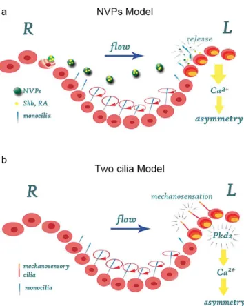
More recently, it was reported the presence of membrane-‐sheathed vesicles in the node that could correspond to the chemical morphogen. These Nodal Vesicular Particles (NVPs), containing Sonic hedgehog (SHH) and Retinoic acid (RA), two molecules involved in LR asymmetry determination (Schilling, Concordet et al. 1999; Tsukui, Capdevila et al. 1999), were shown to be secreted and transported to the left side by the nodal flow. This release of NVPs was shown to be dependent on FGF signalling, and they are transported to the left edge of the node where they fragment and release their contents (Tanaka, Okada et al. 2005) (Fig. 5a).
Figure 5| Establishment of Left-‐Rigth asymmetry in the mouse embryo. In the mouse
embryo two versions of the nodal flow model have been proposed. (a) The NVPs model. NVPs are released from the node cells in an FGF8-‐dependent manner. They are transported to the left side of the node by means of a nodal flow, which is generated by cilia on the ventral surface of the node cells. The content of the NVPs (RA and Shh) is released on the left side of the node inducing an intracellular Ca2+ signal. (b) The two cilia
model. Two types of cilia (monocilia and mechanocilia) were found to be present in the ventral node cells. Monocilia, which show co-‐expression of Pkd2 and Lrd, are motile cilia that drive the nodal flow. In the periphery of the node, mechanosensory cilia are present that express the calcium channel Pkd2 but not Lrd. These cilia are immotile and it is proposed that they are involved in detecting the nodal flow resulting in the generation of an asymmetric intracellular Ca2+ signal that is transduced into asymmetric gene
expression. Adapted from (Schlueter and Brand 2007).
The second model, called physical stimulation, was proposed as a critique of the chemical gradient model. It establishes that there is a second population of nodal cilia that are immotile and can mechanically sense the physical stimulation produced by the nodal flow, the so-‐called
“mechanosensory cilia” (McGrath, Somlo et al. 2003). This model is now commonly known as the “two-‐cilia” model, since one type of cilia generates the flow and the other senses it (Tabin and Vogan 2003).
One class of cilia, found throughout the center of the node, is associated with both Lrd and Polycystin-‐2 protein, while a second class of cilia, present on the node periphery, contains only Polycystin-‐2. Polycystin-‐ 2 is encoded by the gene Pkd2 and functions as a calcium release channel and it was observed that Pkd2 mouse mutants display LR asymmetry defects (Fig. 5b) (Pennekamp et al., 2002).
It has been proposed that immotile mechanosensory cilia sense the fluid flow pressure on the left side of the node triggering an asymmetric intracellular Ca2+ flux which creates a left-‐sided elevation of intracellular of Ca2+ culminating with the left-‐sided nodal expression (McGrath, Somlo et al. 2003). Propagation of the increased Ca2+ concentration through the LPM then breaks LR symmetry, establishing the “leftness” of the LPM and inducing left-‐side specific gene cascades (McGrath, Somlo et al. 2003).
However, the validity of this model was recently questioned (Cartwright, Piro et al. 2004), due to the observation that, even in the presence of directional flow, the magnitude of the shear stresses and flow velocities generated in the mice node would lead to an overall symmetrical effect, and therefore would be unable to asymmetrically bend monocilia.
I.3.2.2
OTHER PLAYERS IN LEFT-RIGHT PATTERNING PRIOR TO THEFORMATION OF CILIA
Although the monocilia in the ventral murine node or its equivalent structures seem to be evolutionarily conserved (reviewed in (Hamada,