Evaluation of the toxicity and hypoglycemic effect of
the aqueous extracts of
Cnidoscolus quercifolius
Pohl
S.M. Lira
1, N.V. Canabrava
1, S.R. Benjamin
1, J.Y.G. Silva
1, D.A. Viana
2, C.L.S. Lima
1,
P.F.M. Paredes
1, M.M.M. Marques
3, E.O. Pereira
1, E.A.M. Queiroz
1and M.I.F. Guedes
1 1Laboratório de Biotecnologia e Biologia Molecular, Universidade Estadual do Ceará, Fortaleza, CE, Brasil 2Laboratório de Patologia Clínica Pathovet, Fortaleza, CE, Brasil 3Laboratório de Parasitologia e Ecologia de Doenc¸as Negligenciadas, Universidade Federal do Piauí, Picos, PI, BrasilAbstract
Diabetes mellitus is one of the most common chronic degenerative diseases, and it is estimated to increase worldwide to around 415 million and to impact 642 million in 2040. Research shows that some plants are sources of bioactive compounds against diabetes. Thus, the objective of this work was to evaluate the oral toxicity and the hypoglycemic effect of the aqueous extract of the leaves ofCnidoscolus quercifoliusPohl. Diabetes was induced in Swiss mice with streptozotocin and the mice were treated with an aqueous extract ofC. quercifoliusleaves for a period of 30 days. Phytochemical analysis showed that the extract was rich inflavonoids, catechins and triterpenoid, which did not show any mortality and behavioral alterations in mice treated with 200, 1000, and 2000 mg/kg body weight of the extract for 14 days. Histopathological analysis of organs (kidney, pancreas, liver) from mice treated with the 2000 mg/kg extract revealed no architectural change. In the present study, we found a 29% reduction in glucose levels in animals receiving 200 mg/kg body weight. These results are very promising because they showed that
C. quercifoliushad a hypoglycemic effect and did not present oral toxicity, thus being a new source of compounds for the control of diabetes.
Key words: Faveleira; Diabetes mellitus; Medicinal plants; Hypoglycemic activity; Aqueous extracts
Introduction
Diabetes mellitus (DM) is a heterogeneous group of metabolic disorders that have in common hyperglycemia as a consequence of failure of insulin action (1). During the early stages of the disease, asymptomatic patients, especially those with type 2 diabetes mellitus (T2DM), can present stupor, coma and death due to ketoacidosis. The severity of the symptoms is related to the type and duration of diabetes (2).
It is estimated that the world population with diabetes is around 415 million and will reach 642 million in 2040 (3). In Brazil, in the period of 1998–2008, the standardized prevalence of DM increased from 2.9 to 4.3%. In 2015, there were 9.1 million people aged between 20 and 79 years with DM, which corresponds to approximately 6.2% of the adult population (4,5). Projections demonstrate that T2DM will be responsible for a remarkable share of the global disease by 2030 (6). The etiology of diabetes is multifactorial, being a product of the interplay between genetic and environmental factors, as well as dietary and lifestyle factors (1).
The use of herbal medicines, medicinal plants and phytonutrients continues to expand rapidly across the world, with an estimated 80% of the world’s population using this type of medication, especially in developing countries (7,8).
The World Health Organization established guidelines for the evaluation of herbal medicines, defining some criteria for assessing the quality, safety and efficacy of plants (9).
Brazil has huge potential for the disclosure of new bioactive substances, standing out amongst countries with the most diverse flora, despite the fact that efficacy of some substances has not been tested pharmacologically (10). The present demand for complementary and alter-native medicine may be in part due to the inadequate knowledge of traditional medicine or the high cost and side effects of manufactured drugs. In the last decades, the use of herbal medicine has been widely embraced and accepted by the public. Herbal medicinal products have been included in healthcare and traditional medical
Correspondence: S.M. Lira:<[email protected]>
practice in developed countries, mainly in UK and Europe (8,11,12).
Several studies have demonstrated the effect of medicinal plants that have shown to be promising in monitoring glycemia, includingCnidoscolus chayamansa, Allium cepa, Psidium guajava, Panax ginseng, Phaseolus
vulgaris, Passiflora Glandulosa, and Copernicia cerifera
(13–19). Among these plants, Cnidoscolus quercifolius Pohl. (Mart Pax et K. Hoffm.) belongs to the Euphorbia-ceae family, popularly known as faveleira. It presents several biological activities such as antitumor effects, through the neofavelone compound obtained from the plant bark (20). The ethanolic extract of bark and leaves show anti-inflammatory activity and antinociceptive activ-ity (21); antimicrobial, antifungal effects were found with acetyl cholinesterase, an antioxidant from the leaves, roots, and barks (22). However, there are no studies demonstrating the hypoglycemic effect ofC. quercifolius. Thus, the objective of this work was to evaluate the toxicity and the hypoglycemic effect of the aqueous extracts of
C. quercifolius leaves in the treatment of
streptozotocin-induced diabetic mice.
Material and Methods
Plant material and preparation of extracts
C. quercifolius Pohl. (Mart. Pax et K. Hoffm.) was
collected from its natural habitat in the city of Fortaleza, CE (Northeastern Brazil), and identified by a botanist of the Prisco Bezerra Herbarium (Universidade Federal do Ceará, UFC), where a specimen voucher was deposited (No. 56043) (22). The dried leaves (100 g) at room temperature, were boiled in water for 5 min. The solution was then filtered through celite and lyophilized. The extract was weighed and stored in a container at 6°C until use.
Phytochemical analysis
The extracts were subjected to phytochemical screen-ing, following the protocols described by Matos (23). Chemical tests were performed using specific reagents, observing color changes or formation of a precipitate, and characteristic for each class of substances. Tests were performed for the detection of phenols, flavones, flavonols, xanthones, catechins, anthocyanins, antho-cyanidins, triterpenoids,flavanones, saponins, alkaloids, and tannins.
Animals
Male and female Swiss mice (Mus musculus) aged between 8 and 12 weeks (25.0–30.0 g), were obtained from the vivarium of the UFC. The animals were kept in polypropylene cages at room temperature between 24° and 25°C in light-dark cycles of 12/12 h. All animals received water and food adlibitum. The Ethical Committee on Animal Research of the Universidade Estadual do
Ceará approved the experimental protocol (No. 1606145-2015).
Acute toxicity
For the acute toxicity study, 28 Swiss male mice and 28 female mice weighing between 25–30 g, were used. Animals were divided into 2 groups (n=7): normal control treated orally with water (NC), and mice treated orally with aqueous extract of faveleira leaves (AEF; 200, 1000, 2000 mg/kg). All animals were fasted for 4 h, and then received food and water again after the administration of the extracts. Following treatment, the mice were observed at 30, 60, 120, 240, and 360 min and every 24-h during a 14-day period. During examination, the following param-eters were assessed: heart rate, respiratory rate, number of deaths and side effects (e.g., piloerection, diarrhea, sialorrhea, hypnosis and seizures) (24). After behavioral observation and at the end of this period all animals were euthanized by cervical dislocation and the kidney, pancreas, and liver were harvested and weighed.
The kidney, pancreas, and liver were used for the histopathological analysis. The isolated fragments were fixed in 10% neutral formalin and placed in paraffin blocks for conventional histological processing (25). Then, 5-mm sections were obtained and stained with hematoxylin-eosin (HE). The slides were examined for the identification of histological alterations with conventional optical micro-scopy (Nikon YS2, Nikon, Japan), and images represen-tative of each organ were captured with a digital camera (Nikon Coolpix L14 7.1 megapixels, Nikon).
Diabetes induction
Diabetes mellitus was induced by a single intraper-itoneal (ip) injection of streptozotocin (STZ; Sigmas, USA) at the high dose of 140 mg/kg in 12-h fasting mice (26). Animals with blood glucose levels equal to or greater than 200 mg/dL were considered to be diabetic.
Experimental protocol
The animals were divided into the following 4 groups (n=6). Normal control group (NC): healthy mice treated with water (0.2 mL waterday–1animal–1); MET 200 group:
diabetic mice treated with metformin at 200 mg/kg body weight diluted in water; AEF 100 group: diabetic mice treated with aqueous extract of faveleira leaves at 100 mg/ kg body weight diluted in water; AEF 200 group: diabetic mice treated with aqueous extract of faveleira leaves at 200 mg/kg body weight diluted in water). Animals received doses orally for a period of 30 consecutive days, as previously reported by Barbosa et al. (27), with some modifications.
Determination blood glucose
23300 version 1.7 device was used, which uses the kinetic method for serum samples.
The serum was subjected to glucose analysis by com-mercial kits using the manufacturer’s technical recom-mendations (Bioclins, Brazil).
Statistical analysis
Data are reported as means±SD. Statistical signifi -cance of differences between groups was assessed using one-way ANOVA, followed by the Tukey test. Po0.05 was considered to be significant.
Results
The phytochemical analysis ofC. quercifoliusaqueous extracts revealed the presence of phenols, flavones, flavonols, xanthones, catechins, triterpenoids, and tannins.
For the acutein vivotoxicity test of theC. quercifolius aqueous extract, doses of 200, 1000, and 2000 mg/kg of animal weight were used. The results of the hippocratic screening showed that there was no motor and sensorial alterations in the animals at the doses tested, and there was no death within 14 days.
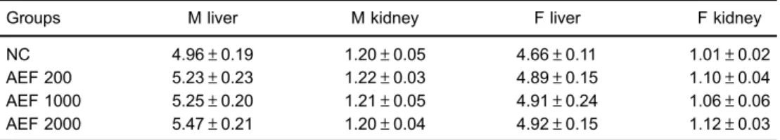
There were no significant differences (P40.05) in liver and kidney weight between the Control group and the groups treated with aqueous extract ofC. quercifoliusat all doses, in both male and female mice (Table 1).
The histopathological analysis of the organs of the animals treated with 2000 mg of the aqueous extract of
C. quercifoliusshowed no architectural alteration (Figure 1A-C).
The streptozotocin-induced diabetes increased the blood glucose levels in the mice to above 200 mg/dL. The hypoglycemic action of the aqueous extract
C. quercifoliussignificantly reduced blood glucose levels
by 29.1% in the diabetic animals treated with 200 mg/kg body weight (Po0.05).C. quercifolius had a hypoglyce-mic effect beginning at the 20th day of treatment, and this effect increased at the end of the treatment (Table 2). There was little hypoglycemic effect at a dose of 100 mg/kg.
Discussion
The phytochemical analysis of the aqueous extract of
C. quercifoliusrevealed the presence of phenols,flavones,
flavonols, xanthones, catechins, triterpenoids, and tan-nins. Studies have shown thatflavonoids, when ingested on a regular basis through diet, can help prevent chronic non-communicable diseases due to their antioxidant, anti-inflammatory, anti-hyperglycemic, anticarcinogenic and antiatherogenic effects, as well as antibacterial and antiviral activities (28,29). Triterpenoids are metabolites that also have an antidiabetic potential, which act to increase the release of insulin, modifying glucose metab-olism, inhibiting hyperglycemic factors, and inhibiting or stimulating enzyme synthesis (30). Antioxidants, such as phenolic acids,flavonoids, and tannins, among others, are present in different parts of plants, and are associated with the reduction of risks of diseases such as diabetes (31).
Paredes et al. (22) conducted a study with methanolic extract of the leaf, root bark and root of C. quercifolius and did not observe the presence of triterpenoids. This difference in results between the aqueous and methanolic extracts ofC. quercifoliuscan be justified by the solubility of the substances, which may or may not be soluble in the solvent used (32).
It is recommended that herbal medicine candidates be tested for their efficacy and safety (33). In the toxicity tests
of C. quercifolius, there were no physical, behavioral or
motor changes, or death of treated animals, as well as no changes in the weights of the organs. This is an important result, since the kidneys are responsible for numerous functions, such as reabsorption, homeostasis, filtration, endocrine and metabolic functions (34). Furthermore, liver weights did not increase, which suggests there was no hepatocellular hypertrophy (35).
According to the histopathological analysis, the aqueous extract of the faveleira did not cause architectural alterations of the liver (Figure 1C). In relation to the pancreas (Figure 1B), there was also no architectural alteration, differently from the study of Kalita et al. (36), which found
Table 1.Effect of aqueous extracts ofCnidoscolus quercifoliuson relative weight of liver and kidneys of male and female mice.
Groups M liver M kidney F liver F kidney
NC 4.96±0.19 1.20±0.05 4.66±0.11 1.01±0.02
AEF 200 5.23±0.23 1.22±0.03 4.89±0.15 1.10±0.04
AEF 1000 5.25±0.20 1.21±0.05 4.91±0.24 1.06±0.06
AEF 2000 5.47±0.21 1.20±0.04 4.92±0.15 1.12±0.03
Figure 1.Histopathological observations of the kidney (A), pancreas (B) and liver (C) of mice treated with the aqueous extracts of faveleira and of the kidney (D), pancreas (E) and liver (F) treated with water. Hematoxylin-eosin staining.
Table 2. Effect of the aqueous extracts of Cnidoscolus quercifoliuson serologic levels of glucose in diabetic mice.
Groups Day 0 Day 10 Day 20 Day 30 %CV
NC 105.71±8.3 107.59±9.3 100.27±12.9 106.15±5.0 +0.41 MET 296.30±24.4 246.65±54.2 280.19±43.8 206.30±24.4 –43.68 AEF 100 295.65±44.1 309.67±61.8 290.99±58.4 265.79±40.1 –10.1 AEF 200 302.22±48.2 253.31±78.5 238.47±41.4* 214.24±54.23*
–29.1
that the methanolic extract of the root ofMusa balbisiana
collaregenerated the pancreatic islets of Langerhans.
These results indicate that the applied dosages were potentially safe. This is the first work that depicts the in vivotoxicity ofC. quercifolius,demonstrating that its use can be safe.
This herbal drug has some advantages, such as efficacy, cost and hypoglycemic effects (37). The aqueous extract of C. quercifolius showed a hypoglycemic effect at a concentration of 200 mg/kg for 20 and 30 days. A previous study by Achia et al. (38), with the leaf extracts
ofC. aconitifoliusin diabetic rats, showed that there was a
reduction in glycemia. These results demonstrate that the genusCnidoscolushas a potential hypoglycemic effect in animals.
In conclusion, the aqueous extract ofC. quercifoliusat a dose of 200 mg/kg body weight presented a hypogly-cemic effect in diabetic mice and showed no toxicity. These results are very promising because they showed
thatC. quercifoliusmight be a new source of compounds
for the treatment of diabetes.
Acknowledgments
The authors would like to thank Prof. Maria Izabel Florindo Guedes, Coordinator of RENORBIO (Rede Nordeste de Biotecnologia), Fortaleza, for technical support. This research was supported by grants from FUNCAP (Fundac¸ão de Amparo a Pesquisa do Estado do Ceará).
References
1. SBD (Sociedade Brasileira de Diabetes). Diretrizes da Sociedade Brasileira de Diabetes. Brasil: Sociedade Brasi-leira de Diabetes; 2014–2015. http://www.diabetes.org.br/ profissionais/images/docs/DIRETRIZES-SBD-2015-2016.pdf. Accessed December 20, 2016.
2. Kharroubi AT, Darwish HM. Diabetes mellitus: The epidemic of the century. World J Diabetes 2015; 6: 850–867, doi: 10.4239/wjd.v6.i6.850.
3. IDF (International Diabetes Federation). Diabetes atlas update. 7th edn., Brussels, Belgium: International Diabetes Federation. 2015. p 9. http://www.diabetesatlas.org/. Accessed December 20, 2016.
4. Freitas LRS, Garcia LP. Evoluc¸ão da prevalência do diabetes e deste associado à hipertensão arterial no Brasil: análise da pesquisa nacional por amostra de domicílios, 1998, 2003 e 2008.Epidemiol Serv Saúde2012; 21: 7–19, doi: 10.5123/S1679-49742012000100002.
5. PNS (Pesquisa Nacional de Saúde).Percepc¸ão do estado de saúde, estilos de vida e doenc¸as crônicas: Brasil, grandes regiões e unidades da federac¸ão; 2013. p 40. http://biblioteca. ibge.gov.br/visualizacao/livros/liv91110.pdf. Accessed December 20, 2016.
6. WHO (World Health Organization).Global status report on noncommunicable diseases, 2010. Geneva: World Health Organization; 2011; http://apps.who.int/iris/bitstream/10665/ 44579/1/9789240686458_eng.pdf. Accessed December 22, 2016.
7. Ong CK, Bodeker G, Grundy CK, Burford G, Shein K.WHO Global atlas of traditional, complementary and alternative medicine. Geneva: WHO. 2005; http://apps.who.int/iris/ bitstream/10665/43108/1/9241562862_map.pdf. Accessed December 22, 2016.
8. Ekor M. The growing use of herbal medicines: issues relating to adverse reactions and challenges in monitoring safety.Front Pharmaco2014; 4: 1–10, doi: 10.3389/fphar. 2013.00177.
9. Ajazuddin, Saraf S. Legal regulations of complementary and alternative medicines in different countries. Pharmacogn Rev2012; 6: 154–160, doi: 10.4103/0973-7847.99950. 10. Mendes SSM, Andrade JA, Xavier MA, Junior JAS,
Pantaleão SM, Estevam CS, et al. Genotoxicity test of
Maytenus rigida andAristolochia birostris in the radicular meristem of the onion,Allium cepa.Rev Bras Farmacogn
2012; 22: 76–81, doi: 10.1590/S0102-695X2011005000180. 11. Braun LA, Tiralongo E, Wilkinson JM, Spitzer O, Bailey M, Poole S, et al. Perceptions, use and attitudes of pharmacy customers on complementary medicines and pharmacy practice. BMC Complement Altern Med 2010; 10: 1–7, doi: 10.1186/1472-6882-10-38.
12. Calapai G. European legislation on herbal medicines: a look into the future.Drug Saf2008; 31: 428–431, doi: 10.2165/ 00002018-200831050-00009.
13. Loarca-Piña G, Mendoza S, Ramos-Gómez M, Reynoso R. Antioxidant, antimutagenic, and antidiabetic activities of edible leaves from Cnidoscolus chayamansa Mc. Vaugh.
J Food Sci2010; 75: 68–72, doi: 10.1111/j.1750-3841.2009. 01505.x.
14. Etxeberria U, La Garza AL, Campión J, Martínez JA, Milagro FI. Antidiabetic effects of natural plant extracts via inhibition of carbohydrate hydrolysis enzymes with empha-sis on pancreatic alpha amylase.Expert Opinion2012; 16: 269–297, doi: 10.1517/14728222.2012.664134.
15. Shen SC, Cheng FC, Wu NJ. Effect of guava (Psidium guajavalinn) leaf solube solids on glucose metabolism in type 2 diabetic rats.Phytother Res2008; 22: 1458–1464, doi: 10.1002/ptr.2476.
16. Luo JZ, Luo L. Ginseng on hyperglycemia: Effects and mechanisms.Evid Based Complement Alternat Med2009; 6: 423–427, doi: 10.1093/ecam/nem178.
17. Barrett ML, Udani JK. A proprietary alpha-amylase inhibitor from white bean (Phaseolus vulgaris): A review of clinical studies on weight loss and glycemic control. Nutrition J
2011; 10: 1–10, doi: 10.1186/1475-2891-10-24.
18. Sousa RVRB, Guedes MIF, Marques MMM, Viana DA, Silva ING, Rodrigues PAS, et al. Hypoglycemic effect of new pectin isolated fromPassiflora Glandulosa Cavin alloxan-induced diabetic mice.World J Pharm Pharm Sci2014; 4: 1571–1586, doi: 10.1186/1477-3155-11-4.
20. Sobrinho TJSP, Tavares EA, Castro VTNA, Filho JV, Militão GCG, Silva TG, et al. Antiproliferative activity of species of the genusCnidoscolusagainst HT-29, Hep-2 and NCI-H292 cells.Mol Clinical Pharm2012; 3: 55–61.
21. Gomes LMA, Andrade TMD, Silva JC, Lima JT, Quintans JLJ, Almeida JRGS. Phytochemical screening and anti-inflammatory activity ofC. quercifolius(Euphorbiaceae) in mice.Pharmacogn Res2014; 6: 345–350, doi: 10.4103/0974-8490.138290. 22. Paredes PFM, Vasconcelos FR, Paim RTT, Marques MMM,
Morais SMM, Lira SM, et al. Screening of bioactivities and toxicity ofCnidoscolus quercifolius Pohl. Evid Based Complement and Alternat Med 2016. 2016: 1–9, doi: 10.1155/2016/7930563.
23. Matos FJA.Introduc¸ão afitoquı´mica experimental. 3rd edn. Fortaleza: Edic¸ões UFC. 2009.
24. Politi FAS, Moreira RRD, Salgado HRN, Pietro RCLR. Preliminary tests on acute oral toxicity and intestinal motility with extract of pulverized bark ofEndopleura uchi(Huber) Cuatrec. (Humiriaceae) in mice.Rev Pan-Amaz Saúde2010; 1: 187–189. 25. Michalany J.Técnica histológica em anatomia patológica.
2nd edn. São Paulo: Michalany; 1990.
26. Cardinal JW, Margison GP, Mynett KJ, Yates AP, Cameron DP, Elder RH. Increased susceptibility to streptozotocin-induced b-cell apoptosis and delayed autoimmune diabetes in alkyl-purine DNA-N-Glycosylase-deficient mice.Mol Cell Biol2001; 21: 5605–5613, doi: 10.1128/MCB.21.16.5605-5613.2001. 27. Barbosa AP, Silveira GO, Menezes IA, Rezende NJM,
Bitencurt JL, Estavam CS, et al. Antidiabetic effect of the
Chrysobalanus icacoL. aqueous extract in rats.J Med Food
2013; 16: 538–543, doi: 10.1089/jmf.2012.0084.
28. Henrique PC, Boas ACV, Lima RAZ, Decarlos AN, Lima LCO. Color, physicochemical parameters and antioxidant potential of whole grape juices subject to different uv-c radiation doses. Ciênc Agrotecnol 2016; 40: 226–234, doi: 10.1590/1413-70542016401035215.
29. Mousavi L, Salleh RM, Murugaiyah V, Asmawi MZ. Hypoglycemic and anti-hyperglycemic study of Ocimum-TenuiflorumL. leaves extract in normal and streptozotocin-induced diabetic rats.Asian Pac J Trop Biomed 2016; 6: 1029–1036, doi: 10.1016/j.apjtb.2016.10.002.
30. Khatun H, Nesa L, Islam R, Ripa FA, Mamum A, Kadir S. Antidiabetic and antidiarrheal effects of the methanolic extract of Phyllanthus reticulatus leaves in mice. Asian Pac J Reproduct2014; 3: 121–127, doi: 10.1016/S2305-0500(14)60015-4.
31. Tukun AB, Shaheen N, Banu CP, Mohiduzzaman MD, Islam S, Begum M. Antioxidant capacity and total phenolic contents in hydrophilic extracts of selected Bangladesh medicinal plants.Asian Pac J Trop Med2014; 7: 568–573, doi: 10.1016/S1995-7645(14)60291-1.
32. Xu DP, Li Y, Meng X, Zhou T, Zhou Y, Zheng J, et al. Natural antioxidants in foods and medicinal plants: extraction, assessment and resources.Int J Mol Sci2017; 18: 1–32, doi: 10.3390/ijms18010096.
33. Castro RA, Albiero ALM. O mercado de matérias primas para indústria defitoterápicos. Rev Fitos2016; 10: 1–93, doi: 10.5935/2446-4775.20160006.
34. Santoro D, Caccamo D, Lucisano S, Buemi M, Sebekova K, Teta D, et al. Interplay of vitamin D, erythropoiesis, and the renin-angiotensin system.Biomed Res Int2015; 2015: 1–11, doi: 10.1155/2015/145828.
35. Asaoka Y, Togashi Y, Mutsuga M, Imura N, Miyoshi T, Miyamoto Y. Histopathological image analysis of chemical-induced hepatocellular hypertrophy in mice. Exp Toxicol Pathol2016; 68: 233–239, doi: 10.1016/j.etp.2015.12.005. 36. Kalita H, Boruah DC, Deori M, Hazarika A, Sarma R,
Kumari S, et al. Antidiabetic and antilipidemic effect ofMusa balbisiana root extract: A potent agent for glucose home-ostasis in streptozotocin-induced diabetic rat. Front Phar-macol2016; 7: 1–11, doi: 10.3389/fphar.2016.00102. 37. Pang B, Zhao LH, Zhou Q, Zhao TY, Wang H, Gu CJ, et al.
Application of berberine on treating type 2 diabetes mellitus.
Int J Endocrinol 2015; 2015: 1–12, doi: 10.1155/2015/ 905749.
38. Achia NK, Ohaeria OC, Ijeha II, Eleazub C. Modulation of the lipid profile and insulin levels of streptozotocin induced diabetic rats by ethanol extract ofCnidoscolus aconitifolius