BJRS
RADIATION SCIENCES
07-2A (2019) 01-18ISSN: 2319-0612 Accepted: 2018-10-27
Establishing adequate conditions for mercury
determination in environmental samples by INAA
C. Perez; E. C. Santos; M. Saiki
Instituto de Pesquisas Energéticas e Nucleares (IPEN - CNEN/SP), 05508-000, São Paulo, SP, Brazil [email protected]
ABSTRACT
Mercury (Hg) is a toxic element released into the environment mainly by anthropic activities. Consequently, the improvement for Hg determination in environmental samples is of great interest. Instrumental Neutron Activa-tion Analysis (INAA) is considered an adequate method to determine several elements. However, Hg determina-tion by INAA is often hampered by its volatility, which causes losses, depending on the local temperature in the reactor where the sample is irradiated. The aim of this study was to establish adequate irradiation conditions in the IEA-R1 reactor for Hg determination in environmental samples by INAA. The following parameters were evaluated: irradiation time, container for irradiation and spectral gamma ray interferences. For the study, ali-quots of certified reference materials (CRMs) and tree bark samples were irradiated together with Hg synthetic standard at the IEA-R1 nuclear research reactor. Gamma ray activities of 197Hg and 203Hg were measured in a spectrometer coupled to a HPGe detector. Obtained results indicated that polyethylene capsules or envelopes can be used as container for sample irradiation and the Hg impurities in these containers were negligible. Irradiation time of one hour was adequate for Hg determination and in long irradiations of 8 h problems of spectral inter-ference of 198Au and 75Se were observed. In addition, Hg loss during the irradiation of 1 h and after irradiation was not observed. Quality control of Hg results, obtained in the CRMs analyses using one hour of irradiation, indicated good precision and accuracy with HORRAT < 2 and |Z score| < 2. The experimental conditions estab-lished in this study were applied to tree bark samples. Detection limits in these analyses were between 0.14 and 1.9 µg g-1.
1. INTRODUCTION
Mercury (Hg) is a toxic element that causes adverse effects on human health and on the environ-ment, arousing much concern everywhere due to its volatility, persistence and bioaccumulation [1]. Its toxicity depends on its chemical form, concentration, route of exposure and the exposed individ-ual's vulnerability [2]. Mercury is emitted into the atmosphere by various anthropic activities, such as coal-fired power plants, mining activities, waste incineration and industrial processes used in foundries and cement production [3]. Consequently, it is of great interest to improve analytical methods for the quantification of this element in environmental samples.
Instrumental neutron activation analysis (INAA) is considered an advantageous technique for ele-ment determination of environele-mental contaminants, since it is a non-destructive technique, that re-quires minimal manipulation of the sample and provides low detection limits [4,5]. However, some elements are not determined with good accuracy by this technique, as in the case of Hg.
In the analysis of Hg by INAA, two radioisotopes that can be measured for its quantification are formed: 197Hg (77.34 keV with half-life of 64.16 h) and 203Hg (279.20 keV with half-life of 46.61 d) [6]. However, the main difficulty in the determination of Hg by INAA is the loss of this element during irradiation due to a combination of electrolytic action creating elemental Hg, recoil due to the (n,γ) reaction, and thermal heating [7]. The use of quartz ampoules as irradiation container would avoid the Hg loss [8], but it is not adequate due to the difficulty in transferring the irradiated sample from the ampoule to a counting container. In addition, the ampoule becomes quite radioac-tive due to the activation of the sodium present in it.
The aim of this study was to establish adequate conditions for Hg determination in environmental samples by INAA in IEA-R1 reactor. In order to obtain reliable results, the following experimental parameters were evaluated: irradiation time, type of container for sample irradiation and the prob-lem of the spectral interferences. For this study, environmental certified reference materials and tree bark samples, used as environmental biomonitor, were analyzed.
2.1. Materials
2.1.1. Certified reference materials
For quality control in relation to the precision and accuracy of the results, certified reference mate-rials (CRMs) INCT M-4 CormTis Cormorant Tissue provided by Institute of Nuclear Chemistry and Technology, IAEA-085 Methylmercury, Total Mercury and Other Trace Elements in Human Hair provided by International Atomic Energy Agency, NRC DOLT-3 Dogfish Liver Certified Ref-erence Material for Trace Metals provided by National Research Council Canada and BCR 186 Trace Elements in Lyophilised Pig Kidney provided by Community Bureau of Reference were ana-lyzed.
Percentages of residual humidity of these CRMs were determined in order to obtain the results on dry basis. For this determination, approximately 250 mg of each material were dried in a Universal oven at a temperature of 85 ºC, for 24 h. The percentages of humidity obtained in this drying pro-cess were 6.1 % for CRM INCT M-4 Cormorant Tissue, 6.6 % for IAEA-085 Human Hair, 14.5% for NCR DOLT-3 Dogfish Liver and 5.7 % for BCR 186 Pig Kidney.
2.1.2. Environmental samples
Tree bark samples of the species Poincianella pluviosa (Sibipiruna) and Tipuana tipu (Tipuana) were collected in several sites of São Paulo Metropolitan Region (SP, Brazil). The collection was carried out at a height from 1.5 to 2.5 m from topsoil using a stainless steel knife. When the bark was wet, it was previously dried at 40 ºC using an oven with forced air circulation. For the analyses, the samples were carefully cleaned using a dental brush with soft nylon bristles in order to remove the foreign materials and then the surface layer of the tree bark, with a thickness less than 3 mm, was grated using a titanium grater. Finally, the samples were ground to a fine powder using a vibra-tory micro mill with an agate mortar (Vibravibra-tory Micro Mill Pulverisette 0, FRITSCH), and then placed in plastic vials that were stored in a desiccator.
Percentages of residual humidity of these powder bark samples, determined as described for CRMs, varied from 4.9 to 12.5 %.
2.2. Procedure for Neutron Activation Analysis 2.2.1. Preparation of synthetic standard of mercury
Certified standard solution of Hg acquired from Spex CertiPrep USA was used to prepare synthetic standard. From the stock standard solution, a diluted solution with Hg concentration of 136.5 µg mL-1 was prepared. To avoid Hg loss in the synthetic standard, thioacetamide solution was also used. Thioacetamide minimizes Hg losses by volatilization, since the sulfur present in its structure reacts with Hg, leading to the formation of HgS [9,10]. A 7.8 mg mL-1 thioacetamide solution was prepared by dissolving 195.7 mg of thioacetamide p.a. from Merck with purified water, and diluting to 25 mL.
Aliquots of 50 μL of thioacetamide solution were pipetted on each small sheet of nº. 40 Whatman filter paper with the size 1.5 cm x 6.0 cm. The sheets were placed in a desiccator for drying the ali-quots at room temperature. Then, aliali-quots 50 μL of diluted Hg solution were pipetted on each sheet, which remained again in a desiccator for drying at room temperature. The sheets were folded and placed in plastic envelopes. The plastic (polyethylene) foils used to prepare these envelopes were previously cleaned using diluted nitric acid solution and purified water, and finally drying at room temperature. The micropipette used was previously checked in relation to its calibration.
To prepare Hg standard in the type W polyethylene capsule (Vrije Univesiteit, Amsterdam, The Nertherlands), a small sheet of Nº. 40 Whatman filter paper was cut and placed inside the capsule. Then, 50 μL of thioacetamide solution were pipetted on the small sheet placed inside the capsule, which remained in a desiccator for drying at room temperature. After that, 50 μL of Hg diluted standard solution were pippeted on this sheet, which remained again in a desiccator for drying at room temperature. The Hg synthetic standards were kept in a refrigerator until its use in the anal-yses.
2.2.2. Irradiation, measurement and calculations
Aliquots of each tree bark sample or certified reference material were weighted (150 to 200 mg) in polyethylene envelopes. Each envelope containing the sample or standard was wrapped with alumi-num foil. This set of samples and standard was placed in an alumialumi-num irradiation device called “rabbit”. Irradiation was performed at the IEA-R1 nuclear research reactor for 1 or 8 h, under a thermal neutron flux varying from 4.2 x 10¹² n cm2 s-1 to 4.6 x 10¹² n cm2 s-1.
In the case of irradiations using polyethylene capsules, aliquots (150-200 mg) of each material were weighed in the capsules, occupying about 2/3 of the total volume of the capsule. Each capsule
con-taining sample or standard was wrapped with aluminum foil and then the set of samples and stand-ard was placed into the “rabbit”.
After adequate decay time of 1 d, for samples irradiated for 1 h, and of 3 d, for samples irradiated for 8 h, samples and standard were placed individually in stainless steel planchets for gamma ray activity measurements. In the case of irradiations using polyethylene capsules, samples and standard were placed individually in plastic counting tubes.
The induced gamma ray activity measurements were carried out using a hyperpure germanium de-tector from Canberra (model GC3020) connected to a digital spectrum analyzer (model DSA 1000). The system used had a resolution (FWHM) of 0.90 keV for 121.97 keV peak of 57Co and of 1.70 keV for 1332.49 keV peak of 60Co. The analysis of gamma spectra was performed using the com-puter program Genie 2000, 3.1 version, also from Canberra. The photopeaks of two radioisotopes of Hg were measured at different decay times: 77.34 keV of 197Hg (t1/2 = 64.16 h) and 279.20 keV of 203
Hg (t1/2 = 46.61 d). The counting times varied from 1800 to 5400 s for the standard, and from 3600 to 36000 s for the samples, depending on their activities. The calculations for obtaining Hg mass fraction were performed by comparative method [11].
2.3. Treatment of data
To evaluate the mass fraction results of Hg in the CRMs in relation to their accuracy and precision, the arithmetic mean of the results, standard deviation, relative standard deviation, HORRAT value, relative error and standardized difference (Z score) were calculated. In addition, detection limits and quantification limits of Hg were obtained for the analyzed environmental samples of tree barks.
2.3.1. Relative standard deviation and HORRAT value
The precision of the results was evaluated according to Wood [12], who considers that this parame-ter is related to the level of concentration (or mass fraction) of the analyte in the sample and it is defined by the Horwitz equation [13] given by:
Where RSDH is the value of Horwitz and c is the analyte's mass fraction. The precision of the meth-od was verified by the HORRAT value given by [12]:
(2)
Where RSD is the relative standard deviation obtained experimentally. The method is considered precise when HORRAT ≤ 2 [12].
2.3.2. Standardized difference or Z score value
To evaluate the accuracy of the CRMs results, Z score values were calculated using Equation 2, where Xlab is the mass fraction obtained in the laboratory, Xref, the mass fraction of the certificate and SDmod, the modified standard deviation, obtained by Equation 3 [14].
(3) (4)
Where SD is the standard deviation of the measurements performed in the laboratory and uXref is the combined uncertainty of the reference value of the certificate. The result is classified as satisfactory or unsatisfactory according to the following criteria [14]:
If | Z score | ≤ 2, the result is considered satisfactory; If 2 <| Z score | <3, the result is considered uncertain; If | Z score | ≥ 3, the result is considered unsatisfactory.
2.3.3. Detection limits and quantification limits
Detection limits and quantification limits were calculated using Currie’s criterion [15], which de-fines the detection limit (DLT) and the quantification limit (QLT) as:
(5)
(6)
Where DLT and QLT are, respectively, detection limit and quantification limit in counting rates, BG is the background and LT is the counting time. Once the DLT and QLT values were obtained,
the detection limits (DL) and quantification limits (QL) in element mass fraction were obtained by the comparative method.
2.4. Preliminary Assays for Establishing Adequate Experimental Conditions
2.4.1. Analysis of Hg impurities in the polyethylene envelopes and in the Nº. 40 Whatman fil-ter paper sheets
To evaluate the presence of Hg in the polyethylene envelopes, used for sample irradiation, empty envelopes were analyzed using the same experimental conditions used in the sample analyses. The same procedure used for determination of Hg in the polyethylene envelopes was applied for the analysis of the Nº. 40 Whatman filter paper sheets, used as support in the preparation of synthetic standard. For this study, filter paper samples were cut in the same size as the sheets used for prepar-ing the synthetic standard.
2.4.2. Irradiation using different polyethylene containers for Hg determination
To choose the most adequate container for irradiation in the Hg determination, aliquots of the CRM IAEA-085 Human Hair and Hg synthetic standard were irradiated for 1 h in polyethylene capsules and in polyethylene envelopes, in order to evaluate if there was Hg loss. Mercury losses during the irradiation were evaluated by comparing the Hg mass fractions obtained and the certified value. To evaluate Hg loss after the irradiation, gamma ray activities of the Hg standards were measured at different decay times and the counting rates obtained were correct to zero decay time, according to the law of radioactive decay (Equation 7) [16].
(7)
Where A and A0 are the counting rates for decay times t and t = 0, respectively and t1/2 is the half-life.
2.4.3. Selection of irradiation time
Mercury losses during the irradiation were evaluated by comparing the Hg mass fractions obtained and the certified value.
Aliquots of the CRM IAEA-085 Human Hair and Hg synthetic standard placed in polyethylene en-velopes were irradiated for 1 and 8 h and the gamma ray activities were measured for different de-cay times. In order to evaluate the Hg loss after irradiation, the counting rates of Hg standard ob-tained for different decay time were corrected for zero decay time. The irradiated Hg standard was kept in the counting room at temperature of 20 ºC throughout the period in which the counts were performed.
3. RESULTS AND DISCUSSION
3.1. Determination of Hg Impurities in the Polyethylene Envelopes and in the Filter Paper Sheets
Mercury impurities in the polyethylene envelopes used in the irradiations were not detected, and detection limits of 0.007 and 0.10 μg per envelope were obtained by measuring 197
Hg and 203Hg, respectively.
The determination of Hg impurity in the Nº. 40 Whatman filter paper sheet (used in the preparation of synthetic standard) indicated that this element is present at very low mass fractions. In the anal y-sis of a filter paper sheet, measuring 1.5 cm x 6.0 cm and weighting 0.12030 g, Hg mass fraction of 0.0702 ± 0.0056 μg g-1
or a quantity of 0.0084 μg per sheet was obtained. Since the Hg mass in the synthetic standard is 6.8 μg, the presence of this element in the filter paper sheet was considered negligible.
3.2. Irradiation Using Different Kinds of Polyethylene Containers for Hg Determination
This study was carried out in order to choose a most adequate container for sample irradiation, avoiding Hg loss by volatilization. The Hg loss in the envelope and in the capsule during 1 h of ir-radiation was verified by the analysis of aliquots of the CRM IAEA-085 Human Hair, with certified value of Hg mass fraction of 23.2 ± 0.8 µg g-1 [17]. By measuring 197Hg and 203Hg for different de-cay times, results of Hg mass fraction were obtained, and they varied from 23.23 ± 0.11 to 26.75 ± 0.40 µg g-1, for sample irradiated in polyethylene capsule, and from 23.58 ± 0.14 to 25.536 ± 0.087 µg g-1, when polyethylene envelope was used. These results indicate that there were no Hg losses
during the irradiations from both containers, since the obtained results for mass fraction are very close to the certified value.
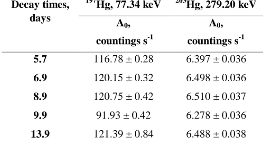
The Hg loss after irradiation was also verified by measuring synthetic standard of Hg for different decay times. The counting rates obtained were corrected for the same decay time (t = 0) from the measurements obtained in different decay times. Tables 1 and 2 show the obtained results for Hg standard irradiated in polyethylene capsule and in polyethylene envelope, respectively. Counting rates with their respective uncertainties were obtained for the photopeaks of 77.34 keV of 197Hg and 279.20 keV of 203Hg.
Table 1: Counting rates of Hg synthetic standard corrected (A0) for decay time t0 = 0. Measure-ments obtained for different decay times. Irradiation time of 1 h in polyethylene capsule.
Decay times, days 197 Hg, 77.34 keV 203Hg, 279.20 keV A0, countings s-1 A0, countings s-1 5.9 108.30 ± 0.27 4.626 ± 0.031 7.0 108.44 ± 0.31 4.611 ± 0.031 8.9 109.77 ± 0.41 4.691 ± 0.031 11.0 108.07 ± 0.75 4.534 ± 0.031 11.9 108.95 ± 0.60 4.601 ± 0.032
Table 2: Counting rates of Hg synthetic standard corrected (A0) for decay time t0 = 0. Measure-ments obtained for different decay times. Irradiation time of 1 h in polyethylene envelope.
Decay times, days 197 Hg, 77.34 keV 203Hg, 279.20 keV A0, countings s-1 A0, countings s-1 5.7 116.78 ± 0.28 6.397 ± 0.036 6.9 120.15 ± 0.32 6.498 ± 0.036 8.9 120.75 ± 0.42 6.510 ± 0.037 9.9 91.93 ± 0.42 6.278 ± 0.036 13.9 121.39 ± 0.84 6.488 ± 0.038
Results of Table 1 show that for the Hg standard irradiated in capsule, the values of the counting rates for the two photopeaks remained approximately constant until about 12 d of decay. In the case of the standard irradiation in polyethylene envelope (Table 2), the counting rates remained approx-imately constant until about 14 d of decay. These results indicate that, for decay times of up to 12 d, there was no Hg loss after irradiation either from the capsule or from the envelope.
Based on these results, polyethylene envelopes were select for sample and standard irradiations for Hg determination due to its ease of acquisition and cheaper cost.
3.3. Mercury Determination Using Different Irradiation Times
The adequate irradiation time for Hg determination without causing its loss was chosen by analyz-ing the CRM IAEA-085 Human Hair, usanalyz-ing irradiation times of 1 and 8 h. Mercury mass fraction obtained with 1 and 8 h of irradiation, by measuring 197Hg and 203Hg for different decay times, pre-sented good agreement with certified value (23.2 ± 0.8 µg g-1) [17]. The results varied from 23.58 ± 0.14 to 25.536 ± 0.087 µg g-1, for sample irradiated for 1 h, and from 25.311 ± 0.020 to 25.316 ± 0.034 µg g-1, for that irradiated for 8 h. These results indicate that there were no Hg losses during the irradiation, since the obtained values of mass fraction are close to the certified value. However, after decay of 24Na in the sample, the radionuclide 75Se was identified in the CRM IAEA-085 Hu-man Hair irradiated for 8 h, becoming impossible to determine Hg by measuring 203Hg, since the gamma ray energies emitted by 75Se and by 203Hg are very close to each other (279.93 keV and 279.20 keV, respectively) [6]. Besides using the irradiation of 8 h for CRM IAEA-085 Human Hair, a complex gamma ray spectrum was obtained in the region of low energy with high counting rates at of 68.89 keV and 411.80 keV of 198Au. According to Zmijewsk [8], an important interference in Hg determination by INAA is that of 198Au, whose photopeaks of energies 70.8 and 80.3 keV are not resolved from 197Hg photopeak. The interference of 198Au in the 77.34 keV photopeak of 197Hg could not be verified by determining life, since these two radionuclides present very close half-lifes (64.16 h for 197Hg and 64.80 h for 198Au) [6].
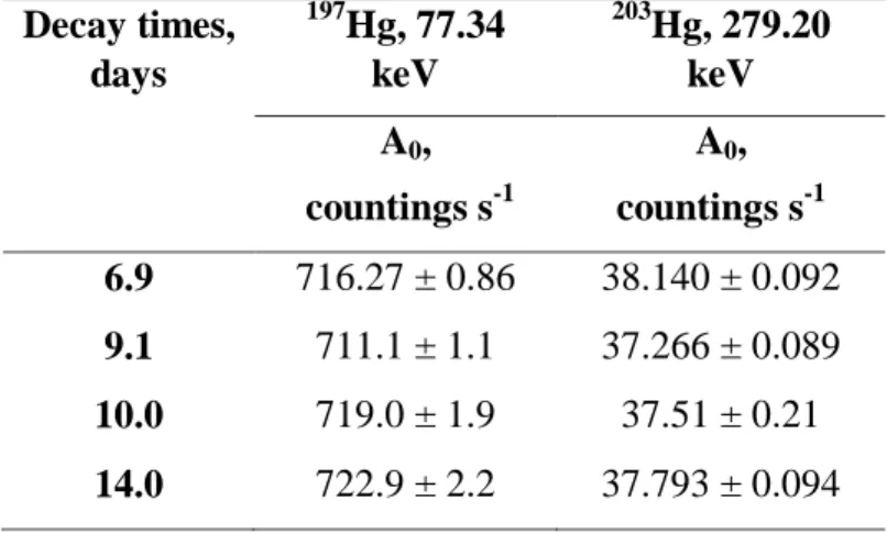
Table 3 shows the results obtained in the study of Hg loss after irradiation for Hg standard irradiat-ed for 8 h in polyethylene envelope. The counting rates with their respective uncertainties correctirradiat-ed for decay time t0 = 0 were obtained for the photopeaks of 77.34 keV of 197Hg and 279.20 keV of
203
Hg. This table shows that the counting rates calculated for zero decay time remained approxi-mately constant until about 14 d of decay, indicating no loss of Hg after irradiation.
Table 3: Counting rates of Hg synthetic standard corrected (A0) for decay time t0 = 0. Measure-ments obtained for different decay times. Irradiation time of 8 h in polyethylene envelope.
Decay times, days 197 Hg, 77.34 keV 203 Hg, 279.20 keV A0, countings s-1 A0, countings s-1 6.9 716.27 ± 0.86 38.140 ± 0.092 9.1 711.1 ± 1.1 37.266 ± 0.089 10.0 719.0 ± 1.9 37.51 ± 0.21 14.0 722.9 ± 2.2 37.793 ± 0.094
Based on the obtained results, irradiation time of 1 h was chosen, in order to avoid gamma ray spec-tral interferences from 198Au and 75Se.
3.4. Analyses of Certified Reference Materials and of Environmental Samples
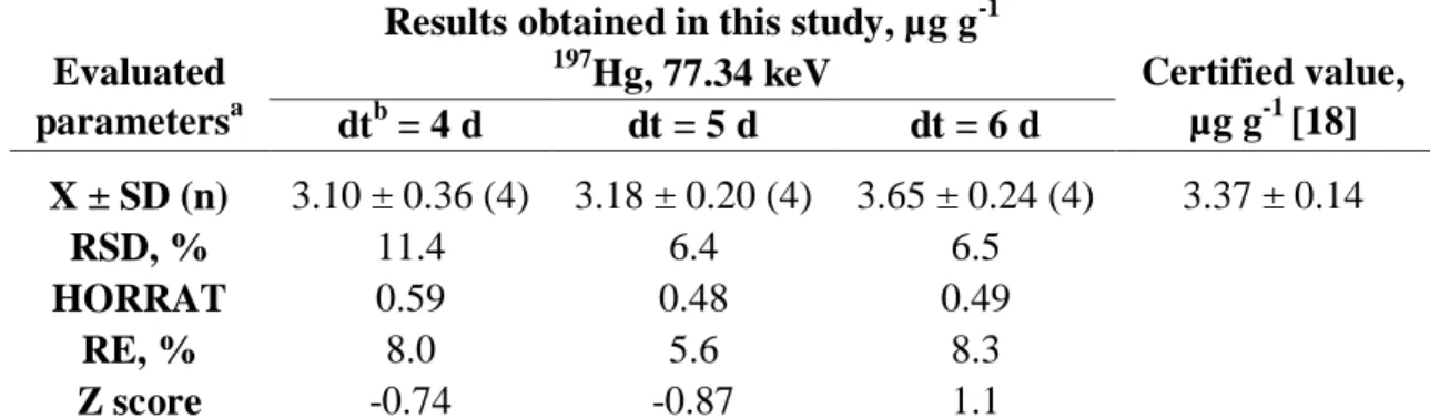
Tables 4 to 7 show the results obtained in the analyses of certified reference materials NCR DOLT-3 Dogfish Liver, BCR 186 Pig Kidney, INCT M-4 Cormorant Tissue and IAEA-085 Human Hair, respectively. In these Tables, results are presented for different decay times used for counting. For CRMs NCR DOLT-3 Dogfish Liver, BCR 186 Pig Kidney and INCT M-4 Cormorant Tissue, it was observed that determinations performed after 4 d of decay presented higher values of relative standard deviation and relative error, probably due to the high activity of 24Na (t1/2 = 14.96 h) that masked the less intense activities of 197Hg. All the obtained results presented satisfactory values of relative error, HORRAT value and Z score for all the materials, except for the measurement of the 197
2. For CRMs NCR DOLT-3 Dogfish Liver and BCR 186 Pig Kidney, Hg was not determined by measuring 203Hg, since 75Se was identified in the spectra of these materials.
Mercury was not detected in the tree bark samples analyzed in this study, so detection limits and quantification limits for Hg determinations were calculated by the measurement of 197Hg and 203Hg radionuclides (Table 8). The bark samples analyzed were probably not collected in regions polluted by Hg emission into the atmosphere. According to Martín et al [21], this element is naturally pre-sent in the environment at very low concentrations (0.005 to 0.06 ng m-3 in air). Results showed in Table 8 indicate that detection limits obtained for 197Hg gamma ray peak are lower than those ob-tained for 203Hg.
Table 4: Mass fractions of Hg obtained in the CRM NCR DOLT-3 Dogfish Liver by measuring the
197
Hg radioisotope. Results obtained for different decay times used for counting.
Evaluated parametersa
Results obtained in this study, µg g-1
Certified value, µg g-1 [18] 197 Hg, 77.34 keV dtb = 4 d dt = 5 d dt = 6 d X ± SD (n) 3.10 ± 0.36 (4) 3.18 ± 0.20 (4) 3.65 ± 0.24 (4) 3.37 ± 0.14 RSD, % 11.4 6.4 6.5 HORRAT 0.59 0.48 0.49 RE, % 8.0 5.6 8.3 Z score -0.74 -0.87 1.1
a. X ± SD: Arithmetic mean and standard deviation; n: number of determinations; RSD: relative standard deviation; RE: relative error; b. dt = decay time.
Table 5: Mass fractions of Hg obtained in the CRM BCR 186 Pig Kidney by measuring the 197Hg radioisotope. Results obtained for different decay times used for counting.
Evaluated parametersa
Results obtained in this study, µg g-1
Certified value, µg g-1 [19] 197 Hg, 77.34 keV dtb = 4 d dt = 5 d dt = 6 d X ± SD (n) 1.58 ± 0.27 (4) 1.65 ± 0.13 (4) 1.78 ± 0.16 (4) 1.97 ± 0.04 RSD, % 17.1 8.2 9.3 HORRAT 1.1 0.55 0.63 RE, % 19.9 16.1 2.7 Z score -1.5 -2.3 -1.2
a. X ± SD: Arithmetic mean and standard deviation; n: number of determinations; RSD: relative standard deviation; RE: relative error; b. dt = decay time.
topes. Results obtained for different decay times used for counting.
a. X ± SD: Arithmetic mean and standard deviation; n: number of determinations; RSD: relative standard deviation; RE: rel-ative error. b. dt = decay time.
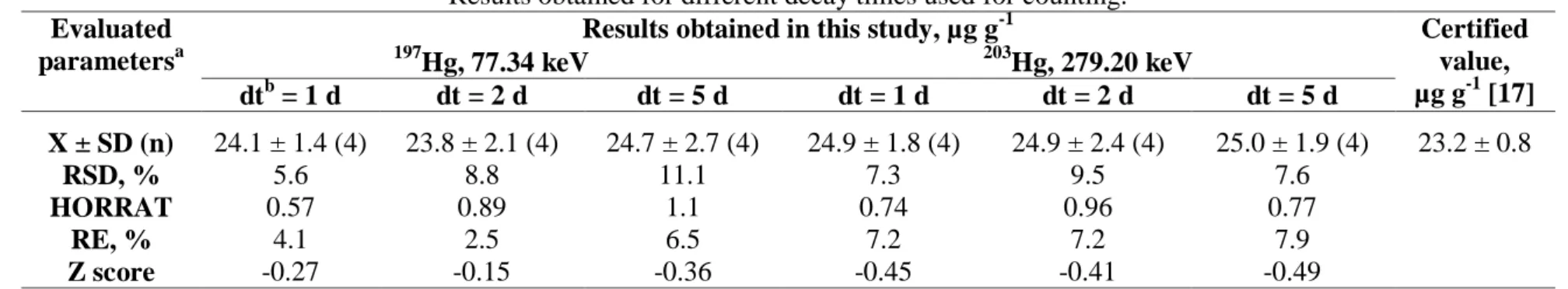
Table 7: Mass fractions of Hg obtained in the CRM IAEA-085 Human Hair by measuring the 197Hg and 203Hg radioisotopes. Results obtained for different decay times used for counting.
Evaluated parametersa
Results obtained in this study, µg g-1 Certified value, µg g-1 [17] 197 Hg, 77.34 keV 203Hg, 279.20 keV dtb = 1 d dt = 2 d dt = 5 d dt = 1 d dt = 2 d dt = 5 d X ± SD (n) 24.1 ± 1.4 (4) 23.8 ± 2.1 (4) 24.7 ± 2.7 (4) 24.9 ± 1.8 (4) 24.9 ± 2.4 (4) 25.0 ± 1.9 (4) 23.2 ± 0.8 RSD, % 5.6 8.8 11.1 7.3 9.5 7.6 HORRAT 0.57 0.89 1.1 0.74 0.96 0.77 RE, % 4.1 2.5 6.5 7.2 7.2 7.9 Z score -0.27 -0.15 -0.36 -0.45 -0.41 -0.49
a. X ± SD: Arithmetic mean and standard deviation; n: number of determinations; RSD: relative standard deviation; RE: rel-ative error. b. dt = decay time.
Evaluated parametersa
Results obtained in this study, µg g-1 Certified value, µg g-1 [20] 197 Hg, 77.34 keV 203Hg, 279.20 keV dtb = 4 d dt = 5 d dt = 6 d dt = 4 d dt = 5 d dt = 6 d X ± SD (n) 1.89 ± 0.24 (4) 2.05 ± 0.11 (4) 2.13 ± 0.11(4) 1.82 ± 0.33 (4) 2.10 ± 0.34 (4) 2.30 ± 0.23 (4) 2.20 ± 0.14 RSD, % 12.6 5.2 5.1 18.1 16.1 10.0 HORRAT 0.87 0.36 0.36 1.2 1.1 0.71 RE, % 14.2 6.7 3.1 17.2 4.6 4.3 Z score 1.2 1.1 0.53 1.1 0.30 -0.40
Table 8: Detection limits (DL) and quantification limits (QL) of Hg obtained in the analysis of the
tree bark samples
Sample codea Radioisotope
DL, μg g-1 QL, μg g-1 A1 197 Hg 0.07 0.22 203 Hg 0.7 2.1 A2 197 Hg 0.08 0.24 203 Hg 0.8 2.3 A3 197 Hg 0.14 0.42 203 Hg 1.9 5.8 A4 197 Hg 0.1 0.3 203 Hg 1.6 4.8 A5 197 Hg 0.07 0.22 203 Hg 1.0 3.0 A6 197 Hg 0.1 0.3 203 Hg 1.4 4.3 a
Samples coded A1 to A5 are from Tipuana tipu (Tipuana) specie. and A6, from
Poincia-nella pluviosa (Sibipiruna) species.
4. CONCLUSION
From the results obtained in the analyses of polyethylene envelope and of the Nº. 40 Whatman filter paper sheet, it can be concluded that Hg is present in these materials at very low levels and it was considered negligible in the analyses.
Results obtained using different containers for irradiation demonstrated that polyethylene capsules and polyethylene envelopes can be used for Hg determination by INAA. Both containers showed no Hg loss. In this study, polyethylene envelope was chosen due to its ease of acquisition and cheaper cost.
Irradiation time of 1 h was chosen due to the problem of gamma ray spectral interference of 198Au and 75Se radionuclides. In addition, high activities of 24Na, formed by the longer irradiation time, may affect the results of Hg determinations. The high activity of 24Na masked the less intense activ-ities of 197Hg. The decay time of 6 d, in general, presented more precise and accurate results.
The results obtained for certified reference materials indicated good agreement with the certificate values, demonstrating the accuracy of the data. Results obtained also presented good precision and accuracy, evaluated by the HORRAT values (HORRAT < 2), and by Z score obtained values (|Z score| < 2). The exception was for Hg determination in the analysis of CRM BCR 186 Pig Kidney using decay time of 5 d.
Mercury was not detected in tree bark samples. The detection limits obtained for tree bark analysis varied from 0.07 and 1.9 μg g-1, and the quantification limits were between 0.22 and 5.8 μg g-1
. Re-sults obtained for Hg determination in tree barks indicated that there is no emission source of this element in the region where they were collected.
5. ACKNOWLEDGMENTS
Authors thank Fundação de Amparo à Pesquisa do Estado de São Paulo (FAPESP) and Conselho Nacional de Desenvolvimento Científico e Tecnológico (CNPq) for financial support. In addition, C. Perez and E.C. Santos are grateful for scholarships from Comissão Nacional de Energia Nuclear (CNEN) and Coordenação de Aperfeiçoamento de Pessoal de Nível Superior (CAPES), respecti-vely.
REFERENCES
1. YANG, J.; ZHAO, Y.; ZHANG, J.; ZHENG, C. Removal of elemental mercury from flue gas by recyclable CuCl2 modified magnetospheres catalyst from fly ash. Part 1. Catalyst characterization and performance evaluation. Fuel, v. 164, p. 419-428, 2016.
2. DRASCH, G. A., Handbook on metals in clinical and analytical chemistry, 1st edition. New York: Marcel Dekker, Inc., 1994.
3. ŠPIRIĆ, Z.; VUČKOVIĆ, I.; STAFILOV, T.; KUŠAN, V.; BAČEVA, K. Biomonitoring of air pollution with mercury in Croatia by using moss species and CV-AAS. Environmental
Monitoring and Assessment, v. 186(7), p. 4357-4366, 2014.
4. BEDREGAL, P. S.; MENDOZA, P. A.; UBILLUS, M. S.; COHEN, I. M.; MONTOYA, E. H. The k0 and relative INAA methods to determine elements in entire archaeological pottery objects. Journal of Radioanalytical and Nuclear Chemistry, v. 300, n. 2, p. 673-678, 2014.
5. ALMEIDA, S. M.; RAMOS, C. A.; MARQUES, A. M.; SILVA, A. V.; FREITAS, M. C.; FARINHA, M. M.; REIS M.; MARQUES, A. P. Use of INAA and PIXE for multipollutant air quality assessment and management. Journal of Radioanalytical and Nuclear
Chemis-try, v.294, n. 3, p 343-347, 2012.
6. IAEA. INTERNATIONAL ATOMIC ENERGY AGENCY. Practical aspects of operating
a neutron activation analysis laboratory, IAEA-TEC-DOC-564, 1990.
7. ANDERSON, D. L. Use of l-cysteine for minimization of inorganic Hg loss during thermal neutron irradiation. Journal of Radioanalytical and Nuclear Chemistry, v. 282, n. 1, p. 11-14, 2009.
8. ŻMIJEWSKA, W. Activation analysis of mercury in environmental samples. Journal of
Radioanalytical and Nuclear Chemistry, v. 35, n. 2, p. 389-418, 1977.
9. TAKEUCHI, T.; SHINOGI, M.; MORI, I. Volatilization losses of mercury in neutron acti-vation analysis. Journal of Radioanalytical Chemistry, v. 53, p. 81-88, 1979.
10. PATTERSON, J. W.; PASSINO, R. Metals speciation separation and recovery (Vol. 1), 1st edition. Chicago: CRC Press, 1987.
11. DE SOETE, D.; GILBELS, R.; HOSTE, J. Neutron activation analysis, 1st edition, New York: Wiley-Interscience, 1972.
12. WOOD, R. How to validate analytical methods. Trends in Analytical Chemistry, v. 18, p. 624-632, 1999.
13. HORWITZ W.; ALBERT, R. The Horwitz ratio (HorRat): A useful index of method per-formance with respect to precision. Journal of AOAC International, v. 89, n. 4, p. 1095-1109, 2006.
14. KONIECZKA, P.; NAMIESNIK, J. Quality assurance and quality control in the
analyti-cal chemianalyti-cal laboratory: a practianalyti-cal approach, 2nd edition. Boca Raton: CRC Press, 2016. 15. CURRIE, L.A. International recommendations offered on analytical detection and
quantifi-cation concepts and nomenclature. Analytica Chimica Acta, v.391, p.127-134, 1999. 16. FRIEDLANDER, G.; KENNEDY, J. W.; MILLER, J. M., Nuclear and radiochemistry,
2nd edition. London: John Wiley & Sons, p. 6, 1964.
17. IAEA. INTERNATIONAL ATOMIC ENERGY AGENCY. Reference sheet, IAEA-085, Methylmercury, total mercury and other trace elements in human hair, 2000.
18. NCR. NATIONAL RESEARCH COUNCIL CANADA. Certificate of analysis, DOLT-3, Dogfish Liver Certified Reference Material for Trace Metals, 2002.
19. BCR. COMMUNITY BUREAU OF REFERENCE. Certificate of analysis, BCR 186, Trace elements in lyophilized pig kidney, 1986.
20. INCT. INSTITUTE OF NUCLEAR CHEMISTRY AND TECHNOLOGY. Certificate of
analysis, MODAS-4 Cormorant Tissue, M-1 CormTis, 2015.
21. MARTÍN, J. A. R.; NANOS, N.; MIRANDA, J. C.; CARBONELL, G.; GIL, L. Volcanic mercury in Pinus canariensis. Naturwissenschaften, v. 100, n. 8, p. 739-747, 2013.