Article
0103 - 5053 $6.00+0.00
*e-mail: [email protected]
Chemiluminescence Determination of Mezlocillin by the Luminol-Potassium Periodate System
Wen Bing Shi* and Ji Dong Yang
Department of Chemisty and Enviroment Science, Fuling Normal University, Yangtze, Chongqing, 408003, P. R. China
Um método novo, simples e sensível foi proposto para a determinação de mezlocilina. Baseia-se na amplificação da emissão de quimiluminescência (CL) gerada a partir da oxidação do luminol em meio alcalino pelo periodato de potássio. A otimização das variáveis experimentais e instrumentais que afetam o efeito da amplificação de CL foi realizada usando-se sistema de injeção em fluxo. Em condições ótimas, o método é eficiente para determinar mezlocilina no intervalo linear de 0,01 a 30 ×10-6 g mL-1 com limite de detecção (3σ) de 3,0×10⁻⁹ g mL-1 e desvio padrão
relativo (RSD) de 1,0% para 1,0×10⁻⁶ g mL-1 de mezlocilina (n=11). O método tem sido aplicado
com sucesso, para determinação de mezlocilina em preparações comerciais, amostras sintéticas e formulações biológicas fluidas.
A new, simple and sensitive method has been proposed for the determination of mezlocillin. It is based on the enhancement of the chemiluminescence (CL) emission generated from the oxidation of luminol in alkaline medium by postassium periodate. The optimization of the experimental and instrumental variables affecting the CL enhancement effect has been carried out using flow-injection system. In the optimum conditions, the method is efficient to determine mezlocillin in the linear range of 0.01-30×10-6 g mL-1 with a detection limit (3σ) of 3.0×10⁻⁹ g mL-1
and the relative standard deviation (RSD) is 1.0% for 1.0×10⁻⁶ g mL-1 mezlocillin (n=11). It has
been successfully applied to the mezlocillin determination in commercial preparations, synthetic samples and biological fluids formulations.
Keywords: chemiluminescence, flow injection, mezlocillin
Introduction
Mezlocillin (Figure 1) is a widely used β-lactam antibiotic belonging to the penicillin group. It is very effective against some bacteria and is used in treating infections of the skin, blood, central nervous system, respiratory tract, sinuses, and urinary tract. It is also is used to treat some gynecological
infections of gynecological.1 Some other antibiotics given
by injection, clavulanic acid, methotrexate and probenecid may interact with mezlocillin.2 Therefore, the safety of using
mezlocillin is very important, and the biological research of mezlocillin is very significant in clinical medicine. The development of a sensitive and simple method for the determination of mezlocillin is important for quality assurance in preparations and for obtaining optimum therapeutic concentrations. For its measurement, several methods have been reported, such as high-performance liquid chromatography (HPLC),3-9 electrochemical10 and atomic
absorption spectrometry.11 The use of chemiluminescence(CL)
detection in pharmaceutical analysis involving the assay of active components in dosage forms represents a fairly selective and sensitive technique, which requires relatively simple and inexpensive instrumentation, and a variety of drugs have been determined by this technique.12,13 Recently, several CL
methods for the determination of penicillin antibiotics have been reported.14-22 These were based on enhanced CL methods
using luminol-hydrogen peroxide,14 enhancing effects on the
luminol CL systems,15,20,21 quenching CL methods using the
luminol CL systems,16,17,22 and direct CL methods based on
potassium superoxide oxidation18 or potassium permanganate
oxidation.19 Moreover, the nature of the reaction mechanism
also has been demonstrated in detail.15
In this work, a new flow-injection CL method is presented for the determination of mezlocillin which is based on the enhancing effect of mezlocillin on the CL emission generated by the oxidation of luminol with postassium periodate (KIO4) in alkaline condition. As far as the authors know, although the CL behaviour of other penicillins has been reported,12-22 this is the first use
of the luminol-KIO4 reaction for the determination of mezlocillin. Compared with previous CL assays for the determination of penicillin,12-22 this method has higher
sensitivity (the detection limit is 3.0 ng mL-1 mezlocillin)
than previously used oxidants, such as hydrogen peroxide,12
potassium ferricyanide,13 iodine,14,15 superoxide16 or
potassium permanganate.17 The method has been applied
to the determination of mezlocillin in pharmaceutical formulations, synthetic samples and biological fluids formulations and the results agreed well with the Pharmacopoeia method.23
Experimental
Reagents
All the reagents used were of analytical reagent grade. Doubly deionized water was used throughout the study. All reagents were obtained from Chongqing Chemical Reagents Company (Chongqing, China) except for luminol, which was purchased from Merck (Darmstadt, Germany) and mezlocillin, which was obtained from Shandong Reyoung Pharmaceutical Co., Ltd., (Shandong, China). A stocked solution of mezlocillin (5×10⁻⁴ g mL-1) was
stored in a refrigerator. Working standard solutions of mezlocillin were prepared daily from the stocked solution by appropriate dilution with doubly deionized water. A 0.01 mol L-1 luminol solution was prepared by dissolving
0.1777 g luminol in 100 mL of 0.01 mol L-1 NaOH solutions.
More diluted solutions were prepared in 0.005 mol L-1
NaOH and used immediately. Potassium periodate stock solution (0.01 mol L-1) was prepared by dissolving 0.5750 g
potassium periodate in water and diluting to 250 mL with water. Potassium periodate working solution (5×10⁻⁵ mol L-1)
was prepared by diluting above stock solution with water.
Apparatus of flow injection system
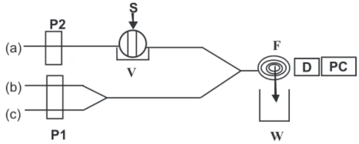
The FIA system (shown in Figure 2) used in this work includes two peristaltic pumps P1 and P2 (Longfang
Instrument Factory, Wenzhou, China). P1 delivered all reagent streams at a flow rate of 2.5 mL min-1 (per tube)
and P2 delivered water carrier stream at a flow rate of 3.6 mL min-1, and PTFE tubing (0.8 mm i.d.) was used
to connect all components in the flow system. A rotary eight-way manual injection valve (Longfang Instrument Factory, Wenzhou, China) was used to inject working and sample solutions into the carrier stream. A volume of 160 µL mezlocillin was injected into the water carrier stream and then mixed with a mixture of luminescent reagents (luminol-KIO4 solution). The emitted CL was collected with a photomultiplier tube (operated at −600V) of the Type IFFL-DD flow-injection chemiluminescence analyzer (Reike, Xi’an,China). The signal was recorded by using an IBM-compatible computer, equipped with a data acquisition interface. Data acquisition and treatment were performed with REMAX software running under Windows 2000. UV-4100 spectrophotometer (Hitachi, Japan) was used to investigate the UV spectra.
Procedure for calibration
Some accurately measured aliquots of mezlocillin working standard solutions (5.0×10⁻⁴ g mL-1) in the range of
0.01-30×10-6 g mL-1 were transferred into a series of 25 mL
volumetric flasks and were diluted with water freshly. The CL signal was measured by injecting 160 µL of working standard solution into the water carrier stream, which then joined the reagent streams (a mixture of 5.0×10⁻⁵ mol L-1
KIO4 and 4.0×10⁻⁵ mol L-1 luminol in 5.0×10⁻³ mol L-1
NaOH solution). Calibration graph was constructed by CL emission intensities versus concentration of mezlocillin and the regression equation was calculated.
Procedure for the pharmaceutical preparations
Injection samples, each with a nominal content of 1.0 g of mezlocillin in 1 vial were diluted to 1000 mL with doubly
Figure 2. Schematic diagram of the flow system for the determination of mezlocillin. (a) H2O; (b) 5.0×10⁻⁵ mol L-1 KIO
4; (c) 4.0×10 -5 mol L-1
luminol in 5.0×10⁻³ mol L-1 NaOH; S, sample; P1, P2, peristaltic pump;
deionized water and further diluted to the working range of the determination of mezlocillin. Proceed as described above.
Procedure for synthetical sample
A certain amount of mezlocillin was put into a 100 mL flask, and then different foreign substances were added and diluted with doubly deionized water for a quantitative analysis.
Procedure for spiked urine
Add an aliquot of standard aqueous solution of mezlocillin (5.0×10⁻⁴ g mL-1) to the urine. Transfer 1
mL of this solution into a 100 mL volumetric flask and dilute to volume with doubly deionized water. Proceed as described above. A blank value was determined by treating mezlocillin-free urine in the same way.
Procedure for real urine samples
Take 10 mL of urine sample from the psychiatric patients suffering from infection of the skin in the local hospital into a clean tube, and then the mixture was diluted ten-fold with doubly deionized water and filtered with 0.45 µm filter (Millex-HA, Millipore). The filtrate was treated and analysed according to the the same procedures as those for pharmaceutical samples. A blank value was determined by treating the deionized water in the same way.
Results and Discussion
Optimization of apparatus parameters
The flow cell reactor (reaction coil) was made by curling a vitreous tube (1.5 mm i.d., 8 cm) and located in front of the photomultiplier; The other apparatus parameters were as follows: flow rates (P1 delivered all reagent streams at a flow rate of 2.5 mL min-1 and P2 delivered water carrier
stream at a flow rate of 3.6 mL min-1); The injected volume
is 160 µL; The distance of valve to flow cell was 8.0 cm; Sampling time was 30 s; The photomultiplier tube was operated at −600V.
Optimization of reaction conditions
It is well known that the luminol CL reaction requires alkaline conditions. Our preliminary experiment showed that among several alkaline buffer solutions tested, including carbonate, phosphate, borate and sodium hydroxide, sodium
hydroxide was the most suitable medium for present CL reaction. It was also observed that the CL reaction has higher CL signal when an appropriate amount of sodium hydroxide was added into the luminol solution than added into the KIO4 solution. Based on these observations, a series of experiments were conducted to select the optimum reaction conditions for the CL determination of mezlocillin using a 1.0×10⁻⁶ g mL-1 mezlocillin standard solution. The
parameters included the concentration of sodium hydroxide in the luminol solution, the concentration of KIO4 and the concentration of luminol.
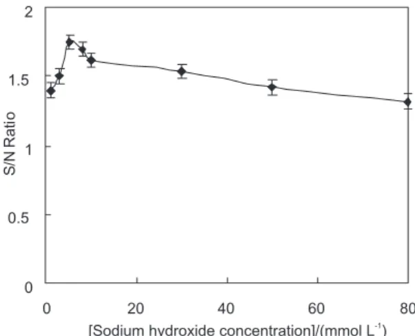
Effect of sodium hydroxide concentration
The effect of sodium hydroxide (as showed in Figure 3) concentration on the CL reaction was studied at different concentrations from 1.0×10⁻³ to 8.0×10⁻² mol L-1 when
the concentrations of KIO4 and luminol were fixed at 5.0×10⁻⁵ mol L-1 and 4.0×10⁻⁵ mol L-1, respectively. The
CL intensity continued to increase with the increase in the concentration of sodium hydroxide concentration from 1.0×10⁻³ to 5.0×10⁻³ mol L-1. When the concentration of
sodium hydroxide was higher than 5.0×10⁻³ mol L-1, CL
intensity decreased. A concentration of 5.0×10⁻³ mol L-1
sodium hydroxide was therefore selected for use in the following experiments.
Effect of luminol concentration
As the luminescence reagent, luminol concentration affected the CL intensity (as showed in Figure 4). The effect of 1.0-120×10-6 mol L-1 luminol on the CL reaction
was examined when the concentration of KIO4 was fixed at 5.0×10⁻⁵ mol L-1 and the concentration of sodium hydroxide
was fixed at 5.0×10⁻³ mol L-1. As the concentration was
increased, both the CL intensity and the background increased. The maximum signal-to-noise(S/N) was obtained at 4.0×10⁻⁵ mol L-1 luminol. Therefore, 4.0×10⁻⁵
mol L-1 luminol was employed in the coming works.
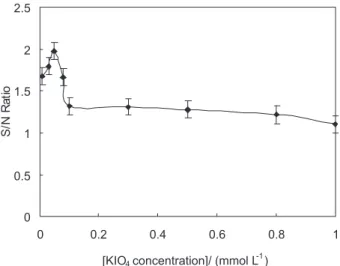
Effect of KIO
4
KIO4 was used as the oxidant in the reaction. The effect of KIO4 concentration (as showed in Figure 5) on the CL reaction was investigated in the range of 0.01 to 1.0×10⁻³
mol L-1 when the concentrations of sodium hydroxide
and luminol were fixed at 5.0×10⁻³ mol L-1 and 4.0×10⁻⁵
mol L-1 respectively. As the concentration was increased,
both the CL intensity and the background increased. The maximum signal-to-noise(S/N) was obtained at 5.0×10⁻⁵
mol L-1 KIO
4. So, 5.0×10
-5 mol L-1 KIO
4 was selected in
the subsequent work.
Performance of the proposed method for mezlocillin measurements
Under the selected conditions given above, the calibration graph of emission intensity versus mezlocillin concentration was linear in the 0.01-30×10-6 g mL-1
range (ΔI =54.576 (mezlocillin) (×10-6 g mL-1) +580.94;
r = 0.9994, n = 11) with a detection limit (3σ) of 3.0× 10⁻⁹ g mL-1. Relative standard deviation (RSD) (n = 11)
was 1.0% for 1.0×10⁻⁶ g mL-1 mezlocillin. Seven replicate
determinations (determinations of precision and accuracy were performed between 7 days and 7 times in 1 day for intra-day and inter-day, respectively) at four concentration levels were carried out to test the accuracy and precision of the proposed method for urine samples and the results listed in Table 1. As can be seen, RSD (precision) of inter-day and intra-inter-day is less than 5%, and accuracy of inter-inter-day and intra-day is satisfactory.
Interference study
The effect of foreign substances was tested by analyzing a standard solution of mezlocillin (1.0×10⁻⁶ g mL-1) to
which increasing amounts of interfering substances was added. The tolerable concentration ratios with respect to 1.0×10⁻⁶ g mL-1 mezlocillin for interference at ± 5% level
were over 1000 for starch, glucose, sucrose, Ca2+; 800 for
Cl−, urea, K+, Br−, Na+, NO 3
−, uric acid, maltose and urea;
600 for SO42−, CO 3
2−; 400 for paracetamol, respectively.
Figure 4. Effect of luminol concentration. Error bars represent one standard deviation for five measurements.
Figure 5. Effect of KIO4 concentration.Error bars represent one standard deviation for five measurements.
Table 1. Accuracy and precision data at four concentrations Concentration Added /
(µg mL-1)
Inter-daya Intra-daya
Found / (µg mL-1) Error (%) RSD (%) Found / (µg mL-1) Error (%) RSD (%)
0.05 0.047 -6.0 4.2 0.046 -0.80 2.5
0.1 0.098 -2.0 3.8 0.097 -3.0 3.2
1 0.96 -4.0 2.6 0.95 -5.0 1.8
5 4.97 -0.6 1.4 4.95 -1.0 3.4
The results show that the proposed method has good selectivity for mezlocillin determination and it can be used in monitoring the mezlocillin in real samples.
Sample analysis
Analysis of mezlocillin in injection samples and synthetic samples
Following the procedure described under experimental section, the proposed method was applied to the determination of mezlocillin in injection samples. Also, a series of synthetic samples were prepared to check the validation of the proposed method. The results are listed in Tables 2 and 3. The recovery tests were carried out on the samples and the obtained recoveries were satisfactory. As illustrated in Tables 2 and 3, the proposed method can be satisfactorily applied to the determination of mezlocillin in injection samples and synthetic samples. The mezlocillin concentrations agree well with those obtained by UV spectrophotometry. The UV spectrophotometric determination of mezlocillin was performed according to the literature.
Analysis of mezlocillin in real urine samples
As stated above, the proposed method gave a detection limit of 3.0×10⁻⁹ g mL-1 for mezlocillin, which is lower
than those given by other methods. For example, Wang et al.11 reported a DL of 7.93×10⁻⁶ g mL-1 by atomic
absorbption spectrometry. When using high-performance liquid chromatography,8 1.0×10⁻⁷ g mL-1 in serum and
1.0×10⁻⁶ g mL-1 in urine of DL were reported, respectively.
So, the high sensitivity of the proposed method allows the determination of mezlocillin in biological fluids. Table 4 shows the results of mezlocillin determination in urine samples from the patients suffering from infection of the skin.
The major urinary metabolite of mezlocillin is penicilloic acid.24 Formation of penicilloic acid decreases
the potential of protonation as compared with the parent compound. The recovery test on the spiked urine samples at different concentration levels demonstrates good results (shown in Table 5). So, the effect of the urinary metabolites of mezlocillin on the chemiluminescence signal is minor. Results obtained by this proposed method demonstrate that the metabolite of mezlocillin did not affect its determination.
Table 2. Results of the determination of mezlocillin in injections
Sample Label / (g) Found/(g) ± RSD(%)
a
Proposed method UV spectrophotometry
Injection 1 1.00 1.02 ± 0.28 0.965 ± 0.36
Injection 2 1.00 1.01 ± 0.46 0.983 ± 0.52
Injection 3 1.00 0.998 ± 0.38 0.978 ± 0.42
aAverage of five measurements.
Table 3. Results of the determination of mezlocillin in synthetic samples
Sample (No.)
Co-existing substances / (µg mL-1) Added /
(µg mL-1)
Found / (µg mL-1)
Recovery / (% ± RSD%, n=3)
1 Glucose (10), starch (10) 0.5 0.49 98.0±3.2
2 Glucose (30), starch (30) 1.0 1.02 102±2.4
3 Glucose (100), starch (100) 0.5 0.48 96.0±3.6
4 Glucose (50), starch (50), urea (50), starch (25) 1.0 0.98 98.0±1.8
5 Starch (100), EDTA (5), CaCl2 (100) 1.0 1.01 101±3.4
6 Glucose (100), starch (100), KCl (50), MgSO4 (10) 1.0 0.99 99.0±2.8
Table 4. Results of the determination of mezlocillin in urine samples Urine sample number Proposed method / (µg mL-1) (RSD %)a
1 1.952 (3.82)
2 1.868 (2.54)
3 1.924 (2.62)
Table 5. Results of the determination of mezlocillin in spiked urine Concentration added / (µg mL-1) Found / (%) (RSD %, n=3)
0.05 93.8 (2.32)
0.1 93.2 (3.26)
0.2 94.4 (1.86)
0.5 92.6 (1.36)
0.8 95.6 (1.80)
1 96.2 (2.36)
10 102 (2.92)
Conclusions
It was found that the reaction medium of the luminol solution, KIO4 and the concentration of mezlocillin affected the CL intensitivity. In alkaline medium, mezlocillin showed strong CL enhancement at 5.0×10⁻³ mol L-1
sodium hydroxide. On this basis, a flow injection method was established for the determination of mezlocillin. This method has the merits of higher sensitivity and wider linear range, being superior to the literature methods. It is applicable to detect mezlocillin in pharmaceutical formulations, synthetic samples and biological fluids formulations.
Acknowledgments
This work is supported by the Chongqing Committee of Education (No. KJ061306).
References
1. Betriu, C.; Sánchez, A.; Gómez, M.; Palau, M. L.; Picazo, J. J.; J. Antimicrob. Chemother. 1999, 43,133.
2. Trissel, L. A.; Martinez, J. F.; Gilbert, D. L.; J. Am. Pharm. Assoc. (Wash) 1999, 39, 514.
3. Haginaka, J. J.; Wakai, H.; Yasuda, U. T.; Anal. Sci. 1985, 1, 73.
4. Fiore, D.; Auger, F. G.; Drusano, G. L.; Dandu, V. R.; Lesko, L. J.; Antimicrob. Agents Chemother. 1984, 26, 775.
5. Haen, E.; Remien, J.; Richter, E.; Frank, U.; Adam, D.; Biopharm. Pharmacokinet, Eur. Congr. 1984, 2, 318. 6. Knoller, J.; Bremm, K. D.; Schoenfeld, W.; Koenig, W.;
Antimicrob. Agents Chemother. 1986, 29, 527.
7. Kames, H. T.; Beightol, L. A.; Farthing, D.; Ther. Drug Monit. 1987, 9, 456.
8. Garca-Glez, J. C.; Mendez, R.; Martn-Villacorta, J.; J. Chromatogr. A 1998, 812, 213.
9. Zhao, Y. C.; Liao, Z. X.; Chin. J. Pharm.2002, 33,139. 10. Ning, M. X.; Cheng, F. L.;Wu, J.; J. Instrum. Anal. 2003,
22,12.
11. Wang, S. Z.; Meng, S. M.; Guo, Z. Y.; J. Yanb. Norm. U. 2003, 19, 46.
12. Calokerinos, A. C.; Deftereos, N. T.; Baeyens, W. R. G.; J. Pharm. Biomed. Anal. 1995, 13,1063.
13. Fuster Mestre, Y.; Lahuerata Zamora, L.; Martinez Calatayud, J.; Luminescence2001, 16, 213.
14. Ros, M.; Karge, E.; Klinger, W.; J. Biolumin. Chemilumi. 1998, 13, 355.
15. Kubo, H.; Saitoh, M.; Murase, S.; Inomata, T.; Yoshimura, Y.; Nakazawa, H.; Anal. Chim. Acta1999, 389, 89.
16. Ventura, S.; Silva, M.; Perez-Bendito, D.; Anal. Chim. Acta 1992, 266, 301.
17. Min, R.W.; Nielsen, J.; Villadsen, I.; Anal. Chim. Acta 1995, 312,149.
18. Sun, J.; Schulman, S. G.; Perrin, J. H.; Anal. Chim. Acta1997, 338, 1.
19. Du, J.; Li, Y.; Lu, J.; Anal. Lett. 2002, 35, 2295.
20. Yinhuan, L.; Yuhai, T.; Hong, Y.; Junmei, F.; Luminescence 2003, 18, 313.
21. Hiroaki, K.; Masatoshi, S.; Anal. Sci. 1999, 15, 919.
22. Gregorio Alaponta, A.; Lahuerta Zamoraa, L.; Martínez Calatayud, J.; J. Pharm. Biomed. Anal. 1999, 21, 311. 23. Pharmacopoeia of People’s Republic of China, part II,
Chemical Industry Press: Beijing, 2000.
24. Kees, F.; Naber, K. G.; Bartoschik-Wich, B.; Stockmann, P.; Arzneimittel forschung1985, 35, 1097.
Received: August 11, 2007