SÍNDROME DE RECONSTITUIÇÃO IMUNE: UM OBSTÁCULO
EM POTENCIAL NO MANEJO DO SARCOMA DE KAPOSI EM
PACIENTES HIV POSITIVOS? – RELATO DE CASO
Felipe Ladeira de Oliveira1, Deborah Maria Brito Sírio2, Gisele Cerutti3, Natallia Barreiros de Natividade4, Vinicius Martins de Menezes5, Alice Miranda6, Luna Azulay-Abulafia7, José Augusto da Costa Nery8
1-4Department of STDs and Leprosy, Instituto de Dermatologia Professor Rubem David Azulay (IDPRDA) 5MD - Fundação Oswaldo Cruz, FIOCRUZ, Rio de Janeiro, Brazil
6MD, PhD Leprosy Laboratory, Oswaldo Cruz Institute, FIOCRUZ, Brazil
7,8MD PhD - Instituto de Dermatologia Professor Rubem David Azulay (IDPRDA), Institution: Instituto de Dermatologia Professor Rubem David Azulay (IDPRDA), Santa Casa da Misericórdia, Rio de Janeiro, Brazil
RESUMO – A diversidade de apresentações clínicas da síndrome de reconstituição imune faz da mesma um desafio clínico, na medida em que é difícil o manejo de infecções oportunistas e outras condições clínicas relacionadas com tal síndrome. A relevância da mencionada síndrome o Sarcoma de Kaposi após o início da terapia antiretroviral é notável, principalmente em países que possuem altos níveis de transmissão de doenças sexualmente transmissíveis e HIV. Desta forma, clínicos e dermatologistas devem estar atentos para identificar sinais e sintomas dessa progressão neoplásica e diferenciá-los do Sarcoma de Kaposi relacionado à síndrome de reconstituição imune de acordo com os critérios de classificação recentes da doença. Vital mencionar que terapia anti-retroviral não deve ser interrompida na maioria dos casos.
PALAVRAS-CHAVE – Infecção por VIH; Sarcoma de Kaposi; HAART.
IMMUNE RECONSTITUTION INFLAMMATORY SYNDROME:
A POTENTIAL PITFALL IN THE MANAGEMENT OF KAPOSI’S
SARCOMA IN HIV POSITIVE PATIENTS? – A CASE REPORT
ABSTRACT – The variety of immune reconstitution inflammatory syndrome’s (IRIS) clinical presentations makes this
syn-drome a challenge, in that it is difficult to manage opportunistic infections and other serious clinical conditions related to the manifestation of this syndrome. The relevance of immune reconstitution inflammatory syndrome – associated with Kaposi sarcoma (IRIS-KS) after initiation of highly active antiretroviral therapy (HAART) is noteworthy, mainly in coun-tries that still have high levels of transmission of sexually transmitted diseases and HIV. Clinicians and dermatologists should be aware to identify signs and symptoms of this neoplasm progression and to differentiate them from KS related IRIS according to the recent classification criteria of this disease and antiretroviral therapy should not be discontinued in the most cases.
KEY-WORDS – Antiretroviral therapy, highly active; HIV infections; Sarcoma, Kaposi.
Conflitos de interesse: Os autores declaram não possuir conflitos de interesse.
No conflicts of interest.
Suporte financeiro: O presente trabalho não foi suportado por nenhum subsídio ou bolsa.
No sponsorship or scholarship granted.
pediram consentimento ao doente para usar as imagens no artigo.
The authors declare that the patient gave written informed consent for the use of its photos in this article.
Recebido/Received – Dezembro/December 2012; Aceite/Accepted – Janeiro/January 2013
Correspondência:
Dr. Felipe Ladeira de Oliveira
Rua Conselheiro Autran 35/805, Vila Isabel 20551-060, Rio de Janeiro, Brazil
Tel.: (55) (21) 97087370
Email: [email protected]
INTRODUCTION
Kaposi’s Sarcoma (KS) is the most frequent neo-plasm in homosexual and bisexual men with AIDS1,2.
An analysis of 107 patients with AIDS-KS in São Paulo – Brazil found it to occur mainly in men (94.4%); one fourth were identified as HIV seropositive after a diag-nosis of KS3, illustrating the high incidence of HIV in our
country and the correlation between AIDS and KS. Another virus is also linked to this neoplasm and plays an etiopathological role: the Human Herpes Virus 8 (HHV-8) often referred to as KS associated herpes virus4. Evidences indicate that HAART containing at least
one inhibitor of HIV protease or a non nucleoside rever-se transcriptarever-se inhibitor can reduce the incidence or in-duce the regression of KS and of other AIDS-associated tumors5-7. This can be attributed to Three factors can
be linked to that action: the reduction in HIV viral load, HIV Tat protein an inflammatory cytokine the improve-ment in CD8+ cytotoxic response against HHV-8; and the anti-inflammatory and anti-angiogenic properties of some drugs included in HAART. All these three actions result in directly inhibiting HIV-KS8.
However, in a few HIV seropositive patients, HAART--induced improvement in immune status (temporally related to an increase in the host’s CD4+ lymphocyte count) paradoxically results in recrudescent cutaneous and/or visceral KS, attributed to an inflammatory reac-tion to an opportunistic pathogen or tumor antigen that occurs early after initiation of HAART, the so called im-mune reconstitution inflammatory syndrome (IRIS)9.
CASE REPORT
A 35-year-old, male, born in Rio de Janeiro - Brazil,
sought medical treatment in July 2010 because of strong pain in the epigastric region and asthenia accompanied by loss of appetite. He had no previous history of sexually transmitted infections or drug use. During the investiga-tion, the patient was diagnosed as HIV positive and in September 2010, small and numerous violaceous no-dules appeared only on both legs. In early December 2010, laboratory investigations showed an HIV viral load of 172,950 copies per millimeter and CD4+ count of 214 cells/mm3 and antiretroviral therapy (HAART)
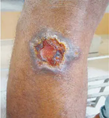
Fig. 1 - Ulcerated Kaposi’s Sarcoma lesion with a clean base
was implemented. The patient was referred to the STDs section of Professor Rubem David Azulay Dermatology Institute, Santa Casa da Misericórdia Hospital, Rio de Janeiro, Brazil.
In January 2011, six weeks after the initiation of HAART (zidovudine, lamivudine, and efavirenz), the patient returned referring worsening of the lesions and fever. Physical examination revealed new violaceous and painful nodules and ulcerated lesions, with a clean base and well-defined borders on the right leg (Fig. 1), chest (Fig. 2) and upper limbs. Neither mucocutaneous KS nor symptoms of visceral involvement were present. VDRL and FTA-abs tests for syphilis were negative.
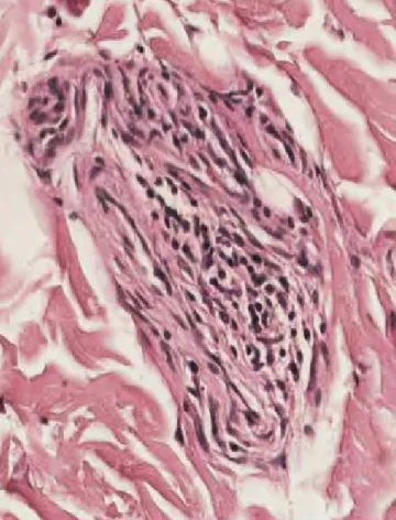
A biopsy was taken from a nodular lesion and histo-logically diagnosed as KS (Figs. 3 and 4). By this time, the HIV viral load was 410 copies per millimeter and CD4+ count was 370 cells per mm3.
Four weeks after the biopsy, the majority of ulcera-ted and nodular violaceous lesions improved and the patient had no further complaints, even without thera-peutic changes.
DISCUSSION
Two important pieces of information lead us to belie-ve that this patient was affected by IRIS and not a simple progression of the Kaposi sarcoma.
The first is that there was no clinical progression of the Kaposi sarcoma lesions (neither in number nor in size) in the month before beginning HAART and the time elapsed between early use of HAART and the worsening of the patient’s lesions (about 6 weeks). According to
Fig 2 - New ulcerated lesions on chest.
Fig 3 - Normal and newly formed blood vessels in superficial
dermis (H&E, 100x).
Fig 4 - Small vascular and slit-like spaces, lined by delicate
endothelial cells; some spaces show evidence of red blood cells infiltration (H&E, 400x).
the literature, the development of new lesions due to IRIS occur within 8 weeks (usually ranging from 3 to 22 weeks) after initiating HAART8. During the immune
status restoration the ability to mount an inflammatory response to a variety of previously indolent antigens is also restored10. This fact can result in clinical signs of
di-sease activity5, triggered by antigenic stimuli which can
be tumor antigens or pathogens present in the body (but clinically latent), rather than development or progres-sion of opportunistic infections11.
In addiction, according to French et al12 our patient
presents a major criteria: atypical presentation of tumor in patients responding to HAART with exaggerated in-flammatory reaction (eg, severe fever or painful lesions) and 2 minor criteria: increased CD4 cell account and spontaneous resolution of disease without specific an-timicrobial therapy or tumor chemotherapy with con-tinuation of HAART. Our case is also consisted with Robertson J, et al case definition of IRIS13.
Most IRIS events are represented by the occurren-ce or recurrenoccurren-ce of dermatological diseases14. KS flare
attributed to IRIS may produce life-threatening visceral progression even if cutaneous nodules regress, with deaths from pulmonary KS5,15. Visceral manifestations
of the disease was not observed in our patient but the. cutaneous nodules enlargement can rapidly and fre-quently ulcerate and lead to secondary infection16,17.
In turn, the diagnosis of KS was confirmed by skin biopsy. The skin lesions of KS can be divided into patch, plaque and nodular stages, but these stages often over-lap clinically and histologically18. In this report, the
le-sions exhibit features of nodular stage with a network of slit-like vascular spaces and erythrocytes within the narrow vascular clefts18,19.
In the medical literature revised till now, there are no specific guidelines for the diagnosis and treatment of IRIS12. However, the antiretroviral therapy should not
be discontinued, except in the severely ill patients14,20. In
our patient, this approach was successful showing the benefit of the continuation of HAART.
We can conclude that the clinician should be aware to identify signs and symptoms of neoplasm progression and to differentiate them from KS related IRIS and thus, to choose the better approach in the patient’s follow-up.
REFERENCES
1. Goedert JJ. The epidemiology of acquired immuno-deficiency syndrome malignancies. Semin Oncol. 2000; 27(4): 390-401.
2. Dourmishev LA, Dourmishev AL, Palmeri D, Sch-wartz RA, Lukac DM. Molecular genetics of Kaposi’s sarcoma associated herpesvirus (human herpesvi-rus-8) epidemiology and pathogenesis. Microbiol Mol Biol Rev. 2003; 67(2):175-212.
3. Yoshioka MC, Alchorne MM, Porro AM, Tomimori--Yamashita J. Epidemiology of Kaposi’s sarcoma in patients with acquired immunodeficiency syn-drome in Sao Paulo, Brazil. Int J Dermatol. 2004; 43(9):643-7.
4. Tamburro KM, Yang D, Poisson J, Fedoriw Y, Roy D, Lucas A, et al. Vironome of Kaposi sarcoma as-sociated herpesvirus-inflammatory cytokine syn-drome in an AIDS patient reveals co-infection of human herpesvirus 8 and human herpesvirus 6A. Virology. 2012; 433(1):220-5.
5. Bower M, Nelson M, Young AM, Thirlwell C, Newsom--Davis T, Mandalia S, et al. Immune reconstitution inflammatory syndrome associated with Kaposi’s sarcoma. J Clin Oncol. 2005; 23(22):5224-8. 6. Murdoch DM, Venter WD, Feldman C, Van Rie A.
Incidence and risk factors for the immune recons-titution inflammatory syndrome in HIV patients in South Africa: a prospective study. AIDS. 2008; 22(5):601-10.
7. Nasti G, Martellotta F, Berretta M, Mena M, Fasan M, Di Perri G, et al. Impact of highly active antire-troviral therapy on the presenting features and ou-tcome of patients with acquired immunodeficiency syndrome-related Kaposi sarcoma. Cancer. 2003; 98(11):2440-6.
8. Feller L, Lemmer J. Insights into pathogenic events of HIV-associated Kaposi sarcoma and immune re-constitution syndrome related Kaposi sarcoma. In-fect Agent Cancer. 2008; 3:1.
9. Hirsch HH, Kaufmann G, Sendi P, Battegay M. Im-mune reconstitution in HIV-infected patients. Clin Infect Dis. 2004; 38(8):1159-66.
10. Lederman MM. Immune restoration and CD4+ T--cell function with antiretroviral therapies. AIDS. 2001; 15 (Suppl 2):S11-5.
11. Connick, E; Kane, MA; White, IE; Ryder, J; Cam-pbell, TB. Immune Reconstitution Inflammatory Syndrome Associated with Kaposi Sarcoma during Potent Antiretroviral Therapy. Clin Infect Dis. 2004; 39(12):1852-5.
12. French MA, Price P, Stone SF. Immune restoration disease after antiretroviral therapy. AIDS. 2004; 18(12):1615-27.
13. Robertson J, Meier M, Wall J, Ying J, Fichten-baum CJ. Immune reconstitution syndrome in HIV:
validating a case definition and identifying clinical predictors in persons initiating antiretroviral thera-py. Clin Infect Dis 2006; 42 (11):1639-46.
14. Papagatsia Z, Jones J, Morgan P, Tappuni AR. Oral Kaposi sarcoma: a case of immune reconstitution inflammatory syndrome. Oral Surg Oral Med Oral Pathol Oral Radiol Endod. 2009; 108(1):70-5. 15. Lehloenya R, Meintjes G. Dermatologic
manifes-tations of the immune reconstitution inflammatory syndrome. Dermatol Clin. 2006; 24(4):549-70, vii. 16. Nguyen S, Giurca C, Melliez H, Dehecq C, Baclet V, Ajana F, et al. Kaposi’s sarcoma in HIV-infected patients: when and how should we evaluate bone involvement? AIDS. 2007; 21(16):2251-2.
17. Von Roenn JH. Clinical presentations and standard therapy of AIDS-associated Kaposi’s sarcoma.
Hematol Oncol Clin North Am. 2003; 17(3):747-62.
18. Calonje, E. Soft-Tissue Tumours and Tumour-like Conditions. In: Burns T, Breathnach S, Cox N, Gri-ffithsC, editors. Rook's Textbook of Dermatology. 8th ed.Oxford: Wiley-Blackwell; 2010. p.
56.35-56.36
19. Buonaguro FM, Tomesello ML, Buonaguro L, Sa-triano RA, Ruocco E, Castello G, et al . Kaposi's sarcoma: aetiopathogenesis, histology and clini-cal features. J Eur Acad Dermatol Venereol. 2003; 17(2):138-54.
20. Loke WC, Spittle MF, Mitchell S, Kulasegaram R. Timing of highly active antiretroviral therapy and chemotherapy for Kaposi’s sarcoma in patients with HIV infection. Int J STD AIDS. 2006; 17(8):565-6.