Confocal Scanning Optical Microscopy Analysis
∗NILSON NUNES-TAVARES1, NARCISA LEAL CUNHA-E-SILVA2 and AÍDA HASSÓN-VOLOCH1
1Laboratório de Físico-Química Biológica
2Laboratório de Ultraestrutura Celular Hertha Meyer, Instituto de Biofísica Carlos Chagas Filho
Universidade Federal do Rio de Janeiro, Centro de Ciências da Saude – 21491-590 Rio de Janeiro, RJ, Brasil
Manuscript received on April 18, 2000; accepted for publication on April 24, 2000; contributed byAída Hassón-Voloch∗∗
ABSTRACT
Acetylcholine is the neurotransmitter responsible for the transmission of impulses from cholinergic neurons to cells of innervated tissues. Its biosynthesis is catalyzed by the enzyme Choline acetyltransferase that is considered to be a phenotypically specific marker for cholinergic system. It is well known that the reg-ulation of Choline acetyltransferase activity under physiological and pathological conditions is important for development and neuronal activities of cholinergic functions. We observed the distribution of Choline acetyltransferase in sections from the normal and denervated main electric organ sections of Electropho-rus electricus(L.) by immunofluorescence using a anti-Choline acetyltransferase antibody. The animals were submitted to a surgical procedure to remove about 20 nerves and after 30 and 60 days, they were sacrificed. After 30 days, the results from immunohistochemistry demonstrated an increase on the Choline acetyltransferase distribution at denervated tissue sections when compared with the sections from the normal contralateral organ. A very similar labeling was observed between normal and denervated tissue sections of the animals after 60 days. However, Choline acetyltransferase activity (nmolesACh/ min/ mg of protein) in extracts obtained from electrocyte microsomal preparation, estimated by Fonnun’s method (Fonnun 1975), was 70% lower in the denervated extracts.
Key words:choline acetyltransferase, electrocyte, denervation, immunohistochemistry.
INTRODUCTION
Choline acetyltransferase (ChAT; EC 2.3.1.6) is the enzyme responsible for the biosynthesis of the neu-rotransmitter acetylcholine (ACh) which was de-scribed by Nachmansohn and Machado in 1943. Many authors have pointed out that choline
acetyl-∗Invited paper
∗∗Member of the Academia Brasileira de Ciências
Correspondence to: Aída Hassón-Voloch E-mail: [email protected]
transferase is directly related to many types of neuro-logical disorders such as Alzheimer disease, myas-thenia gravis, multiple sclerosis, and Huntington’s disease (Rossier 1977, Molenaaret al. 1981, Kato
1989). Thereafter, either purification as the study of the role of this enzyme in biological models has been object of considerable attention (Hersh & Wu 1994, 1995, Randet al. 1994, Salvaterraet al.1995).
The electrogenic tissue fromE. electricus(L.)
endings and differentiated histophysiologically in three electric organs: main, Hunter and Sachs (Cou-ceiro & Ackerman 1948, Esquibelet al.1971). The
electrocyte is the functional unit of the electric tissue (Schwartzet al. 1975) which, in turn, is essentially
formed by a non innervated (anterior) and an in-nervated (posterior) face with numerous synapses, myelin fibers, and it is also rich in acetylcholine re-ceptor molecules (Chagaset al. 1957, Couceiro &
Ackerman 1948, Couceiroet al.1953).
Previous studies carried out by Esquibelet al.
(1971) have demonstrated the structural organiza-tion of the electric organ from newbornE. electricus
(L.) by using both optical and electron microscopy, resulting in the observation of myofibril-like struc-tures in the cytoplasm of the developing electro-cyte. However, in elder E. electricus (L.) these
structures were not observed (Machadoet al.1976).
An analogy between the electric organ and striated muscles has also been carried out since the elec-tric organ presents important proteins of the ener-getic metabolism, found in muscle, such as creatine phosphokinase (Hazan-Carneiro & Hassón-Voloch 1983), lactate dehydrogenase (Torres Da Mattaet al.
1975) as well as cytoskeletal proteins like myosin and desmin (Costaet al.1986, 1988).
Acetylcholine was the first neurotransmitter de-scribed (Loewi 1921) and plays a central role in fundamental processes as learning, memory, sleep (Bartus & Beer 1982, Aigner & Mishkin 1986) and muscle contraction in mammals. The electric organ has cholinergic neurons that innervate electrocytes growing from myotubes and have about 1,000 fold ACh receptors than muscle cells (Whittaker 1987). Recently, we described that most of ChAT found in the electrocyte is mainly present as cytosolic pro-tein with two isoforms (Nunes-Tavares & Hasson-Voloch 1998). Earlier studies showed that denerva-tion of the main electric organ fromE. electricus(L.)
produced increases on RNA contents, RNA/DNA and RNA/protein ratio, which were observed by his-tochemistry studies, and aminoacid incorporation, although the total protein did not showed significant alteration (Ribeiro & Chagas 1975,
Falcato-Ribeiroet al. 1977). Denervation is a technique
that can be used when we aim to study the inter-action between muscles and nerves. After denerva-tion, the electric organ shows a late and slow atro-phy which allows the study of nerve control upon its structure (Martins-Ferreira & Couceiro 1951). Histochemical and biochemical studies of dener-vated electrocytes, after 30 and 60 days, showed the disappearance of myelin sheath as well as the increase on the activity of some important enzymes of the energetic metabolism (Falcato-Ribeiroet al.
1977, 1980, Torres DaMattaet al. 1985). Other
studies concerning neurodegenerative diseases, as Alzheimer and Parkinson, has demonstrated that the vesicular acetylcholine transporter (VAChT), and the high-affinity choline transporter (HAChT), may re-main unchanged or, in some circumstances, increase in cortex and hippocampus over the control (Ruberg
et al.1990, Kuhlet al.1996). Lesion paradigms in
animals have been used to directly study the effects of the loss of cholinergic input and the response of cholinergic neurons to injury.
The present study describes the histochemical investigation of choline acetyltransferase (ChAT) distribution in normal and denervated electric organ by immunofluorescence under confocal microscopy (CSLM).
MATERIAL AND METHODS
Electrophorus electricus(L.). were provided to us
by “ Museu Emílio Goeldi, Belém do Pará - Brasil” and kept in an aquarium with fresh filtered water.
Immunochemicals
The Anti-ChAT polyclonal antibody to Human pla-cental (AB143) enzyme was purchased from Chemi-con Co (USA) and anti rabbit IgG (H+L) - FITC conjugated was obtained from GIBCO (USA);
[3H]AcCoA (4.0Ci/mmol) from Amersham
(Eng-land); AcCoA from Sigma Chemical Co (USA). All other chemicals were of analytical grade.
An animal (0.8 −1 m long) was anesthetized in
ice-cold water containing 2.0% urethane, and sub-mitted to a surgical denervation as previously de-scribed (Nesralla-Lobãoet al. 1980). Briefly, this
procedure consists of a longitudinal incision on the dorsolateral anterior part of the fish by sectioning of the nerves (about 20 nerves) that innervate the electric organ coming from the spinal cord. Dur-ing the surgery, the fish was maintained on artificial breathing and preserving the metabolic activities. After 30 and 60 days, the eels were anesthetized and then killed by decapitation. Both denervated and contralateral normal main organs were immediately removed.
Choline Acetyltransferase Immunohistochemistry
Fragments of the main electric organ (normal and denervated) were prepared for immunohistochem-istry. They were incubated for 1 hour in a fixative solution containing 4% paraformaldehyde in 0.1 M phosphate buffer, pH 7.4 (PB), and then in 0.1 M phosphate buffer, with 0.015 M NaCl (PBS), con-taining 20% sucrose, for 2 hours, and finally, in 30% sucrose PBS solution overnight. Sections of 10µm
thickness were obtained after cutting the tissue frag-ments on a freezing ultramicrotome (Ultracut Re-ichert). The sections were preincubated with 50 mM
N H4Cl, for 1 h, washed with PBS-T (PBS plus 0.2%
Tween −20), and subsequently incubated with: a
polyclonal anti-ChAT (1:500 dilution) overnight at 40◦C, washed with PBS-T plus 10% normal goat
serum, and with a FITC-labeled goat anti-rabbit IgG (1:20 dilution) for 2 hours at room temperature. The specimens were then mounted onto slides. Controls were made omitting the primary antiserum for each experiment. Normal and denervated sections were examined in a Zeiss 310 Confocal Laser Scanning Microscope (CSLM), by using a 488 argon laser. All images were obtained under the same conditions of brightness and contrast and uniformly processed us-ing Adobe PhotoShop.
Protein Assay
Protein concentration was determined by the Folin-phenol method of Lowryet al.(1951) using bovine
serum albumin as a standard.
Enzyme Assay
The total extracts and supernatants used in enzy-matic assays were obtained by the method of Somló
et al. (1977) modified. Using the radiochemical
method of Fonnun (1975), with [3H] AcCoA as sub-strate, we performed the assay for choline acetyl-transferase activity. The substrate medium was as follows: 64 mM sodium phosphate buffer, pH 7.6; 384 mM NaCl; 10mM choline chloride; 25 mM NaEDTA; 10 mM eserine sulfate and 12µMAcCoA.
To the assay tube containing 10µl of substrate
mix-ture, 20µl[3H]AcCoA (0.1µCi) was added plus
20µg of protein, at intervals of 30 seconds, at 37◦C,
for 15 minutes. The reaction was stopped by ad-dition of 50 mM NaEDTA plus 2 ml of Kalignost solution (5mg/ml tetraphenylborate in acetonitrile). The synthesis of[3H]ACh was measured in a liquid
scintillation Packard apparatus (Model 1600 TR), and the activity is expressed as nmoles ACh formed per minute per mg of protein. Each assay was made in triplicate.
RESULTS
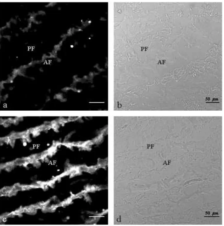
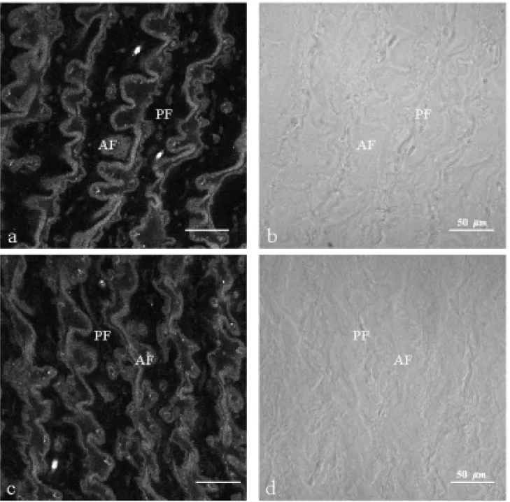
The presence of choline acetyltransferase (ChAT) was observed by immunofluorescence in the cyto-plasm of the electrocyte from electric organ ofE. electricus(L.) either in normal (Figs. 1a,b and 2a,b)
Fig. 1a-b – Thin section (10µm) of normal contralateral electric tissue; a: immunolabelled with policlonal antibody anti-ChAT. Immunoreactivity is essentially cytoplasmatic; b: phase contrast. AF; anterior face, PF; posterior face. Fig. 1c-d – Thin section (10µm) of denervated electric tissue (30 days); c: immunolabelled with policlonal antibody
anti-ChAT. A high increase of labelling in the cytoplasm is observed; d: phase contrast. AF; anterior face, PF; posterior face.
of ChAT seems to be highly increased on the cy-toplasm of denervated electric tissue section of 30 days (Fig.1c), when compared with the normal (Fig. 1a). 60 days after denervation, the ChAT distribu-tion showed almost the same pattern in both normal and denervated tissue sections (Figs 2a, c).
Fig. 2a-b – Thin section (10µm) of normal contralateral electric tissue a: immunolabelled with policlonal antibody
anti-ChAT. Immunoreactivity is essentially cytoplasmatic; b: differential interferencial contrast. AF; anterior face, PF; posterior face. Fig. 2c-d - Thin section (10µm) of denervated electric tissue (60 days), c: immunolabelled with
policlonal antibody anti-ChAT, The same pattern of labelling in cytoplasm is observed when compared to normal contralateral tissue; d: differential interferencial contrast. AF; anterior face, PF; posterior face.
ranged from 3.80 to 4.50 nmoles ACh/ min/ mg of protein after 30 days. In animals of 60 days after denervation the ChAT activity showed a decrease from 0,40 to 0,38 nmoles ACh/ min/ mg of protein, respectively. After denervation these values were of 70% lower, on both denervated whole organ ho-mogenate and the resulting supernatant.
DISCUSSION
The study of choline acetyltransferase is of partic-ular interest due to its importance on the choliner-gic system (Waseret al. 1989), and also because
its deficiency has been correlated with many phys-iopathological diseases (Kato 1989, Wurtmanet al.
differ-TABLE I
ChAT activity in Normal and Denervated main electric organ.
30 days∗ 60 days∗
Normal 3.80±0.65 0.40±0.03
Total
Extracts denervated 0.81±0.14 0.11±0.09
Normal 4.50±0.87 0.38±0.25
Supernatants
Denervated 0.71±0.31 0.14±0.09
∗ChAT activity is given in nmolesACh. min−1. mg of protein−1. All values
represent means±SEM of 3 separate measurements.
ent aims by several authors, as molecular proper-ties, while others were interested in investigate its cellular localization (Malthe-Sørenssen & Fonnun 1972, Polsky & Shuster 1976, Uyedaet al. 1974,
Docherty & Bradford 1986, Dochertyet al. 1987,
Leveyet al. 1998). In the last years most of the
studies on ChAT have been improved, mainly those focusing molecular biology aspects, in mammals (Berrardet al. 1987, Toussaint et al. 1992,
Shi-mojo et al. 1998). Some studies carried out in
fishes by using anti-ChAT antibodies have brought detailed information on the cholinergic system (Zot-toliet al. 1987, 1988, Brantley & Bass 1988).
Al-though catalytic activity ascribed to this enzyme has been observed in non-neural tissues, including pla-centa (Hersh & Peet 1978), spermatozoa (Bishop
et al. 1976), plants (Barlow & Dixon 1973) and
bacteria (White & Cavallito 1970), its functional significance in such tissues is unknown. As ChAT appears to be present in the cholinergic neuron, it has generally been accepted that the enzyme does not represent the rate-limiting step for ACh biosyn-thesis. The detection of choline acetyltransferase by antibody is very specific as recently described by Nunes-Tavares and Hassón-Voloch (1998) who observed through western blotting analysis a sin-gle band with ChAT purified from electric organ. During the past 10 years, research efforts have fo-cused on the regulatory control of this enzyme. The main characteristic of ChAT regulation is centered
on the temporal and spatial control of transcription (Hersh & Wu 1994). The content of ACh in tissues with cholinergic innervation is very stable. If the release of ACh is augmented as a consequence of increased functional activity of mammalian cholin-ergic neurons, the ACh is replaced almost immedi-ately by new synthesis (MacIntosh & Collier 1976, Tucek 1982). However, the resynthesis is slower in piscine electric organ (Dunantet al. 1974, 1977).
The denervation of the electric organ from Elec-trophorus electricus(L.) demonstrated a differential
distribution, either qualitatively as quantitatively, of choline acetyltransferase when compared with the normal organ, which in turn may have been caused by the loss of electrogenic stimulus in the tissue, as demonstrated in striated muscles from mammals (Lebherz 1984). Generally, a structural degenera-tion occurs, and the resynthesis of enzymes that are involved in specific tasks increases (Falcato-Ribeiro
et al. 1980). Other authors demonstrated a
het-erologous expression for ChAT in fishes and other species (Falcato-Ribeiroet al. 1980, Gundersenet al. 1985, Berrardet al. 1986, 1987, McCamanet al. 1988, Brice et al. 1989). This mechanism of
is markedly stronger. However, after 60 days of denervation there is a significant similarity of la-beling between the normal and denervated tissues showing recovery of metabolism; other authors have also observed this feature for other important en-zymes of the metabolic pathway (Falcato-Ribeiro
et al. 1980, Torres-DaMatta et al. 1985,
Hazan-Carneiro & Hassón-Voloch 1983). We have also compared the enzymatic activity of ChAT with its immunohistochemical staining. Biochemical alter-ations produced by denervation were observed on to-tal electric organ extracts and supernatants from mi-crosomal membrane preparations. The comparative analysis between normal and denervated organ sec-tions demonstrated that changes due to denervation might increase ChAT mRNA level in 30 days. Like-wise, the immunohistochemistry assay here used showed that the spatial distribution of the enzyme molecules is maintained. Our results strongly sug-gest that many cholinergic neurons survive the injury intact but shrink as recently described by Naumann
et al. (1994). Studies made by Falcato-Ribeiro and
Chagas Filho (1975) on denervated muscles showed modifications in the protein profiles. Our results re-inforce the idea of alterations caused by denervation on the binding between ChAT antigen and antibody used. The observed increase of binding capacity in denervated tissue sections of 30 days and its re-covery after 60 days could possibly be explained as a result of an alteration of electroplate structure, evoked by the a long absence of stimulation by the nerve, which is reflected in the protein synthesis.
The paradigm observed after denervation, i.e., an increase of expression of choline acetyltrans-ferase after 30 days of denervation accompanied by a decrease of activity, and the similarity of labeling be-tween the tissues sections (normal and denervated) after 60 days, are probably easier to identify in the electric tissue due to the slow process of atrophy fol-lowing the lesion. It is possible that these changes probably represent a readjustment to maintain the basic conditions of performance of the denervated electric organ.
ABBREVIATIONS
ChAT = Choline acetyltransferase; ACh = Acetyl-choline; NaEDTA = Tetrasodium ethylene-diami-netetraacetate; PB = Phosphate-buffer; PBS = Phos-phate-buffered saline; PBS-T = PhosPhos-phate-buffered saline plus Tween-20; NaCl = Sodium Chloride; Ac-CoA = Acetyl coenzyme A.
ACKNOWLEDGEMENTS
This work is dedicated to the memory of Dr Carlos Chagas Filho founder of Institute of Biophysics at UFRJ.
This work was supported by grants from Con-selho Nacional de Desenvolvimento Científico e Tecnológico (CNPq), Conselho de Ensino para Gra-duados da UFRJ (CEPEG-UFRJ); Financiadora de Estudos e Projetos (FINEP) and Fundação Carlos Chagas Filho de Amparo à Pesquisa do Estado do Rio de Janeiro (FAPERJ).
We thank Célia M.B. Silva for helpfull com-ments; Dr. Elaine Gomes Quintana and Dr Fernando Costa e Silva for critical reading of the manuscript, Dr Renato A. DaMatta for helpful assistence and Sérgio L. Carvalho for technical assistance.
REFERENCES
Aigner TG & Mishkin M.1986. The effects of
phys-iostigmine and scopolamine on recognition memory in monkeys. Behav Neural Biol45:81-87.
Barlow RB & Dixon RO.1973. Choline acetyltrans-ferase in the NettleUrtica dioicaL.Biochem J132:
15-18.
Bartus RT& Beer RD.1982. Cholinergic hypothesis of geriatric memory dysfunction. Science217:
408-414.
Berrard S, Biguet NF, Gregoire D, Blanot F, Smith J & Mallet J.1986. Synthesis of catalitically ac-tive choline acetyltransferase inXenopusoocytes
Berrard S, Brice A, Lottspeich F, Braun A, Bard YA & Mallet J.1987. cDNA cloning and complete se-quence of porcine choline acetyltransferase;in vitro
translation of the corresponding RNA yields an active protein. Proc Natl Acad Sci USA84:9280-9284.
Bishop MR, Sastry BV, Schmidt DE & Harbison RD. 1976. Ocurrence of Choline and other quar-tenary ammonium compounds in mammalian sper-matozona.Biochem Pharmacol25:1617-1622.
Brantley RK & Bass AH.1988. Cholinergic neurons in the brain of teleost fish (Porichthys notatus) located
with a monoclonal antibody to choline acetyltrans-ferase. J Comp Neurol275(1): 87-105.
Brice A, Berrard S, Raynaud B, Ansieau S, Copolla T, Weber MJ & Mallet J. 1989. Complete se-quence of cDNA encoding an active rat choline acetyltransferase: a tool to investigate the plasticity of cholinergic phenotype expression.J Neurosci Res 23:266-273.
Chagas Filho C, Penna-Franca E, Hassón-Voloch A, Crocker C, Nishie K & Garcia E.1957. Studies of the mechanism of curarization. An Acad Bras Ci 29:53-58.
Costa ML, Mermelstein CS, Moura Neto V & Cha-gas Filho C.1988. Desmin expression in the
elec-tric organs ofElectrophorus electricus(L.). J Cell Biochem(Supp).12c:323-329.
Costa ML, Oliveira MM, Alberti JR O, Moura Neto V & Chagas Filho C.1986. Characterisation de la desmine dans l’organe électrique de l’Electrophorus electricus(L.). C R Acad Sci Paris,303(série III):
547-550.
Couceiro A & Ackerman M.1948. Sur quelques as-pects du tissu électrique de l’Electrophorus electricus
(L.).An Acad Bras CiXX,4:383-395.
Couceiro A, de Almeida DF & Freire JR.1953. Local-isation histochimique de l’Acetylcholinesterase dans le tissue électrique de l’Electrophorus electricus(L.). An Acad Bras Ci25:205-214.
Docherty M & Bradford HF.1986. A cell-surface
antigen of cholinergic nerve terminals recognized by antisera to choline acetyltransferase. Neurosci Lett 70:234-238.
Docherty M, Bradford HF & Wu JY.1987. The prepa-ration of highly purified GABAergic and cholinergic synaptosomes from mammalian brain.Neurosc Lett 81:232-238.
Dunant Y, Gautron J, Israel M, Lesbats B & Ma-naranche R.1974. Evolution de la décharge de l’organe électrique de la Torpille et variations simul-tanées de l’acetylcholine au cours de la stimulation.
J Neurochem23:635-643.
Dunant Y, Israel M, Lesbats B & Manaranche R.
1977. Oscillation of acetylcholine during nerve ac-tivity in theTorpedoelectric organ. Brain Res125:
123-140.
Esquibel MA, Alonso I, Meyer H, Oliveira Castro G & Chagas Filho C.1971. Quelques aspects de l’histogénèse et de l’ontogénèse des organes elec-triques chez l’Electrophorus electricus (L.). C R Acad Sci Paris, (Série D)273:196-199.
Falcato-Ribeiro AF & Chagas Filho C.1975. Effect
de la dénérvation sur le métabolisme macromolecu-laire chez le tissu éléctrique de l’Electrophorus elec-tricus(L.).C R Acad Sci Paris, (Série D)281:
1425-1430.
Falcato-Ribeiro AF, Chagas Filho C & Couceiro A. 1977. Evidência histoenzimológica de
modifi-cações na atividade de algumas enzimas em tecido eletrogênico desnervado.Rev Bras Biol37(4):
791-798.
Falcato-Ribeiro AF, Chagas Filho C & Couceiro A.
1980. Histoenzymological evidence for the modifi-cation in the activity of some enzymes in the dener-vated electrogenic tissue ofElectrophorus electricus
(L.).IRCS Medical Science8:776-777.
Fonnun FA.1975. Rapid radiochemical method for the determination of Choline Acetyltransferase. J Neurochem24:407-409.
Gundersen CB, Jenden DJ & Miledi R.1985. Choline acetyltransferase and acetylcholine in Xenopus
oocytes injected with mRNA from the electric lobe ofTorpedo. Proc Natl Acad Sci USA82:608-611.
111-114.
Hersh LB & Peet M.1978. Effect of salts on the phys-ical and kintic properties of human placental choline acetytransferase.J Neurochem30:1087-1093.
Hersh LB & Wu D.1994. Choline Acetyltransferase: Celebrating its fiftieth year.J Neurochem62:
1653-1663.
Hersh LB & Wu D.1995. Activation of Choline
Acetyl-transferase by limited proteolysis.J Biol Chem270:
19395-19401.
Kato T.1989. Choline Acetyltransferase activities in single spinal motor neurons from patients with Amy-otrophic Lateral Sclerosis. J Neurochem52:
636-640.
Kuhl de, Minoshima S, Fessler JA, Frey KA, Foster NL, Ficaro EP, Wieland DM & Kooeppe RA.1996.
In vivo mapping of cholinergic terminals in normal aging, Alzeheimer’s disease and Parkinson’s disease.
Ann Neurol40:399-410.
Lebherz HG.1984. Neuronal control of synthesis of spe-cific proteins in muscle fibers. Trends in Biochem. Sci.. August, 351-354.
Levey AI, Gilmor ML, Count SE & Wiley RG.1998.
Coordinate expression of the vesicular acetycholine transporter and choline acetyltransferase following septohippocampal pathway lesions. J Neurochem 71:2411-2420.
Loewi O.1921. Uber humorale ubertragarkeit der hirzen-wickung.Pflugers Arch189:239-242.
Lowry OH, Rosenbrough NJ, Farr AL & Randall RJ.1951. Protein measurement with the Folin phenol
reagent.J Biol Chem193:265-275.
Machado RD, de Souza W, Cotta-Pereira G & Oliveira Castro G.1976. On the fine structure of the electrocyte ofElectrophorus electricus(L.).Cell Tiss Res174:355-366.
MacIntosh FC & Collier B.1976. Neurochemistry of cholinergic terminals in Neuromuscular junction. Handbook of Experimental Pharmacology, New se-ries, (ed. Zaimis), Springer-Verlag, Berlin. 42: 99-228.
McCaman RE, Carbini L, Maines V & Salvaterra PM.1988. Single RNA species injected inXenopus
oocyte directs the synthesis of active choline acetyl-transferase.Mol Brain Res3:107-114.
Malthe-Sørenssen D & Fonnun F.1972. Multiple
forms of Choline Acetyltransferase in several species demonstrated by Isoelectric Focusing. Biochem J 127:229-236.
Martins-Ferreira H & Couceiro A.1951. Comporte-ment du tissue électrique de l’Electrophorus electri-cus(L.) en conséquence de la dénervation.An Acad Bras Ci23(4): 376-385.
Molenaar PC, Newsom-Davis J, Polak L & Vicent A.
1981. Choline Acetyltransferase in skeletal muscle from patients withMyasthenia Gravis. J Neurochem 37(5): 1081-1088.
Nachmansohn D & Machado AL.1943. The formation of acetylcholine. A new enzyme choline acetylase.J Neurophysiol6:397-403.
Naumann T, Kermer P & Frotcher M.1994. Fine
structure of rat septohippocampal neurons. III. Re-covery of choline acetyltransferase immunoreactivity after fimbria-fornix transection.J Comp Neurol350:
161-170.
Nesralla-Lobão H, Gomes-Quintana E, Machado RD & Chagas Filho C.1980. Cholinergic
mam-branes from normal and denervated electric organ of Electrophorus electricus(L.). IRCS Med. Sci Biochem8: 450-451.
Nunes-Tavares N & Hassón-Voloch A.1998. Choline acetyltransferase from electric organ of Electropho-rus electricus(L.). Physicochemical
characteriza-tion and immunochemical identificacharacteriza-tion. Zeits Naturf 53c:407-415.
Polsky R & Shuster L.1976. Preparation and charac-terization of two isozymes of Choline Acetyltrans-ferase from Squid Head Ganglia. (II- Self-Association, Molecular weight determinations and Studies with inactivating antisera.Biochem Biophys Acta445:43-66.
Rand JB, Alfonso A, Grundahl K & McManus JR.
elegans. J Neurosci14(4): 2290-2300.
Rossier J.1977. Choline Acetyltransferase: A review with special reference to its cellular and subcellular localization.Int Rev Neurobiol20:284-337.
Ruberg M, Mayo W, Brice A, Duyckaerts C, Hauw JJ, Simon H, Lemoal M & Agid Y.1990. Choline
Acetyltransferase activity and [3h] Vesamicol bind-ing in the temporal cortex of patients with Alzheimer’s disease. Parkinson 1s disease and rats with basal forebrain lesions.Neurosci35:327-333.
Salvaterra PM, Kouji Yasuyama & Toshihiro Kita-moto.1995. Localization of Choline
acetyltransfe-rase-expressing neurons in the larval visual system of Drosophila melanogaster. Cell Tissue Res282:
193-202.
Schwartz IR, Pappas GD & Bennet MVL.1975. The structure of electrocytes in weakly electric teleosts.
J Neurocytol4:87-114.
Shimojo M, Wu D & Hersh LB.1998. The cholinergic gene locus is coordinately regulated by protein kinase A II in PC12 cells.J Neurochem71:1118-1126.
Somló C, Souza W, Machado RD & Hassón-Voloch A.1977. Biochemical and Cytochemical localisa-tion of ATPases on the membranes of the electrocyte ofElectrophorus electricus(L.). Cell Tiss Res185:
115-128.
Torres da Matta J, Hassón-Voloch A & Hargreaves AB.1975. Lactate deshidrogenase from the electric
organ ofElectrophorus electricus(L.) - Isoenzyme analysis.Comp Biochem Physiol52B:451-454.
Torres da Matta J, Silva CB, da Matta AN & Hassón-Voloch A.1985. Effect of denervation on the glycolitic metabolism of the main electric or-gan ofElectrophorus electricus(L.).Comp Biochem Physiol81B:969-973.
Toussaint JL, Geoffroy V, Scmitt N, Werner A, Gar-nier JM, Simoni P & Kempf J.1992. Human choline acetyltransferase (ChAT): partial gene sequence and potencial control regions.Genomics12:412-416.
Tucek S.1982. The synthesis ofAcetylcholine in skeletal
muscles of the rat.J Physiol322:53-69.
Uyeda CT, Eng LF, Chao LP & Wolfgran F.1974.
Antibody to bovine choline acetyltransferase and im-munofluorescent localisation of the enzyme in neu-rons.Nature250:243-245.
Waser G, Riggio G & Raeber AJ.1989. Purification and isolation of choline acetyltransferase from the electric organ of Torpedo marmorata by affinity
chromatography.Eur J Biochem186:487-492.
White HL & Cavallito CJ.1970. Inhibition of bac-terial and mammalian choline acetyltransferases by styrylpyridine analogues. J Neurochem17:
1579-1589.
Whittaker VP. 1987. Cholinergic synaptic vesicles from the electromotor nerve terminal of Torpedo.
Composition and life cycle. Ann N Y Acad Sci493:
77-91.
Wurtman RJ, Blusztajn JK, Ulus IH, Coviella IL, Buyukuysal RL, Growdon JH & Slack BE.1990. Choline metabolism in cholinergic neurons: implica-tions for the pathogenisis of neurodegenerative dis-eases.Adv Neurol51:117-125.
Zottoli SJ, Rhodes KJ & Mufson EJ.1987.
Com-parison of acetylcholinesterase and choline acetyl-transferase staining patterns in the optic tectum of the goldfishCarassius auratus. A histochemical and
im-munocytochemical analysis.Brain Behav Eval30
(3-4): 143-159.