Multiple Sclerosis Journal 2017, Vol. 23(9) 1258 –1267 DOI: 10.1177/
1352458516674367 © The Author(s), 2016. Reprints and permissions: http://www.sagepub.co.uk/ journalsPermissions.nav MULTIPLE SCLEROSIS MSJ JOURNAL Background
Cognitive impairment (CI) is known to be present in all stages of multiple sclerosis (MS); however, the prevalence estimates vary considerably between stud-ies, ranging from 40% to 65%.1 The profile of CI in the
overall MS population is now relatively well known, involving mainly complex attention, information pro-cessing speed, episodic memory, and executive func-tions.1,2 Therefore, brief neuropsychological batteries
for MS3 and newly developed assessment tools4
mainly focus on the assessment of these functions. However, few studies investigated the differences in the prevalence and profile of CI between the different MS disease subtypes, providing heterogeneous results.5–9 Many of these studies included small
clini-cal samples and focused mainly on relapsing remitting
(RR) or progressive forms. Moreover, the association of CI with several clinical features, such as physical disability, sex, and disease duration, is not well estab-lished, since inconsistent results have been reported in the literature.10–13 The heterogeneity of the published
literature could be, at least in part, attributable to small sample size and dissimilarities in the clinical charac-teristics of the studies’ samples. Exploring the inde-pendent effects of age, physical disability, disease duration, and disease subtype could prove central to provide a better understanding of the potential role and interaction of cognitive reserve, brain aging, and dis-ease severity for determining CI in MS.
The aims of this collaborative, nationwide, cross- sectional study were to describe the prevalence and
Age and disability drive cognitive impairment
in multiple sclerosis across disease subtypes
Luis Ruano, Emilio Portaccio, Benedetta Goretti, Claudia Niccolai, Milton Severo, Francesco Patti, Sabina Cilia, Paolo Gallo, Paola Grossi, Angelo Ghezzi, Marco Roscio,Flavia Mattioli, Chiara Stampatori, Maria Trojano, Rosa Gemma Viterbo and Maria Pia Amato Abstract
Background: There is limited and inconsistent information on the clinical determinants of cognitive
impairment (CI) in multiple sclerosis (MS).
Objective: The aim of this study was to compare the prevalence and profile of CI across MS disease
subtypes and assess its clinical determinants.
Methods: Cognitive performance was assessed through the Brief Repeatable Battery and the Stroop test
in consecutive patients with MS referred to six Italian centers. CI was defined as impairment in ⩾2 cogni-tive domains.
Results: A total of 1040 patients were included, 167 with clinically isolated syndrome (CIS), 759 with
relapsing remitting (RR), 74 with secondary progressive (SP), and 40 with primary progressive (PP) disease course. The overall prevalence of CI was 46.3%; 34.5% in CIS, 44.5% in RR, 79.4% in SP, and 91.3% in PP. The severity of impairment and the number of involved domains were significantly higher in SP and primary progressive multiple sclerosis (PPMS) than in CIS and RR. In multivariable logistic regression analysis, the presence of CI was significantly associated with higher Expanded Disability Status Scale (EDSS) and older age.
Conclusion: CI is present in all MS subtypes since the clinical onset and its frequency is increased in the
progressive forms, but these differences seem to be more associated with patient age and physical dis-ability than to disease subtype per se.
Keywords: Multiple sclerosis, cognitive impairment, disease course, epidemiology Date received: 6 July 2016; revised: 17 September 2016; accepted: 20 September 2016
Correspondence to: Luis Ruano Department of Clinical Epidemiology, Predictive Medicine and Public Health, University of Porto Medical School, Alameda Prof. Hernâni Monteiro, 4200-319 Porto, Portugal.
Luis Ruano
EPIUnit, Institute of Public Health, University of Porto, Porto, Portugal/Department of Clinical Epidemiology, Predictive Medicine and Public Health, University of Porto Medical School, Porto, Portugal/Entre Douro e Vouga Hospital Centre, Santa Maria da Feira, Portugal
Emilio Portaccio Benedetta Goretti Claudia Niccolai Maria Pia Amato
University of Florence, Florence, Italia
Milton Severo
EPIUnit, Institute of Public Health, University of Porto, Porto, Portugal/Department of Clinical Epidemiology, Predictive Medicine and Public Health, University of Porto Medical School, Porto, Portugal Francesco Patti Sabina Cilia University of Catania, Catania, Italia Paolo Gallo
Paola Grossi University of
Padova, Padova, Italia
Angelo Ghezzi Marco Roscio
Gallarate Hospital, Gallarate, Italia
Flavia Mattioli Chiara Stampatori
Spedali Civili Brescia, Brescia, Italia
Maria Trojano Rosa Gemma Viterbo
University of Bari, Bari, Italia
profile of CI in a large sample of patients with MS, with a specific focus on prevalence and neuropsycho-logical profiles across different disease subtypes, and to assess the association between CI and the main demographic and clinical features.
Methods
Study design and setting
We invited all consecutive MS patients attending their regular clinical follow-up visits in six Italian MS Centres during the study period (January–October 2010) to participate in the study. The inclusion criteria were (a) diagnosis of MS based on the 2001 McDonald criteria14 and (b) being between 18 and 70 years old.
The exclusion criteria were as follows: (a) presence of current or past neurological disorder other than MS; (b) active major psychiatric illness (such as schizo-phrenia, bipolar disorder, and major depressive disor-der); (c) history of learning disability; serious head trauma, alcohol or drug abuse; and (d) relapse and/or corticosteroid use within 4 weeks preceding the neu-ropsychological assessment. The classification of dis-ease subtypes was based on the 1996 Lublin’s definition.15 All the participants provided informed
consent and the study was approved by the ethics committees of the different institutions.
Clinical and neuropsychological assessment
Patient data were collected using a common data-base shared among the participating centers and included disease course, age at onset, disease dura-tion, relapses in the previous year, current treatment with disease modifying drugs, and education (com-plete years of formal schooling). Physical disability was assessed using the Expanded Disability Status Scale (EDSS),16 a scale validated to monitor disease
progression in MS.17 Fatigue was assessed using the
fatigue severity scale (FSS), a scale developed and validated for MS18 that is composed of nine items
with a score range of 9–63. Depression was assessed using the Montgomery and Asberg Depression Scale (MADRS), a standardized measure of mood disor-der, with scores ranging from 0 to 60.19 The FSS and
MADRS scales were not part of the initial study pro-tocol; nevertheless, they were routinely used in sev-eral of the study centers, resulting in FSS being applied in 728/1040 and MADRS in 356/1040 patients at the time of the study assessment.
A neuropsychological evaluation was performed using the Brief Repeatable Battery (BRB)3 and the
Stroop test.20 The BRB incorporates tests of verbal
memory acquisition and delayed recall (Selective Reminding Test (SRT)); visual memory acquisition and delayed recall (10/36 Spatial Recall Test (SPART)); attention, concentration, and speed of information processing (Paced Auditory Serial Addition Test (PASAT); Symbol Digit Modalities Test (SDMT)); and verbal fluency on semantic stimu-lus (Word List Generation (WLG)). The neuropsy-chologists involved in the study had participated in a common training session in which test administration and scoring procedures had been clarified and agreed upon. Test failure was defined as a score below the 5th or above the 95th percentile, when appropriate, according to age, sex, and education-adjusted Italian norms.21 Impairment in a given cognitive domain was
defined as failure in at least one test assessing that domain, namely, SRT for Verbal Learning, SPART for Visuospatial Learning, SDMT and PASAT for Information Processing Speed, and WLG and the Stroop tests for Executive Function. CI was defined as impairment in at least two cognitive domains.
Statistical analysis
Group comparisons were performed using Student’s
t-test for independent samples, the non-parametric
Kruskal–Wallis test or χ2 test with z-test adjusted for
multiple comparisons (Bonferroni method), where appropriate. The tests were two-sided, with a signifi-cance level of 0.05. To confirm the theoretical cogni-tive domains assessed by the cognicogni-tive tests, we performed principal component analysis.
To measure the association between the presence of CI and the different clinical and demographic varia-bles, we calculated crude and adjusted odds ratio (OR), using simple and multivariate logistic regres-sion. We built an a priori model (Model 1), including the demographic variables and education, and esti-mated the adjusted OR of the other variables. In Model 2, we adjusted to all variables that in Model 1 had a p-value lower that 0.1. Finally, we fitted in Model 3 the two variables that remained significant in Model 2 (age and EDSS), and estimated the adjusted OR of the other main clinical variables (disease dura-tion and clinical course). We also assessed the pres-ence of interactions between the variables in Model 2 and Model 3, and tested the inclusion of quadratic factors for each continuous variable, to check the presence of a non-linear relation between the inde-pendent variables and the log odds. The presence of multi-collinearity was assessed by calculating the cor-relation matrix between the main variables, and the inflation (VIF) and generalized variance-inflation factors (GVIF) for logistic regression. The
goodness of fit of the models was assessed using the Hosmer–Lemeshow test and the discrimination power was using the C-statistic. The same steps were repli-cated to fit logistic regression models for impairment in each cognitive domain (final models shown). Statistical analysis was performed using IBM SPSS Statistics Version 23.0.
Results
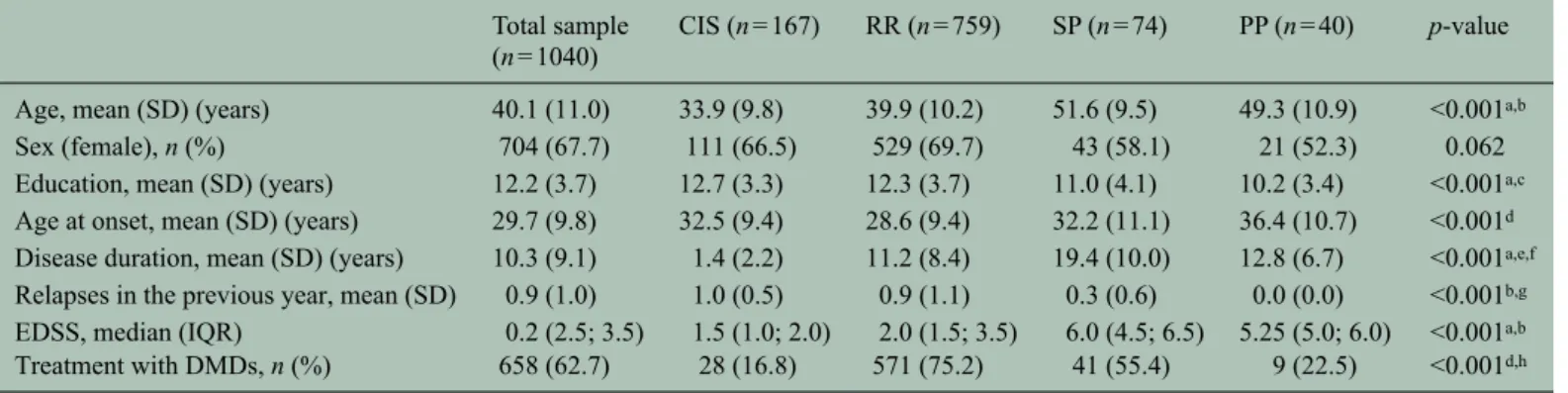
The study sample consisted of 1040 patients, 167 clinically isolated syndrome (CIS), 759 RR, 74 sec-ondary progressive (SP) and 40 primary progressive (PP) MS patients. The main demographic and clinical characteristics of the sample are depicted in Table 1. The refusal rate in the largest study center (Florence) was 14.5%. Although exact records of refusals are not available for the other centers, the feedback was that the vast majority of the patients agreed to participate. In the principal component analysis of the items from the neuropsychological evaluation, the variance explained by the four retained components was 69% (Supplementary Table 1). For component 1 (23% var-iance), the items with a high factor loading corre-sponded to SRT test; for component 2 (17% variance), to the PASAT test; for component 3 (15% variance), to the WLG and Stroop tests; and for component 4 (14% variance), to the SPART test (Supplementary Table 1), while that of the SDMT presented a moder-ate loading factor for both components 2 (0.44) and 3
(0.59). These components corresponded approxi-mately with the theoretical cognitive domains: com-ponent 1 to verbal learning, comcom-ponent 2 to information processing speed, component 3 to execu-tive function, and component 4 to visuospatial learn-ing; based on these results and on the previous literature, we retained the theoretical construct for the cognitive domains, including the SDMT in the infor-mation processing speed domain.
In the whole study sample, the prevalence of CI was 46.3%; 34.5% in CIS, 44.5% in RR MS, 79.4% in SP, and 91.3% in patients with PP. The differences in prevalence were statistically significant in the com-parisons of CIS versus SP, CIS versus PP, RR versus SP, and RR versus PP (p < 0.001) (Table 2). Overall, information processing speed was the most com-monly affected cognitive domain (47.9%). There were no significant differences between patients with CIS and RR regarding the frequency of impairment in the different domains (Table 2). On the whole, in patients with SP and PP courses, the presence of CI, as well as impairment on different cognitive domains, was approximately twofold increased when compared to CIS and RR (Table 2). There were no significant differences between the prevalence of impairment by domain between SP and PP patients.
Considering the whole sample, patients with CI were older, had a longer disease duration, higher disability levels on the EDSS, and an older age at MS onset.
Table 1. Clinical and demographic characteristics of the study patients. Total sample
(n = 1040)
CIS (n = 167) RR (n = 759) SP (n = 74) PP (n = 40) p-value
Age, mean (SD) (years) 40.1 (11.0) 33.9 (9.8) 39.9 (10.2) 51.6 (9.5) 49.3 (10.9) <0.001a,b
Sex (female), n (%) 704 (67.7) 111 (66.5) 529 (69.7) 43 (58.1) 21 (52.3) 0.062
Education, mean (SD) (years) 12.2 (3.7) 12.7 (3.3) 12.3 (3.7) 11.0 (4.1) 10.2 (3.4) <0.001a,c
Age at onset, mean (SD) (years) 29.7 (9.8) 32.5 (9.4) 28.6 (9.4) 32.2 (11.1) 36.4 (10.7) <0.001d
Disease duration, mean (SD) (years) 10.3 (9.1) 1.4 (2.2) 11.2 (8.4) 19.4 (10.0) 12.8 (6.7) <0.001a,e,f
Relapses in the previous year, mean (SD) 0.9 (1.0) 1.0 (0.5) 0.9 (1.1) 0.3 (0.6) 0.0 (0.0) <0.001b,g
EDSS, median (IQR) 0.2 (2.5; 3.5) 1.5 (1.0; 2.0) 2.0 (1.5; 3.5) 6.0 (4.5; 6.5) 5.25 (5.0; 6.0) <0.001a,b
Treatment with DMDs, n (%) 658 (62.7) 28 (16.8) 571 (75.2) 41 (55.4) 9 (22.5) <0.001d,h
CIS: clinically isolated syndrome; RR: relapsing remitting; SP: secondary progressive; PP: primary progressive; SD: standard deviation; EDSS: Expanded Disability Status Scale; IQR: interquartile range; DMDs: disease modifying drugs.
Superscript letters denote significant differences between groups, adjusted for multiple comparisons with the Bonferroni method (adjusted p-value = 0.008):
aCIS versus RR, SP, and PP. bRR versus SP and PP. cRR versus PP.
dRR versus CIS, SP, and PP. eRR versus SP.
fSP versus PP. gPP versus CIS and SP. hSP versus RR, CIS, and PP.
There were no significant differences in sex, educa-tion, and relapses in the previous year between cogni-tively preserved and impaired patients (Table 3). In the univariate logistic regression, there was a signifi-cant association between the presence of CI and older age (OR (10 years) = 1.75; p < 0.001), longer disease duration (OR (10 years) = 1.68; p < 0.001), and higher disability levels on the EDSS (OR (2 points) = 1.99;
p < 0.001). There were no significant differences regarding sex (OR = 1.08; p = 0.59), education (OR = 0.97; p = 0.12), and clinical disease activity (OR = 0.76; p = 0.05). In the subset of patients with fatigue data (n = 728), there was a significant associa-tion between higher FSS score and CI (OR (5 points) = 1.05; p = 0.03), while in the subset with depression data (n = 356), no association was found between the MADRS score and CI (OR (5 points) = 1.02;
p = 0.07). When adjusting for the effect of the
demographic variables in the a priori model, disease duration, EDSS, clinical course, and relapses in the previous year presented an association of p < 0.1 and were fitted in Model 2. In this model, the presence of CI was significantly associated only with older patient age, while the association with other variables was non-significant (Table 4). When adjusting the OR of disease duration and clinical course to age and EDSS (Model 3), the association with CI is non-significant (p = 0.47 and p = 0.30, respectively). It is important to note the decrease in the OR of disease duration and dis-ease course when they are fitted in the model with EDSS and age, while the OR for these two latter vari-ables stays approximately the same (Table 4). The VIF and GVIF for the variables (Table 4) are well below the conservative cut point of 5.0,22 indicating a relatively
low multi-collinearity. There was no significant effect of the quadratic terms of the continuous variables or of interaction factors between the variables.
Table 2. Prevalence and profile of cognitive impairment in the study sample. Total sample (n = 1040) CIS (n = 167) RR (n = 759) SP (n = 74) PP (n = 40) p-value
Cognitive impairment (⩾2 domains) 46.3% 34.5% 44.5% 79.4% 91.3% <0.001a
Verbal learning 31.1% 27.1% 28.7% 57.7% 46.2% <0.001a
Visuospatial learning 20.5% 14.5% 19.9% 35.3% 31.6% <0.001a
Information processing speed 47.9% 41.2% 45.7% 79.4% 66.7% <0.001a
Executive function 40.8% 41.8% 36.2% 76.4% 92.3% <0.001a
Number of impaired domains (impaired patients), mean (SD)
2.6 (0.7) 2.7 (0.7) 2.5 (0.7) 2.8 (0.8) 2.5 (0.6) 0.056
Number of impaired domains (all patients), mean (SD)
1.4 (1.2) 1.2 (1.2) 1.4 (1.2) 2.4 (1.1) 2.3 (0.7) <0.001a
CIS: clinically isolated syndrome; RR: relapsing remitting; SP: secondary progressive; PP: chronic progressive; SD: standard devia-tion.
Superscript letters denote significant differences between groups, adjusted for multiple comparisons with the Bonferroni method (adjusted p-value = 0.008):
aCIS versus SP; CIS versus PP; RR versus SP; and RR versus PP.
Table 3. Comparison of clinical and demographic characteristics between impaired and non-impaired patients. Without cognitive
impairment (n = 486)
With cognitive impairment (n = 422)
p-value
Age, mean (SD) (years) 36.9 (9.8) 43.2 (11.2) <0.001
Sex (female), n (%) 334 (68.7%) 283 (67.1%) 0.320
Education, mean (SD) (years) 12.52 (3.3) 12.12 (4.0) 0.109
Age at onset, mean (SD) (years) 28.5 (8.9) 30.7 (10.5) 0.001
Disease duration, mean (SD) (years) 8.4 (7.8) 12.5 (10.0) <0.001
Relapses in the previous year, mean (SD) 0.93 (0.99) 0.82 (0.99) 0.128
EDSS, mean (SD) 2.1 (1.4) 3.0 (1.8) <0.001
Treatment with DMDs, n (%) 289 (59.5%) 266 (63.0%) 0.276
T
able 4.
Logistic regression models of the prevalence of cognitive impairment in patients with MS.
Univariate regression Model 1 a Model 2 b Model 3 c OR (95% IC) p-value OR (95% IC) p-value OR (95% IC) p-value OR (95% IC) p-value Age (10 years) 1.75 (1.75; 2.00) <0.001 1.76 (1.53; 2.01) <0.001 1.49 (1.25; 1.77) <0.001 1.62 (1.42; 1.86) <0.001 Education (years) 0.97 (0.94; 1.01) 0.12 1.06 (0.79; 1.42) 0.92 1.02 (0.98; 1.06) 0.42 Sex (female) 1.08 (0.82; 1.43) 0.59 1.06 (0.79; 1.42) 0.69 0.94 (0.68; 1.30) 0.72 Disease duration (10 years) 1.68 (1.44; 1.97) <0.001 1.28 (1.07; 1.53) 0.08 1.17 (0.95; 1.45) 0.14 1.08 (0.89; 1.30) 0.47 EDSS (2 points) 1.99 (1.68; 2.36) <0.001 1.84 (1.53; 2.21) <0.001 1.75 (1.39; 2.20) <0.001 1.80 (1.51; 2.15) <0.001 Clinical course <0.001 <0.001 0.34 0.30 CIS vs RR 1.52 (1.06; 2.17) 0.02 1.18 (0.81; 1.71) 0.38 0.98 (0.61; 1.58) 0.93 0.91 (0.62; 1.34) 0.63 CIS vs SP 7.29 (3.66; 14.52) <0.001 3.59 (1.73; 7.46) 0.001 1.34 (0.39; 3.27) 0.53 1.29 (0.56; 2.97) 0.56 CIS vs PP 19.89 (4.50; 87.88) <0.001 10.66 (2.35; 48.40) 0.002 3.77 (0.76; 18.79) 0.11 3.30 (0.69; 15.89) 0.14
Relapses in the previous year
0.76 (0.58; 1.00) 0.05 1.17 (0.87; 1.59) 0.09 1.07 (0.76; 1.49) 0.71 FSS (5 points) 1.05 (1.00; 1.10) 0.03 1.00 (0.95; 1.05) 0.87 MADRS (5 points) 1.12 (0.99; 1.26) 0.07 1.07 (0.03; 1.22) 0.34
Current treatment with DMDs
1.16 (0.89; 1.52)
0.27
1.24 (0.94; 1.64)
0.14
MS: multiple sclerosis; OR: odds ratio; IC: Interval of Confidence; EDSS: Expanded Disability Status Scale; FSS: Fatigue Severity Scale; MADRS: Montgomery and
Asber
g Depression Scale; DMDs:
disease modifying drugs; CIS: clinically isolated syndrome; RR: relapsing remitting; SP: secondary progressive; PP: primary progressive. p-value for Hosmer–Lemeshow goodness of fit test: Model 1
= 0.47; Model 2 = 0.10; Model 3 = 0.08. C-statistic: Model 1 = 0.66; Model 2 = 0.71; Model 3 = 0.70. V ariation inflation factors: age = 1.39; disease duration = 1.68; EDSS = 1.34; disease subtype = 1.70.
Generalized variation inflation factors: age
= 1.18; disease duration = 1.29; EDSS = 1.16; disease subtype = 1.09. aV
ariables in the model adjusted for sex, education, and age.
bV
ariables in the model adjusted for sex, education, age, disease duration, EDSS,
clinical course, and relapses in the previous year
.
cV
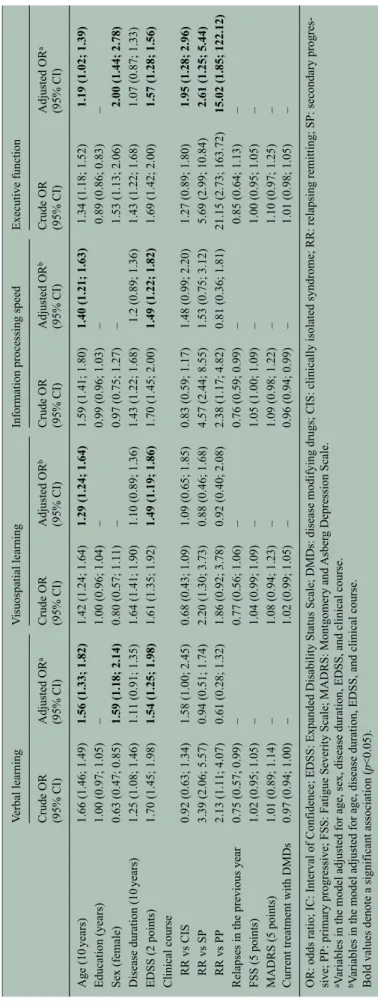
Moreover, in an analysis focusing on single cognitive domains, both higher physical disability on the EDSS and older age were associated with increased preva-lence of impairment, even after adjusting for the other variables of interest (Table 5). Executive function was the only cognitive domain in which impairment remained associated with disease subtype (Table 5) after adjusting for the other variables in the model (PP and SP > CIS > RR).
Discussion
In this large, collaborative study, we assessed the cog-nitive performance of MS patients using a neuropsy-chological battery specifically developed and validated for the disease. Although the study was clinic-based rather than population-based, it involved the main national MS centers, thus providing a rea-sonably good representation of the population of MS patients in the country.
The prevalence of CI in our study was found to be 46.3%, a figure in line with what has been reported in the recent literature.1,2,8 The overall profile of CI was
also consistent with what has been described,2
partic-ularly concerning the frequent impairment in infor-mation processing speed and episodic memory. However, the prevalence of impairment in executive function was higher than what has been reported in some of the previous literature.1,2 The two tests used
for assessing aspects of executive functions in this study were the Stroop test and the WLG test: notably, a component of speed in information processing can-not be ruled out in these tests. To address this issue, we performed principal component analysis to con-firm the theoretical cognitive domains. We found four main components, with the WLG and the Stroop tests having a high factor loading for the same component (0.78 and 0.66, respectively). Additionally, using healthy controls from a previously published norma-tive sample,21 we performed an exploratory logistic
regression analysis to determine if the differences in the Stroop and WLG scores between patients and con-trols remained significant after adjusting for the SDMT. We found that adjusting for SDMT did not change the OR of the associations between these test scores and patient status (Stroop: crude OR = 1.32 (p < 0.001); adjusted OR = 1.23 (p < 0.001); WLG: crude OR = 0.31 (p < 0.001); adjusted OR = 0.37 (p < 0.001)). The results from this analysis indicate that the ST and the WLG tests have an ability to dis-tinguish between patients and controls that is not greatly reduced after controlling for the processing speed component assessed by the SDMT, suggesting they have a potential value in assessing executive
function in MS. Overall, these findings suggest the importance of assessing executive function in patients with MS and advocate for an inclusion and further evaluation of tools such as the WLG test in future studies of CI in MS.
CI was more frequent in patients with RR than CIS (44.5% vs 34.5%); however, the difference was not statistically significant. Patients with RR and CIS pre-sented a similar cognitive profile, with a more fre-quent involvement of information processing speed and executive function compared with other cognitive domains. In comparison with CIS and RR, the preva-lence of CI was significantly higher in the progressive forms, as was the number of affected cognitive domains. Indeed, when compared with patients with CIS and RR, our patients with PP and SP had an approximately twofold higher prevalence of impair-ment in the distinct cognitive domains, with no par-ticular domain disproportionately represented. There is some controversy in the literature regarding the prevalence of CI in the secondary compared with the PP forms, with different authors reporting patients with SP as more, equally, or less affected than patients with PP.1,5,7 As for the neuropsychological profile,
efforts to define distinct cognitive profiles between SP and PP patients have revealed only subtle, often inconsistent, differences.1,5,7 In this study, patients
with SP and PP presented similar prevalence and pro-file of CI: several cognitive domains were affected in a sizeable proportion of patients, with higher preva-lence of impairment in information processing speed and executive function followed by verbal learning. It should be acknowledged, however, that a potential under-representation of the PPMS subtype in our study population can suggest some selection of study participants, since patients with PPMS—for whom no disease modifying drugs are available—may be less likely referred to specialized MS centers.
In the multivariable analysis, we found that the main determinants of overall CI were increased physical disability on the EDSS and older patient age, rather than disease duration or subtype per se.
Additionally, the multivariable analysis by cognitive domain confirmed increased physical disability and older age as the two main determinants of impair-ment, the effect of disease subtype only remaining significant in the executive function domain. These findings support a prominent effect on cognitive func-tioning of aging and disease severity, rather than of different pathogenetic mechanisms related to each disease subtype. It is interesting to note that agreeing results have been found in a large single center study,
T
able 5.
Logistic regression models of the prevalence of impairment by cognitive domain in patients with MS.
V
erbal learning
V
isuospatial learning
Information processing speed
Executive function Crude OR (95% CI) Adjusted OR a (95% CI) Crude OR (95% CI) Adjusted OR b (95% CI) Crude OR (95% CI) Adjusted OR b (95% CI) Crude OR (95% CI) Adjusted OR a (95% CI) Age (10 years) 1.66 (1.46; 1.49) 1.56 (1.33; 1.82) 1.42 (1.24; 1.64) 1.29 (1.24; 1.64) 1.59 (1.41; 1.80) 1.40 (1.21; 1.63) 1.34 (1.18; 1.52) 1.19 (1.02; 1.39) Education (years) 1.00 (0.97; 1.05) – 1.00 (0.96; 1.04) – 0.99 (0.96; 1.03) – 0.89 (0.86; 0.83) – Sex (female) 0.63 (0.47; 0.85) 1.59 (1.18; 2.14) 0.80 (0.57; 1.11) – 0.97 (0.75; 1.27) – 1.53 (1.13; 2.06) 2.00 (1.44; 2.78) Disease duration (10 years) 1.25 (1.08; 1.46) 1.11 (0.91; 1.35) 1.64 (1.41; 1.90) 1.10 (0.89; 1.36) 1.43 (1.22; 1.68) 1.2 (0.89; 1.36) 1.43 (1.22; 1.68) 1.07 (0.87; 1.33) EDSS (2 points) 1.70 (1.45; 1.98) 1.54 (1.25; 1.98) 1.61 (1.35; 1.92) 1.49 (1.19; 1.86) 1.70 (1.45; 2.00) 1.49 (1.22; 1.82) 1.69 (1.42; 2.00) 1.57 (1.28; 1.56)
Clinical course RR vs CIS
0.92 (0.63; 1.34) 1.58 (1.00; 2.45) 0.68 (0.43; 1.09) 1.09 (0.65; 1.85) 0.83 (0.59; 1.17) 1.48 (0.99; 2.20) 1.27 (0.89; 1.80) 1.95 (1.28; 2.96) RR vs SP 3.39 (2.06; 5.57) 0.94 (0.51; 1.74) 2.20 (1.30; 3.73) 0.88 (0.46; 1.68) 4.57 (2.44; 8.55) 1.53 (0.75; 3.12) 5.69 (2.99; 10.84) 2.61 (1.25; 5.44) RR vs PP 2.13 (1.11; 4.07) 0.61 (0.28; 1.32) 1.86 (0.92; 3.78) 0.92 (0.40; 2.08) 2.38 (1.17; 4.82) 0.81 (0.36; 1.81) 21.15 (2.73; 163.72) 15.02 (1.85; 122.12)
Relapses in the previous year
0.75 (0.57; 0.99) – 0.77 (0.56; 1.06) – 0.76 (0.59; 0.99) – 0.85 (0.64; 1.13) – FSS (5 points) 1.02 (0.95; 1.05) – 1.04 (0.99; 1.09) – 1.05 (1.00; 1.09) – 1.00 (0.95; 1.05) – MADRS (5 points) 1.01 (0.89; 1.14) – 1.08 (0.94; 1.23) – 1.09 (0.98; 1.22) – 1.10 (0.97; 1.25) –
Current treatment with DMDs
0.97 (0.94; 1.00) – 1.02 (0.99; 1.05) – 0.96 (0.94; 0.99) – 1.01 (0.98; 1.05) –
OR: odds ratio; IC: Interval of Confidence; EDSS: Expanded Disability Status Scale; DMDs: disease modifying drugs; CIS: clinically isolated syndrome; RR: relapsing remitting; SP: secondary progres
-sive; PP: primary progres-sive; FSS: Fatigue Severity Scale; MADRS: Montgomery and
Asber
g Depression Scale.
aV
ariables in the model adjusted for age, sex, disease duration, EDSS, and clinical course.
bV
ariables in the model adjusted for age, disease duration, EDSS, and clinical course.
Bold values denote a significant association (
where clustering by disease subtype did not show any differences in the cognitive profile of CI.23
Regarding the relation between physical disability and CI, there is some heterogeneity in the published literature.10–12 The results from this study clearly
imply an association between increasing degrees of physical and cognitive disability, that is also sup-ported by the few available longitudinal series, with smaller sample size.24,25 The observed relationship
may be an effect of disease severity, progression, and biological changes associated with aging, with increasing burden of lesions in the brain, atrophy, and diffuse changes in the white and gray matter, as depicted by imaging and pathological studies.26 A
recent study has also suggested the existence of iso-lated cognitive relapses that can be detected only through periodic cognitive assessment and may con-tribute to the burden of CI in the long run.27
The absence of an independent effect of disease dura-tion in overall CI is another noteworthy finding from this study. On one hand, age and disease duration are correlated and it may be difficult to disentangle the effect of these two variables. However, the correlation between patient age and disease duration in this patient sample is not particularly strong (r = 0.54; Supplementary Table 2), resulting in low multi-collin-earity between the variables (Table 4). On the other hand, it is interesting to note the parallel between our cross-sectional cognitive findings and what has been reported in large natural history studies on disease prognosis, where physical disability and disease pro-gression are more related to patient age than to the duration of the disease or the clinical phenotype at onset,28,29 suggesting, as in this study, that disease
duration is not an accurate predictor of disease pro-gression. Overall, these results support the hypothesis that in MS the shift from a predominantly inflamma-tory phase, dominated by clinical relapses, to a pre-dominantly neurodegenerative phase, dominated by irreversible progression of neurological disability, may be mainly driven by biological factors related to aging. Furthermore, the results concur with the hypothesis of cognitive reserve, as aging has previ-ously been associated with decreased plasticity and capability of functional reorganization in MS that probably results from the interaction between cerebral aging and the accumulation of structural brain damage.30
As for the role of sex, the published research usually points to an overall worse functional prognosis in males with MS when compared to females.31 Some
previous studies have suggested this also applies to
cognitive outcomes,32 but the issue is controversial
in the literature, as most recently published large series have found no significant differences in the prevalence of overall CI.8,12,13,23 In our sample, in
spite of a higher physical disability level in males, we were not able to confirm any significant effect of sex in the prevalence of overall CI, neither as a first order association nor when adjusting for other pre-dictors. Nevertheless, sex-related differences were found in the verbal learning and executive function domains. The better performance of women in ver-bal learning tests had already been reported, and could perhaps contribute to explain the higher preva-lence of CI in males in some of the published litera-ture, as tests designed to evaluate executive functions, in which females performed worst in this study, are not always used to assess patients with MS. Nevertheless, the presence of sex-related differ-ences in some cognitive domains could hint at an interaction between sexual hormones, disease activ-ity, and neurodegeneration, as hypothesized by some authors.32
There was also no association of CI with the use of disease modifying drugs. This may be accounted for by the discontinuation or absence of treatment in the older and more disabled patients with the progressive phenotypes. It is also possible that patients with RR with more active and severe disease are more likely to be treated, which renders it difficult to determine the impact of disease modifying drugs on cognition. Longitudinal, controlled studies are needed to shed some light on this score.
As for the association of progressive course and higher impairment in executive function, this is mainly driven be the Stroop test results. We can spec-ulate that this relationship is due to increased frontal dysfunction33 and frontotemporal lobe atrophy34 in
patients with progressive forms compared with patients with RR. However, the higher impairment in executive function found in CIS patients was mainly driven by a worse performance on the WLG test, which is consistent with findings obtained in a small clinical series.8
One limitation of our study is the partial data on depression and fatigue that are well-known potential confounders for cognitive performance in MS.1
However, performing a sensitivity analysis in the sub-sets of patients with available data we found that fatigue and depression scores were not retained in the multivariable analysis. These results suggest that fatigue and depression were not major contributors to MS-related CI in these patients.
The model using age and physical disability alone (Model 3) presented an accuracy of 70% to classify patients as having CI, implying that there are other factors that could explain the remaining variability in the subject cognitive outcome, such as genetic deter-minants, environmental factors, comorbidities, as well as different individual resilience to brain damage due to intellectual enrichment and cognitive reserve.12,35 Indeed, previous studies have found an
association between CI and measures of cognitive reserve, such as the cognitive reserve index,35 which
is composed of education and an assessment of pre-morbid IQ and prepre-morbid leisure activities. The use of these measures should probably be expanded in future studies, as education alone is probably not a good enough surrogate of cognitive reserve in many populations, as suggested by the results from the pre-sent and several of the previous studies, which have reported no direct association of CI and education.8,13
In conclusion, the findings obtained from this large clinical series strongly imply that the presence of CI is more related to patient age and disease severity than to disease duration or subtype per se. Furthermore, this study clearly documents a significant presence of CI since the earlier stages of MS, which increases in frequency and severity in the progressive stages. It also adds evidence to previous clinical studies5–9 and
therapeutic trials in CIS,36 pointing to the need for
systematic neuropsychological assessment since the beginning of MS and monitoring throughout the dis-ease course, suggesting that prompt diagnosis and management strategies should ideally be pursued at a younger patient age, when compensatory abilities, brain plasticity, and cognitive reserve may better miti-gate the effects of pathological damage in the brain.
Author Contributions
The first two authors contributed equally to the manuscript.
Declaration of Conflicting Interests
The author(s) declared no potential conflicts of inter-est with respect to the research, authorship, and/or publication of this article.
Funding
The author(s) received no financial support for the research, authorship, and/or publication of this article.
References
1. Amato MP, Zipoli V and Portaccio E. Multiple sclerosis-related cognitive changes: A review of cross-sectional and longitudinal studies. J Neurol Sci 2006; 245: 41–46.
2. Chiaravalloti ND and DeLuca J. Cognitive
impairment in multiple sclerosis. Lancet Neurol 2008; 7: 1139–1151.
3. Bever CT Jr, Grattan L, Panitch HS, et al. The brief repeatable battery of neuropsychological tests for multiple sclerosis: A preliminary serial study. Mult
Scler 1995; 1: 165–169.
4. Langdon DW, Amato MP, Boringa J, et al. Recommendaations for a Brief International Cognitive Assessment for Multiple Sclerosis (BICAMS). Mult Scler 2012; 18: 891–898.
5. Huijbregts SC, Kalkers NF, de Sonneville LM, et al. Differences in cognitive impairment of relapsing remitting, secondary, and primary progressive MS.
Neurology 2004; 63: 335–339.
6. Ruet A, Deloire M, Charre-Morin J, et al. Cognitive impairment differs between primary progressive and relapsing-remitting MS. Neurology 2013; 80: 1501–1508.
7. Planche V, Gibelin M, Cregut D, et al. Cognitive impairment in a population-based study of patients with multiple sclerosis: Differences between late relapsing-remitting, secondary progressive and primary progressive multiple sclerosis. Eur J Neurol 2016; 23: 282–289.
8. Potagas C, Giogkaraki E, Koutsis G, et al. Cognitive impairment in different MS subtypes and clinically isolated syndromes. J Neurol Sci 2008; 267: 100–106. 9. Zipoli V, Goretti B, Hakiki B, et al. Cognitive
impairment predicts conversion to multiple sclerosis in clinically isolated syndromes. Mult Scler 2010; 16: 62–67.
10. Lynch SG, Parmenter BA and Denney DR. The association between cognitive impairment and physical disability in multiple sclerosis. Mult Scler 2005; 11: 469–476.
11. Fischer JS, Foley FW, Aikens JE, et al. What do we really know about cognitive dysfunction, affective disorders, and stress in multiple sclerosis? A practitioner’s guide. Neurorehab Neural Re 1994; 8: 151–164.
12. Martins Da Silva A, Cavaco S, Moreira I, et al. Cognitive reserve in multiple sclerosis: Protective effects of education. Mult Scler 2015; 21: 1312–1321. 13. Borghi M, Cavallo M, Carletto S, et al. Presence and
significant determinants of cognitive impairment in a large sample of patients with multiple sclerosis. PLoS
ONE 2013; 8: e69820.
14. McDonald WI, Compston A, Edan G, et al. Recommended diagnostic criteria for multiple sclerosis: Guidelines from the International Panel on the diagnosis of multiple sclerosis. Ann Neurol 2001; 50: 121–127.
15. Lublin FD and Reingold SC. Defining the clinical course of multiple sclerosis: Results of an international survey. National Multiple Sclerosis Society (USA) Advisory Committee on Clinical Trials of New Agents in Multiple Sclerosis.
Neurology 1996; 46: 907–911.
16. Kurtzke JF. Rating neurologic impairment in multiple sclerosis: An expanded disability status scale (EDSS).
Neurology 1983; 33: 1444–1452.
17. Meyer-Moock S, Feng YS, Maeurer M, et al. Systematic literature review and validity evaluation of the Expanded Disability Status Scale (EDSS) and the Multiple Sclerosis Functional Composite (MSFC) in patients with multiple sclerosis. BMC Neurol 2014; 14: 58.
18. Krupp LB, LaRocca NG, Muir-Nash J, et al. The fatigue severity scale. Application to patients with multiple sclerosis and systemic lupus erythematosus.
Arch Neurol 1989; 46: 1121–1123.
19. Montgomery SA and Asberg M. A new depression scale designed to be sensitive to change. Br J
Psychiatry 1979; 134: 382–389.
20. Stroop JR. Studies of interference in serial verbal reactions. J Exp Psychol 1935; 18: 643–662. 21. Amato MP, Portaccio E, Goretti B, et al. The Rao’s
Brief Repeatable Battery and Stroop Test: Normative values with age, education and gender corrections in an Italian population. Mult Scler 2006; 12: 787–793. 22. Stine RA. Graphical interpretation of variance
inflation factors. Am Stat 1995; 49: 53–56. 23. Achiron A, Chapman J, Magalashvili D, et al.
Modeling of cognitive impairment by disease duration in multiple sclerosis: A cross-sectional study. PLoS
ONE 2013; 8: e71058.
24. Moccia M, Lanzillo R, Palladino R, et al. Cognitive impairment at diagnosis predicts 10-year multiple sclerosis progression. Multi Scler 2016; 22: 659–667. 25. Amato MP, Ponziani G, Siracusa G, et al. Cognitive
dysfunction in early-onset multiple sclerosis: A reappraisal after 10 years. Arch Neurol 2001; 58: 1602–1606.
26. Rocca MA, Amato MP, De Stefano N, et al. Clinical and imaging assessment of cognitive dysfunction in multiple sclerosis. Lancet Neurol 2015; 14: 302–317. 27. Pardini M, Uccelli A, Grafman J, et al. Isolated
cognitive relapses in multiple sclerosis. J Neurol
Neurosurg Psychiatry 2014; 85: 1035–1037.
28. Confavreux C and Vukusic S. Natural history of multiple sclerosis: A unifying concept. Brain 2006; 129: 606–616.
29. Cossburn M, Ingram G, Hirst C, et al. Age at onset as a determinant of presenting phenotype and initial relapse recovery in multiple sclerosis. Multi Scler 2012; 18: 45–54.
30. Schoonheim MM, Geurts JJ and Barkhof F. The limits of functional reorganization in multiple sclerosis. Neurology 2010; 74: 1246–1247. 31. Shirani A, Zhao Y, Kingwell E, et al. Temporal
trends of disability progression in multiple sclerosis: Findings from British Columbia, Canada (1975– 2009). Mult Scler 2012; 18: 442–450.
32. Savettieri G, Messina D, Andreoli V, et al. Gender-related effect of clinical and genetic variables on the cognitive impairment in multiple sclerosis. J Neurol 2004; 251: 1208–1214.
33. Aviv RI, Francis PL, Tenenbein R, et al. Decreased frontal lobe gray matter perfusion in cognitively impaired patients with secondary-progressive multiple sclerosis detected by the bookend technique. AJNR
Am J Neuroradiol 2012; 33: 1779–1785.
34. Riccitelli G, Rocca MA, Pagani E, et al. Cognitive impairment in multiple sclerosis is associated to different patterns of gray matter atrophy according to clinical phenotype. Hum Brain Mapp 2011; 32: 1535–1543.
35. Amato MP, Razzolini L, Goretti B, et al. Cognitive reserve and cortical atrophy in multiple sclerosis: A longitudinal study. Neurology 2013; 80: 1728–1733. 36. Penner IK, Stemper B, Calabrese P, et al. Effects
of interferon beta-1b on cognitive performance in patients with a first event suggestive of multiple sclerosis. Multi Scler 2012; 18: 1466–1471.
Visit SAGE journals online journals.sagepub.com/ home/msj