Treatment of short bowel syndrome in children. Value of the
Intestinal Rehabilitation Program
UENIS TANNURI1*, FABIODE BARROS2, ANA CRISTINA AOUN TANNURI3
1Full Professor, Department of Pediatric Surgery and Liver Transplant, Faculdade de Medicina, Universidade de São Paulo (FMUSP). Head of the Service of Pediatric Surgery and Liver Transplant at Instituto da Criança, Hospital das Clínicas, and of the Pediatric Surgery Research Laboratory, FMUSP. Head of the Laboratory of Pediatric Surgery at Hospital das Clínicas, FMUSP, São Paulo, SP, Brazil
2Physician, Service of Pediatric Surgery, Instituto da Criança, Hospital das Clínicas, FMUSP, São Paulo, SP, Brazil
3Associate Professor, Department of Surgical Technique and Experimental Surgery, FMUSP. Physician, Service of Pediatric Surgery, Instituto da Criança, Hospital das Clínicas, and the Pediatric Surgery Research Laboratory (LIM-30), FMUSP, São Paulo, SP, Brazil
S
UMMARYStudy conducted at Serviço de Cirurgia Pediátrica e Transplante Hepático, Instituto da Criança, Hospital das Clínicas, Faculdade de Medicina, Universidade de São Paulo (HC-FMUSP), São Paulo, SP, Brazil
Article received: 6/29/2016
Accepted for publication: 7/26/2016
*Correspondence:
Address: Av. Dr. Arnaldo, 455, sala 4106 São Paulo, SP – Brazil Postal code: 01246-903 [email protected]
http://dx.doi.org/10.1590/1806-9282.62.06.575
The main cause of acute intestinal failure is short bowel syndrome, generally as a result of resection of extensive segments of small intestine. As a result, the main symptoms are watery diarrhea, malabsorption syndrome, chronic malnutrition, and death, if the patient is not properly treated. If the length of the remaining intestine is greater than 30 cm, complete adaptation is possible and the patient may not require parenteral nutrition. The currently recommended treatment in-cludes the use of prolonged parenteral nutrition and enteral nutrition, always aimed at constant weight gain, in conjunction with surgeries aimed at elongat-ing the dilated bowel. This set of procedures constitutes what is called an Intes-tinal Rehabilitation Program. This therapy was used in 16 children in periods ranging from 8 months to 7.5 years, with survival in 75% of the cases. Finally, the last resort to be used in children with complete resection of the small bow-el is an intestinal transplant. However, to date there is no record of a Brazilian child that has survived this procedure, despite it being attempted in seven pa-tients. We conclude that the results of the intestinal rehabilitation program are encouraging for the continuation of this type of treatment and stimulate the creation of the program in other pediatric care institutions.
Keywords: short bowel syndrome, child, intestinal failure, prolonged parenteral nutrition, intestinal rehabilitation.
I
NTRODUCTIONAcute changes in intestinal function may be related to various clinical or surgical diseases in children. However, short bowel syndrome is the most important and serious cause of acute intestinal failure. The designation “short” stems from the fact that the disease is acquired after a surgical procedure, where resection of extensive segments of the intestine is necessary due to non-viable intestinal structures. This results in a clinical condition, character-ized by fast intestinal transit leading to diarrhea and nu-trient malabsorption. It may also be caused by partial re-sections of the intestine if the remaining portions function poorly. The clinical manifestations depend on the length of the remaining portions of the jejunum and ileum, the presence of an intestinal bypass, the presence or absence of the ileocecal valve, the remaining length of the colon and the persistence of intestinal disease. The most
fre-quent causes in newborns are necrotizing enteritis, mid-gut volvulus and other congenital malformations such as intestinal atresia, intestinal aganglionosis, and gastros-chisis. In older children, the most important causes are intestinal volvulus and resections of extensive intestinal segments in repeated abdominal surgeries, indicated by the presence and complications caused by intestinal ad-hesions. Other important causes of intestinal function failure with dificult clinical treatment refer to intestinal neuronal dysplasia and all other forms of chronic intes-tinal pseudo-obstruction.1
days after intestinal resection and/or remaining length of the small intestine from the angle of Treitz of less than 50 cm for premature infants, less than 75 cm for term new-borns and less than 100 cm for a 1 year old child.2
Until the beginning of the 1970s, children with SBS died early as a result of malnutrition that occurred quick-ly due to the lack of effective nutritional treatment. The introduction of prolonged parenteral nutrition by then, together with appropriate dietary measures, has substan-tially changed the prognosis of children subjected to ma-jor small intestine resections. More recently, hospital pro-grams with groups of professionals speciically designated and trained for the treatment of these patients have been responsible for the recovery of a large number of patients. This prevents the last surgical resort, that is, intestinal transplant, which may be indicated in children with com-plete resection of the small intestine or those in which the vascular access routes for intravenous administration of nutrients have been exhausted.
I
NCIDENCE,
ETIOLOGY,
AND PATHOLOGYIn the Brazilian literature there are no publications on the prevalence of SBS. According to international publi-cations, it is estimated at around 3 to 5 cases per 100,000 births per year. However, it is known that the occurrence has increased in recent years as a result of improved ser-vices for newborns in neonatal intensive care units.
In our country, most SBS cases occur in the neona-tal period, as a result of vascular changes to the intestine during intrauterine development. The second cause con-sists of acquired intestinal problems requiring resection of the large intestine, in which case necrotizing enteritis and intestinal volvulus are the most important causes, as quoted previously.3
Soon after intestinal resection, the main digestive symptom is diarrhea, due to poor absorption of nutri-ents and water. However, the intensity of symptoms will depend on the absorption capacity of the remaining in-testine. Adaptive phenomena that are intended to increase the absorption of luids and nutrients occur, such as: di-lation and elongation of the bowel loops, thickening of the wall, hyperplasia of the mucosal villi, increased depth of crypts, acceleration of proliferation rates of enterocytes, and hyperplasia of the ibers in the muscular layer. This adaptation phase can last from 1 to 2 years, during which intestinal absorption is inadequate, therefore requiring parenteral nutrition.
The ability to absorb water and nutrients depends on the length of the small intestine, but also on the presence of the colon and the ileocecal valve. In general, resection of
the jejunum, even if of extensive segments, only causes a transitory reduction in absorption of nutrients, due to the large absorptive capacity of the ileum, the enterohepatic circulation of bile salts and of vitamin B12. Generally, the resection of large sections of the ileum may cause conse-quences that are more harmful to intestinal function. Af-ter a meal with hypertonic content, there is usually a large supply of water into the lumen of the irst jejunal loops, in order to make the content isotonic and allow absorption of nutrients. After resection of the ileum, the jejunal con-tent comes directly to the colon, which has to increase its absorptive capacity. Another consequence of resection of the ileum is inadequate absorption of vitamin B12 and bile acids, which causes diarrhea if they reach the lumen of the colon. Also, the loss of the ileum causes depletion of the “pool” of bile salts, which in turn causes a reduction in the absorption of fat and fat-soluble vitamins. Under normal conditions, hydrolyzed bile acids bind with calcium to form calcium stearate and prevent the absorption of this ion, which also binds with oxalates, preventing their absorption. Thus, in children with short bowel syndrome, the reduced “pool” of bile acids will be responsible for increased absorp-tion of oxalates, with consequent oxaluria and formaabsorp-tion of urinary calculi containing oxalates. Similarly, due to the reduction of calcium in the intestinal lumen, promoted by the increased absorption, unconjugated bilirubin remains in solution, increasing its content in the enterohepatic cir-culation, changing the constitution of the bile and leading to a tendency for the formation of gallstones.
Adaptation phenomena occurring after resection of extensive portions of the intestine are responsible for nor-malization of the digestive function, with oral nutrition in normal conditions in approximately 90% of children with short bowel syndrome. These phenomena consist of increased blood low to the remaining intestine, intesti-nal growth and dilation, increased absorption surface, growth of the intestinal villi and of the depth of the crypts. The pathophysiology of these adaptive mechanisms is not well understood, although it is universal knowledge that the presence of nutrients in the intestinal lumen with increased digestive secretions is the most important stim-ulus for intestinal growth.
tropism and growth, given that, according to investiga-tions, the administration of growth hormone promotes increased uptake of glutamine after intestinal resection.4
T
REATMENTThe clinical evolution of patients with SBS is divided into three phases: the initial acute phase following the surgical procedure is characterized by intense diarrhea and consequential electrolyte disturbances, gastric acid hypersecretion and gastrinemia, generally lasting around a month. In this period, electrolytes must be replaced, and acid-base balance disturbances must be corrected. Next is the adaptation phase, which can last from a few months to a year. In this period, parenteral nutrition and enteral nutrition with adequate diets are adminis-tered, diarrhea is less intense and the nutritional state tends to stabilize, as long as the patient is properly treat-ed. Eventually, there comes the maintenance phase, in which, even though poor absorption is still present, it often becomes possible to interrupt parenteral nutrition and keep the patient on exclusive oral nutrition. Gains in weight and height will depend on the absorptive ca-pacity of the remaining intestine. It is known that the adaptation of the remaining small intestine always de-pends on adequate maintenance of the nutritional state, with growth and somatic development, even at the ex-pense of parenteral nutrition and enteral nutrition with artiicial diets. In general, when the length of the remain-ing small intestine is greater than 30 cm, the patient can be adapted in intervals of up to 1 year and be free of par-enteral nutrition with exclusive oral nutrition.
From a surgical point of view, a child with SBS may depend on the following procedures:
1. Obtaining venous access for prolonged parenteral nutrition.
2. Surgical procedures to lengthen the remaining intes-tine and improve its absorptive ability.
3. Being treated by a group of professionals trained in intestinal rehabilitation programs.
Venous access
The most important aspect in patients with SBS refers to how both the pediatrician and the surgeon will obtain and maintain venous access for the administration of nutri-ents for as long as possible. Whenever there is no possibil-ity of recovering the intestine, the survival time of the pa-tient depends on the durability of the central catheters.
The placement of a central venous catheter should always be carried out in the operating room under
gen-eral anesthesia. The catheters must be put in place by puncturing the central veins, with venous dissections as the second option. As such, the veins of the superior vena cava should preferably be punctured, namely the inter-nal jugular and subclavian veins. The choice between these depends on the experience of the surgeon, remembering that puncture of the subclavian vein is associated with an increased risk of pneumothorax or hemothorax. The femoral veins, although associated with absence of pneu-mothorax or hepneu-mothorax and less puncture accidents, are left as the second choice due to greater dificulties in the handling of dressings. After the vein is punctured, the catheter is introduced using a classic and widely used technique called Seldinger, in which the catheter is thread-ed over a guide wire.
In young children or in the presence of blood dys-crasias, one may opt for venous dissection. The irst choice for access to the central veins is based on tribu-tary veins of the superior vena cava system: the basilic or axillary veins in the axilla and the internal or exter-nal jugular veins or facial vein in the neck. In older chil-dren or teenagers, the cephalic vein in the deltopectoral sulcus may present good caliber and provide another option for venous access. Tributary veins of the inferi-or cava system may be used when the veins referred to above are not available due to previous dissections or thrombosis of the superior vena cava. In these cases, the saphenous arch or inferior deep epigastric veins are the most used. The choice of the inferior vena cava system is valid and justiied by the low rate of complications and also by the fact that thrombosis of iliac veins or in-ferior cava are often asymptomatic, unlike thrombosis of superior vena cava.
Regardless which vein is used, radiographic control of the correct position of the catheter is essential to con-trast via radiography or radioscopy carried out in the op-erating room before closing the incision of the phlebot-omy. The tip of the catheter should be positioned at the entrance to the right atrium or approximately 1 cm in-side it. Incorrect positions must be promptly corrected, as they may lead to venous thrombosis.
The catheter should be externalized through a coun-ter-opening, at a distant location from the entry point of the vein after traveling through a subcutaneous tunnel. The dressings must be meticulously changed every 2 or 3 days or at any time if there is an extravasation of the so-lution, using antiseptic solutions.
silicone instead of polyurethane or polyvinyl because they are less thrombogenic.
Catheters are responsible for the occurrence of re-peated infections and thrombosis of the superior and in-ferior vena cava, which are the most frequent long-term complications in children with SBS. The repeated ex-change of catheters may lead to exhaustion of all the con-ventional access routes, requiring alternative methods to obtain access to central vein. Therefore, dissection of the azygos vein or its branches (intercostal veins) has been reported and standardized in our service through right thoracotomy, with placement of a port-a-cath and ixa-tion of the chamber on the anterior surface of the chest. Another option is to make a purse-string suture in the
atrial appendage and insertion of the catheter directly into the right atrium. Furthermore, transhepatic punc-tures have been described for access to the suprahepatic veins that drain directly into the inferior vena cava.5
Prolonged parenteral nutrition and enteral nutrition
Prolonged parenteral nutrition should be indicated ear-ly, in the irst few days after surgery, shortly after hemo-dynamic stabilization of the child and correction of elec-trolyte disorders. This should at irst be undertaken in hospital environment; and later, during the maintenance phase, it may be performed in the patient’s home. Anoth-er possibility is the administration of nutrients in an out-patient hospital regime for daily periods of 6 to 8 hours, in addition to oral nutrition.
At the patient’s home, the solutions can be adminis-tered during the day or night. The advantage of the irst option is its greater safety, which makes the use of infu-sion pumps expendable. If the administration is done at night, the child may have normal activity during the day; however, the use of an infusion pump with an alarm sys-tem is essential in case of air entrapped in the infusion system. Some children do not tolerate rapid administra-tion of large volumes of hypertonic soluadministra-tions and require continual infusions over 24 hours daily.
The most serious and frequent complication for chil-dren with SBS on a parenteral nutrition regimen is sys-temic infection, stemmed from the presence of a central venous catheter, as previously mentioned. It is impor-tant to investigate any noncompliance with the rules of the protocol for handling the catheter and solutions. Failure to ind a speciic source of infection and the per-sistence of fever results in intravenous administration of broad-spectrum antibiotics. On the other hand, the situation is potentially serious when the blood culture
reveals Staphylococcus aureus or S. epidermidis. Speciic an-tibiotics for a three-week period are recommended in these cases. Fungal infections must be treated with med-ication and removal of the catheter. Last, catheter ob-struction caused by blood clots is a frequent problem and often involves exchange of the catheter, using the same vein.
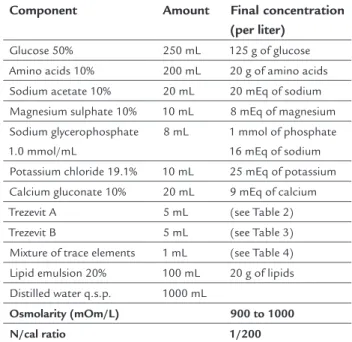
The solutions to be administered containing all of the nutrients mixed into one bottle are speciied in Table 1. The mixture contains a caloric source in the form of glucose and lipid emulsion, a solution of crystalline ami-no acids, electrolytes, vitamins, and trace elements.
TABLE 1 Standard solution with a mixture of all nutrients in a single vial.
Component Amount Final concentration (per liter)
Glucose 50% 250 mL 125 g of glucose Amino acids 10% 200 mL 20 g of amino acids Sodium acetate 10% 20 mL 20 mEq of sodium Magnesium sulphate 10% 10 mL 8 mEq of magnesium
Sodium glycerophosphate 1.0 mmol/mL
8 mL 1 mmol of phosphate 16 mEq of sodium
Potassium chloride 19.1% 10 mL 25 mEq of potassium Calcium gluconate 10% 20 mL 9 mEq of calcium Trezevit A 5 mL (see Table 2) Trezevit B 5 mL (see Table 3)
Mixture of trace elements 1 mL (see Table 4) Lipid emulsion 20% 100 mL 20 g of lipids Distilled water q.s.p. 1000 mL
Osmolarity (mOm/L) 900 to 1000 N/cal ratio 1/200
TABLE 2 Composition of vitamin mixture.
Trezevit A Adult Pediatric
Vitamin A (retinyl palmitate) 3300 IU 2300 IU
Vitamin D3 (cholecalciferol) 200 IU 400 IU Vitamin E (alpha tocopherol acetate) 10 IU 7 IU Vitamin K1 (phytomenadione) 150 mcg 200 mcg
Vitamin B1 (thiamine hydrochloride) 6 mg 1.2 mg Vitamin B2 (ribolavin sodium phosphate) 3.6 mg 1.4 mg Vitamin B3 (nicotinamide) 40 mg 17 mg Vitamin B5 (dexpanthenol) 15 mg 5 mg
Vitamin B6 (pyridoxine hydrochloride) 6 mg 1 mg Vitamin C (ascorbic acid) 200 mg 80 mg
TABLE 3 Composition of the second vitamin mixture.
Trezevit B Adult Pediatric
Vitamin B7 (biotin) 60 mcg 20 mcg Vitamin B9 (folic acid) 600 mcg 140 mcg
Vitamin B12 (cyanocobalamin) 5 mcg 1 mcg Water for injection q.s.p. 5 mL 5 mL
TABLE 4 Composition of the mixture of trace elements.
Component Amount (g) Concentration (mcg/mL)
ZnSO4.7H2O 1.9 zinc-400
CuSO4.6H2O 0.9 copper-200
NaF 0.22 luorine-10
NaI 0.069 iodine-59
MnSO4.H2O 0.62 manganese-200
The concomitant supply of nutrients orally or through a nasogastric tube is always recommended. Delivering nutri-ents directly into the stomach is particularly advantageous when using chemically deined diets with a disagreeable la-vor, which are not very acceptable to children when orally ingested. On the other hand, the catheter enables diets to be infused continuously, which allows better absorption.
Diets
The introduction of diets should be undertaken in the initial phase, when there is hemodynamic and electrolyte stabiliza-tion and the child has overcome the paralytic ileus period, with the intestinal transit reestablished. The introduction should be made with elemental diets, with some degree of prior digestion of nutrients. In general, the diet should con-tain carbohydrates, fats and proteins in the ratio of 5:3:2, elec-trolytes, vitamins, and trace elements. After full dilution, the diet’s osmolarity should be no higher than the plasma’s (300 mOsm/L). Remember that children with SBS tolerate any type of hyperosmolar diet poorly, even when introduced into the stomach. The different types of diets are presented below:
1. Natural diets. In the preparation, natural elements without any prior manipulation are used: full fat or skimmed cow’s milk, soy, meat iber, meat broth, egg, butter, which may be mixed with certain commercial formulas (Isolac®, Karo®, Nidex®, Casec® etc). The
main advantage of these diets is easy handling, with preparation possible in the child’s own home. The biggest disadvantage is that they usually require a gastrointestinal tract whose length is not greatly com-promised, because they are not digested in advance and must be administered via the stomach.
2. Dairy diets. These are widely used. It must be remem-bered that human milk, due to its protective proper-ties and stimulation of digestive maturation, should be preferred for newborns in any clinical situation, whenever possible. This begins with small doses and gradual increases, as an exclusive food or associated with other types of milk.
3. Dietary supplements. These are generally synthetic preparations, used only to enrich existing formulas. They may be simple (TCM®, Casec® etc) or compounds,
in which several nutrients are mixed (Meritene®,
Sus-tacal® etc).
4. Elemental and semi-elemental diets containing pre-viously hydrolyzed proteins and carbohydrates. The Brazilian market offers various types of diets, most com-monly the Alfaré®, Pregestimil®, and Neocate® brands.
They should initially be offered in diluted form and in small quantities, and progressively increased in volu-me and concentration, depending on the acceptance and tolerance, which are basically represented by the emergence or worsening of diarrhea and dehydration. 5. Modular diets. These are widely used in practice, par-ticularly in hospitals and in patients with few resour-ces. The natural and low cost nutrients are added gra-dually one at a time, in variables lengths of time, depending on adaptation to the nutrient introduced beforehand. These diets, which are easy to prepare and easily available, contain: carbohydrates (cream of rice, potato starch or glucose), fat (medium chain triglycerides, babassu or corn oil), protein (chicken iber or crystalline amino acids), mineral salts, vita-mins A, D, E, K, B12, and biotin.
In the irst week runs, a mixture of 3% cream of rice, 5% glucose, and a small portion of kitchen salt are admin-istered. If there is good acceptance, without diarrhea, in the following week 10% chicken broth is added, followed by 20% and, at the end of the week, 1% medium chain triglycerides are added. In the third week, 10% chicken iber is added with a few drops of lemon with the ob-jective of supplying vitamins, especially vitamin C. In the fourth week, iber concentration is increased
to 20% and medium chain triglycerides to 2 and 3%, progressively. In this phase, minerals begin to be sup-plied as a mixture of sodium chloride, potassium chloride, calcium gluconate, potassium phosphate, and magnesium sulfate or through the addition of leaf vegetable broth.
re-placed by corn oil at an equal percentage. Finally, cooked egg yolk is introduced, half at the beginning and then the entire unit. This diet is maintained for 2 to 3 months and, if there is no diarrhea, the transi-tion to soy milk can be undertaken.
It is important to note that during the administration of one of the enteral diet types, weight loss, dehydration, and loss of electrolytes occur in most cases, which always require the administration of parenteral nutrition as con-comitant support.
As stressed above, the introduction of enteral diets or oral diets is slow and subject to regressions and/or suspen-sions, depending on the child’s response. Only after stool volume and consistency are clearly stable other less digest-ed and more natural foods can be introducdigest-ed. In the main-tenance and stabilization phases, the gradual introduction of the diets is also an excellent clinical assessment test in re-lation to the adaptation of the intestine. Laboratory assess-ments with complicated intestinal absorption tests have lit-tle value and meaning from a therapeutic point of view. Also, the interpretation of a particular test is of little practical ap-plicability. Sometimes, the attempted introduction of a cer-tain diet, followed by proper and reasonable assessment of the clinical response of the child, has greater value.
Certain medications can complement the clinical treatment. Thus, the gastric hypochlorhydria in the ear-ly stage can be reversed with the administration of H2 re-ceptor antagonists or proton-pump inhibitors. Watery diarrhea and fast intestinal transit can be controlled with the administration of opioids or loperamide. Other drugs cited in scientiic investigations are: octeotride acetate, an analogue of somatostatin that inhibits gastrointestinal secretions, and whose current use is limited due to the high cost; cholestyramine, which by binding to bile salts can have a beneicial effects in cases of diarrhea induced by the high content of bile salts in the colon; and urso-deoxycholic acid, which inhibits the uptake of bile acid metabolites that cause harmful effects to the liver paren-chyma. In children with resection of the ileum, oxaluria levels should be monitored and, if high, dietary restric-tion of oxalate intake should be recommended, with cal-cium supplementation. Metabolic bone disease can be prevented with the administration of calcium, vitamin D and other fat-soluble vitamins, and vitamin B12.
Surgical procedures to lengthen the remaining intestine and im-prove its absorptive ability
In general, clinical treatment must be maintained for a period of one year. Cases in which keeping the child
ex-clusively on enteral nutrition during this period is not possible are labeled as SBS refractory to clinical treatment, making the child a candidate for surgical treatment. Clas-sically, most children with more than 20 cm of small in-testine and ileocecal valve, or over 40 cm without the valve, have a high chance of survival without parenteral nutri-tion and without the need for surgical treatment.
Surgical treatment may be indicated for those chil-dren who cannot achieve functional adaptation of the re-sidual intestine or those that present any bowel obstruc-tion or major dilaobstruc-tions during the evoluobstruc-tion of treatment. Below, we describe the surgical techniques proposed by different authors with the intention of improving the ab-sorptive capacity of the intestine. They can be used alone or combined with each other. In practice, these surgical techniques are not responsible for increasing the absorp-tive surface of the short intestine and therefore do not enable withdrawal from parenteral nutrition without the occurrence of spontaneous adaptation of the remaining intestine, with prolonged parenteral nutrition.
Valves
These have the objective of impeding the transit and pre-venting the relux of bacteria from the colon. The valves partially obstruct the transit of small intestine, causing progressive dilatation of the intestine, muscular hyper-trophy, and proliferation of the crypt cells.
Enteroplasty
This involves surgical procedures to reduce the dilated loops, with the goal of decreasing stasis and bacterial hyper-colo-nization, as well as eventual bacterial translocation.
Intestinal lengthening
The irst technique was described by Bianchi. It consists of dividing the dilated intestine in two parallel intestinal segments, with the use of a stapler, dividing the irriga-tion equally between both segments. The separated seg-ments are anastomosed in continuity, doubling the length of the intestine.6
in-testine to absorb macronutrients and reduces occurrence of bacterial translocation, enabling the withdrawal of par-enteral nutrition. However, the greatest limitation of the clinical studies is the absence of controls groups.7
T
HEI
NTESTINALR
EHABILITATIONP
ROGRAM ATI
NSTITUTO DAC
RIANÇA(C
HILDREN’
SI
NSTITUTE),
HC-FMUSP
Experience in the treatment of children with SBS at our institution began in the late 1980s after the introduction and standardization of parenteral nutrition as a routine and effective method. In 1982, we undertook the irst home-based, prolonged parenteral nutrition in the coun-try.8 In the irst study period, 19 children with resection
of more than 75% of the intestinal length were initially treated at the hospital and then in their own homes. The total periods of nutritional therapy ranged from 4 months to 4.5 years, and the children remained on parenteral nu-trition at home for periods ranging from 1 week to 4 years. Weight gain, growth, and satisfactory development were found in all cases, and seven children (36.8%) survived free from parenteral nutrition.3
By the end of 2010, the introduction of certain chang-es and technical standardization produced encouraging results. These modiications, in conjunction with pro-longed treatment based on enteral and parenteral nutri-tion aimed always at constant weight gain for the patient, constitute what is known as an Intestinal Rehabilitation Program.9 These standardizations include the following:
1. Formation of a team of physicians, nurses, and nutri-tionists for the treatment of patients, with physicians with good experience and surgical skill in the place-ment and replaceplace-ment of central venous catheters. Obtainment of a central venous access route is vital and indispensable to the continuity of treatment. 2. Constant use of parenteral nutrition and introduction
of enteral nutrition, as early as possible, whenever the-re is intestinal transit. After the patient’s stabilization period, usually after 4 to 6 months, parenteral nutri-tion can be administered in cyclic form, with the infu-sion of nutrients interrupted for periods of 6 to 8 hours. 3. Rigorous caution with the central catheters, always bea-ring in mind that all therapeutic resources should be used before replacing the catheter, in case of infection or occlusion of the lumen due to blood coagulation. 4. Introduction of a new technique for prevention of
ca-theter-related infection, which consists of infusion of ethanol locks. A daily injection of 2 to 3 mL of 70% ethanol solution is given, enough to ill the lumen of
the catheter, which is maintained for the period du-ring which the administration of the nutrient solu-tion is interrupted, generally from 2 to 6 hours. Af-ter this period, the ethanol is aspirated and the catheter is treated with saline for use.10
5. Use of lipid emulsions containing ish oil (rich in omega-3) for the prevention of liver disease caused by prolonged parenteral nutrition. Studies have shown that lipid emulsions containing omega-3 pro-mote improvement of the bile low, reduce the levels of circulating phytosterols, regulate the levels of in-lammatory prostaglandins responsible for reduc-tion of the bile low, and act directly on the hepa-tocyte membrane composition, facilitating the transport of bile.11-13
6. Indication of intestinal lengthening surgeries based on the serial transverse enteroplasty technique.
To date (May 2016), 16 children with SBS have been treat-ed according to the protocol describtreat-ed above. All of the children had a small intestine with less than 30 cm in length, and in four of them there was complete resection of the small intestine. The conditions responsible for the short intestine were: gastroschisis (ive patients), midgut volvulus (four patients), necrotizing enteritis (three pa-tients), multiple intestinal atresias (three papa-tients), and giant teratoma at the root of the mesentery with involve-ment of the superior mesenteric artery (one patient). Treat-ment periods ranged from 8 months to 7.5 years. Eight intestinal lengthening surgeries (STEP) were performed in ive children.
As a result, four children remain hospitalized, two children are being treated at home, three receive parenter-al nutrition in an outpatient regimen and four children died as a result of septicemia (75% survival) (Figure 1). Fi-nally, three children were able to withdraw from parenter-al nutrition and currently maintain exclusive orparenter-al nutri-tion. The comparison with the group of children treated in the irst period of the study shows statistically signii-cant results (p=0.03, Fisher’s test).
S
MALL INTESTINE TRANSPLANTATIONIntestine transplantation is the inal surgical option for the treatment of SBS. It is indicated in the following sit-uations:14,15
• Patients with total resection of the small intestine.
esopha-geal varices, coagulopathy, bleeding stomas, liver ibro-sis or cirrhoibro-sis.
• Presence of thrombosis in the main central venous ac-cess points, with more than two thromboses in the subclavian, femoral or jugular veins.
Absolute contraindications for intestinal transplant in-clude: progressive neurological dysfunction, active sep-sis, intractable disease affecting extra-intestinal organs, malignancy, and serious psychosocial problems. Relative contraindications are: patient hospitalized in intensive care, immunodeiciency, drug addictions, loss of conven-tional venous access, benign neoplasm of doubtful prog-nosis, and children weighing less than 10 kg.
Types of intestinal grafts: depending on the needs of the patient with intestinal failure, the grafts can be transplant-ed as small intestine alone, or a compound graft that may include the liver, duodenum, pancreas, and/or stomach. Compound grafts are designated as liver/intestine or mul-tivisceral grafts. Obtaining various forms of grafts is based on preservation of the blood low through the celiac artery and superior mesenteric artery and the venous drainage through the superior mesenteric vein in the isolated intes-tinal graft, or the hepatic veins in compound grafts. The indications for the different types of graft are:
1. Intestine alone: Used in patients with intestinal fai-lure with no evidence of terminal liver disease. 2. Liver/intestine: Indicated in patients with intestinal
failure and terminal liver disease induced by prolon-ged parenteral nutrition.
3. Multivisceral (liver, stomach, duodenum, pancreas, small intestine): Used in patients with intestinal fai-lure whose etiology affects the gastrointestinal tract (intestinal pseudo-obstruction, thromboembolic vas-cular events, and tumors). A modiication can be per-formed with the exclusion of the liver, if the liver func-tion of the receiver is preserved.
Current situation of Brazil in relation to intestine transplantation in children
To date, there is no record of a Brazilian child having sur-vived intestine transplant surgery, alone or combined with the liver. Three children have undergone transplants in our country, with two passing away and the third sur-viving, although the graft was lost after 24 hours, as a re-sult of arterial thrombosis. The patient remains on pro-longed parenteral nutrition. Five other children were subjected to the procedure at the Jackson Memorial Hos-pital in Miami (USA), at an average cost of 1 million to 1.5 million dollars, all without success. Three of the chil-FIGURE 1 Child aged 10 years with short bowel syndrome. Presented acute abdomen at the age of 4 years, with necrosis of the small
intestine and colon. Subjected to massive bowel resection, leaving only the duodenum and left colon (A – contrast radiography). The patient was admitted with a weight of 13.5 kg and skin lesions suggestive of zinc deiciency (B). After 5 years of exclusive parenteral nutrition (C), clinical status became excellent, without any signs of nutritional deiciency and a inal weight of 34 kg. It should be stressed that the liver function is normal, without changes to the hepatocellular or canalicular enzymes. Despite considering the hypothesis of a small intestine transplant, it should be remembered that the patient survived with excellent results and good quality of life compared to other children treated with intestinal transplantation.
dren died as a result of the procedure and two other are awaiting a second transplant, now multivisceral due to the irreversible liver damage.
Although the current literature records that the sur-vival of intestine transplants in children is 50 to 70%,16,17
our study shows that the Brazilian experience is differ-ent. We found that rehabilitation of the small intestine is possible, even in the case of small remaining segments, such as minimal extensions of 10 to 15 cm. Two of the children from the series submitted to full resection of the small intestine and part of the colon have survived for more than 5 years under a parenteral nutrition regimen, with good quality of life. Thus, the results of the Intesti-nal Rehabilitation Program encourage us to continue this type of treatment and stimulate the creation of the pro-gram in other pediatric care institutions.
R
ESUMOTratamento da síndrome do intestino encurtado na criança: valor do Programa de Reabilitação Intestinal
A principal causa da falência intestinal aguda é a síndro-me do intestino encurtado, decorrente, em geral, de res-secção de extensos segmentos de intestino delgado. Em consequência, os principais sintomas são diarreia aquosa, síndrome de má absorção, desnutrição crônica e óbito, caso o paciente não seja adequadamente tratado. Se o com-primento do intestino remanescente for superior a 30 cm, poderá haver adaptação completa e o paciente poderá i-car livre da nutrição parenteral. O tratamento atualmen-te preconizado inclui a utilização de nutrição parenatualmen-teral prolongada e de nutrição enteral, objetivando sempre o ganho ponderal constante, em paralelo a cirurgias que vi-sem ao alongamento do intestino dilatado. Esse conjun-to de procedimenconjun-tos constitui o que se denomina Progra-ma de Reabilitação Intestinal. Essa terapia foi utilizada em 16 crianças, em períodos que variaram de 8 meses a 7 anos e meio, com sobrevida em 75% dos casos. O último recurso utilizado em crianças com ressecção completa do intestino delgado é o transplante intestinal. Até o momen-to, não há registro de criança brasileira que tenha sobre-vivido a esse procedimento, a despeito de sete pacientes terem sido submetidos a ele. Os resultados do Programa
de Reabilitação Intestinal nos anima a continuar com esse tipo de tratamento e estimular a criação do programa em outras instituições de atendimento pediátrico.
Palavras-chave: síndrome do intestino curto, criança, fa-lência intestinal, nutrição parenteral prolongada, reabi-litação intestinal.
R
EFERENCES1. Olieman JF, Tibboel D, Penning C. Growth and nutritional aspects of infantile short bowel syndrome for the past 2 decades. J Pediatr Surg. 2008; 43(11):2061-9.
2. Heiji HA, Meijers-IJsselstijn H, Olieman JF, Rings EHHM, Sleeboom C, Taminiau JAJM. National Protocol Short Bowel Syndrome. National Committee Short Bowel Syndrome; 2005.
3. Tannuri U. Short bowel syndrome in children--treatment with home parenteral nutrition. Rev Assoc Med Bras. 2004; 50(3):330-7.
4. Guo M, Li Y, Li J. Effect of growth hormone, glutamine, and enteral nutrition on intestinal adaptation in patients with short bowel syndrome. Turk J Gastroenterol. 2013; 24(6):463-8.
5. Tannuri U, Tannuri AC, Maksoud JG. The second and third right posterior intercostal veins: an alternate route for central venous access with an implantable port in children. J Pediatr Surg. 2005; 40(11):e27-30. 6. Bianchi A. Intestinal lengthening: an experimental and clinical review. J R
Soc Med. 1984; 77 Suppl 3:35-41.
7. Wester T, Borg H, Naji H, Stenström P, Westbacke G, Lilja HE. Serial transverse enteroplasty to facilitate enteral autonomy in selected children with short bowel syndrome. Br J Surg. 2014; 101(10):1329-33.
8. Brito IA, Mathias AL, Tannuri U, Bastos JC. Sobrevida prolongada e nutrição parenteral domiciliar em criança submetida à ressecção total do intestino delgado e ceco. J Ped (Rio de J). 1982; 52:223-8.
9. Stanger JD, Oliveira C, Blackmore Cr, Avitzur Y, Wales PW. The impact of multi-disciplinary intestinal rehabilitation programs on the outcome of pediatric patients with intestinal failure: A systematic review and meta-analysis. J Pediatr Surg. 2013; 48(5):983-92.
10. Wales PW, Kosar C, Carricato M, Silva N, Lang K, Avitzur Y. Ethanol lock therapy to reduce the incidence of catheter-related bloodstream infections in home parenteral nutrition patients with intestinal failure: preliminary experience. J Pediatr Surg. 2011; 46(5):951-6.
11. Barros F, Moreira DAR, Bueno MG, Monteriro RF, Redondo AC, Falcão MC, et al. Tratamento da doença hepática associada à nutrição parenteral prolongada com ômega 3: experiência inicial de três casos. Pediatria (São Paulo). 2011; 33(3):184-90.
12. Diamond IR, Pencharz PB, Wales PW. Omega-3 lipids for intestinal failure associated liver disease. Semin Pediatr Surg. 2009; 18(4):239-45. 13. Diamond IR, Sterescu A, Pencharz PB, Kim JH, Wales PW. Changing the
paradigm: omegaven for the treatment of liver failure in pediatric short bowel syndrome. J Pediatr Gastroenterol Nutr. 2009; 48(2):209-15. 14. Kaufman SS, Atkinson JB, Bianchi A, Goulet OJ, Grant D, Langnas AN, et al.
Indications for pediatric intestinal transplantation: a position paper of the American Society of Transplantation. Pediatr Transplant. 2001; 5(2):80-7. 15. Okumura M, Mester M. The coming of age of small bowel transplantation:
a historical perspective. Transplant Proc. 1992; 24(3):1241-2.
16. Ganousse-Mazeron S, Lacaille F, Colomb-Jung V, Talbotec C, Ruemmele F, Sauvat F, et al. Assessment and outcome of children with intestinal failure referred for intestinal transplantation. Clin Nutr. 2015; 34(3):428-35. 17. Martinez Rivera A, Wales PW. Intestinal transplantation in children: current