Noninvasive ventilation in acute respiratory failure
from respiratory syncytial virus bronchiolitis
Ventilação não invasiva na insuficiência respiratória aguda na
bronquiolite por vírus sincicial respiratório
INTRODUCTION
Bronchiolitis is the most common lower respiratory tract infection in infants,(1) and the respiratory syncytial virus (RSV) is its most common agent.(2,3) here is generally a positive outcome: from 1 to 5% of cases are admitted to inpatient care, and 1.8 to 2.7% of cases are admitted to intensive care units.(4) In its most severe form, bronchiolitis leads to respiratory failure, requires mechanical ventilation(1) and is an important cause of morbidity and mortality.
he treatment options for bronchiolitis are scant.(5,6) With the exception of support therapy, including oxygen delivery and mechanical ventilation, there is no Zahara Nizarali1, Marta Cabral1, Catarina
Silvestre2, Clara Abadesso2, Pedro Nunes2,
Helena Loureiro2, Helena Almeida2
1. Department of Pediatrics, Hospital Professor Doutor Fernando Fonseca - Amadora, Portugal. 2. Pediatric Intensive and Special Care Unit, Hospital Professor Doutor Fernando Fonseca - Amadora, Portugal.
ABSTRACT
Objectives: he present study focused on respiratory syncytial virus bronchiolitis with respiratory failure. he aim of the study was to determine whether noninvasive ventilation reduces the need for endotracheal intubation or slows the clinical progression of acute respiratory syncytial virus bronchiolitis by reducing the incidence of infectious complications.
Methods: he present study was a retrospective cohort study. Cohort A was comprised of children who were admitted to the pediatric intensive and special care unit from 2003-2005 before starting noninvasive ventilation; cohort B was comprised of children who were admitted to the pediatric intensive and special care unit from 2006-2008 after starting noninvasive ventilation. With the exception of noninvasive ventilation, the therapeutic support was the same for the two groups. All children who were diagnosed with respiratory syncytial virus bronchiolitis and respiratory failure between November 2003 and March 2008 were included in the cohort. Demographic, clinical and blood gas variables were analyzed.
Results: A total of 162 children were included; 75% of the subjects were less than 3 months old. Group A included 64 children, and group B included 98 children. In group B, 34 of the children required noninvasive ventilation. he distributions of the variables age, preterm birth, congenital heart disease, cerebral palsy and chronic lung disease were similar between the two groups. On admission, the data for blood gas analysis and the number of apneas were not signiicantly diferent between the groups. In group B, fewer children required invasive ventilation (group A: 12/64 versus
group B: 7/98; p=0.02), and there was a reduction in the number of cases of bacterial pneumonia (group A: 19/64 versus group B: 12/98; p=0.008). here was no record of mortality in either of the groups.
Conclusion: By comparing children with the same disease both before and after noninvasive ventilation was used for ventilation support, we veriied a reduction in infectious complications and cases requiring intubation.
Keywords: Bronchiolitis; Respiratory syncytial virus, human; Continuous positive airway pressure
This study was conducted at Hospital Professor Doutor Fernando Fonseca - Amadora, Portugal.
Conflicts of interest: None.
Submitted on August 2, 2012 Accepted on December 3, 2012
Corresponding author:
Zahara Nizarali
Department of Pediatrics, Hospital Professor Doutor Fernando Fonseca
scientiic evidence to support the use of any treatment.(6,7) In recent years, noninvasive ventilation (NIV) has become an important alternative to respiratory support treatment for children with acute respiratory failure.(8,9) In the United States, NIV was used in children beginning in the early 1990s, and the use of NIV has subsequently spread.(10) In theory, the use of NIV in respiratory failure allows for the recruitment of collapsed or non-ventilated alveoli, increases the functional residual capacity, improves the ventilation/perfusion ratio, optimizes respiratory dynamics, reduces the respiratory load and improves gas exchange.(11)
McNamara et al. described an improvement in RSV bronchiolitis-related apneas with the use of continuous positive airway pressure (CPAP).(12) Using such clinical parameters as respiratory rate, heart rate and blood gas analysis, Nunes et al. veriied the efectiveness of NIV beginning at the irst hour of use.(13) In two recent French studies, the success rate of NIV in bronchiolitis varied from 75 to 83%.(14,15)
While several studies have demonstrated the eicacy of NIV in bronchiolitis, the present study evaluated the eicacy of NIV in a speciic disease group: RSV bronchiolitis. he exclusion of the remaining disease agents allowed the comparison of cohesive and overlapping samples.
he objective of this study was to determine whether NIV reduces the need for endotracheal intubation or facilitates the clinical progression of children with acute RSV bronchiolitis by reducing the incidence of infectious complications.
METHODS
his study was conducted in a multidisciplinary pediatric intensive and special care unit (UCIEP) at the Hospital Professor Doutor Fernando Fonseca in Portugal. he UCIEP is a mixed medical and surgical unit that admits patients from the irst weeks of life to 18 years of age. Patients are typically transferred from the emergency room, pediatric ward, operating room or from other hospitals. he unit has a maximum capacity of 11 patients.
Between November 2003 and March 2008, all children who were admitted to the UCIEP with the diagnosis of RSV bronchiolitis and respiratory failure were included in the study. At the time of the study, ethics committee approval was not required for retrospective studies in Portugal.
he study relied on a longitudinal retrospective design. Two cohorts were used to compare treatment protocols:
cohort A was comprised of children who were admitted between January 2003 and December 2005 when NIV was not used, while cohort B was comprised of children who were admitted between November 2006 and March 2008 when the treatment protocol included the use of NIV as the irst choice for ventilation support.
Treatment in both groups included oxygen and aerosol therapy with bronchodilators (albuterol) and/or saline. In agitated patients, chloral hydrate (30 to 50 mg/ kg) and midazolam (0.1 mg/kg) were used for sedation. Corticosteroids were not used. Chest physiotherapy was conducted to assist in the elimination of respiratory secretions. When possible, nutritional support was provided through enteral feeding with a nasojejunal or orojejunal feeding tube (Table 1).
Bronchiolitis was deined as respiratory signs and symptoms consisting of coughing, tachypnea, chest retraction and wheezes or crackles on pulmonary auscultation, with no consolidation in a chest radiography. It may or may not include fever.
Respiratory failure was deined as a transcutaneous oxygen (O2) saturation lower than 92% when breathing air or carbon dioxide (CO2) with a partial pressure of less than 55 mmHg in a capillary blood gas analysis.
To estimate oxygenation, the ratio between pulse oximetry (SatO2) and the fraction of inspired oxygen
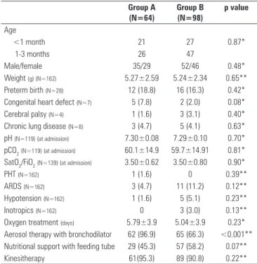
Table 1 - Characterization of the sample, severity criteria (at admission and during inpatient care) and treatment
Group A (N=64)
Group B (N=98)
p value
Age
<1 month 21 27 0.87*
1-3 months 26 47
Male/female 35/29 52/46 0.48*
Weight (g) (N=162) 5.27±2.59 5.24±2.34 0.65**
Preterm birth (N=28) 12 (18.8) 16 (16.3) 0.42*
Congenital heart defect (N=7) 5 (7.8) 2 (2.0) 0.08*
Cerebral palsy (N=4) 1 (1.6) 3 (3.1) 0.40*
Chronic lung disease (N=8) 3 (4.7) 5 (4.1) 0.63*
pH (N=119) (at admission) 7.30±0.08 7.29±0.10 0.70* pCO2(N=119) (at admission) 60.1±14.9 59.7±14.91 0.81* SatO2/FiO2(N=139) (at admission) 3.50±0.62 3.50±0.80 0.90*
PHT (N=162) 1 (1.6) 0 0.39**
ARDS (N=162) 3 (4.7) 11 (11.2) 0.12**
Hypotension (N=162) 1 (1.6) 5 (5.1) 0.23**
Inotropics (N=162) 0 3 (3.0) 0.13**
Oxygen treatment (days) 5.79±3.9 5.04±3.9 0.23*
Aerosol therapy with bronchodilator 62 (96.9) 65 (66.3) <0.001** Nutritional support with feeding tube 29 (45.3) 57 (58.2) 0.07**
Kinesitherapy 61(95.3) 89 (90.8) 0.22**
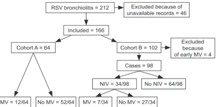
RSV bronchiolitis = 212
Included = 166
Cohort A = 64 Cohort B = 102
Excluded because of early MV = 4
Cases = 98
MV = 12/64 No MV = 52/64 MV = 7/34 No MV = 27/34
NIV = 34/98 No NIV = 64/98
Excluded because of unavailable records = 46
(FiO2) was calculated. his ratio provides a noninvasive alternative to the oxygenation index (arterial oxygen pressure, PaO2/FiO2).(16)
RSV infection was diagnosed with an immunochromatography test (Coris®) by identifying the virus in respiratory secretions.
Acute respiratory failure or apneas were indications of NIV.
To establish NIV, the unit protocol was used based on the NIV protocol that was published by the Respiratory Group at the Spanish Society of Pediatric Intensive Care.(17)
For NIV, conventional ventilators (Drager Babylog 8000 plus®) and speciic ventilators (Respironics BiPAP Vision®, Respironics BiPAP Harmony®, Infant Flow Driver® and Infant Flow Advance®) were used. NIV was applied in CPAP or BiPAP (Bilevel Positive Airway Pressure) modes according to the child’s age and the availability of a nasal mask, short binasal plug and nasopharyngeal tube (a cut endotracheal tube that is positioned in the nasopharynx).
When CPAP was used, the treatment began at 4 cmH2O and was progressively increased according to the patient’s need and tolerance. With BiPAP, an inspiratory pressure of 8 cmH2O was added and was progressively increased at 2-cmH2O intervals according to the patient’s need and tolerance. FiO2 was maintained at the level necessary to maintain saturations of greater than 94%. he variation in expiratory positive airway pressure (EPAP) and inspiratory positive airway pressure (IPAP) was maintained according to the clinical and blood gas analysis progression. A minimum respiratory frequency of 10 to 30 cycles per minute was maintained according to the child’s age.
In group A, the indications for invasive mechanical ventilation were acute respiratory failure or apneas. In group B, mechanical ventilation was used when NIV failed. NIV failure was deined as a lack of improvement or deterioration in the blood gas analysis parameters or the clinical state.
he diagnosis of bacterial co-infection was based on the presence of at least two of the following criteria: a fever of more than 38ºC, an increased C-reactive protein of greater than 5 mg/dL and a white blood cell count of greater than 15,000 mm3. hese criteria were also used to begin antibiotic treatment.
Bacterial pneumonia was considered when the diagnosis of bacterial co-infection involved consolidation in a chest radiography.
Demographic, clinical and blood gas variables were analyzed. For the statistical analysis, quantitative variables were expressed as means and standard deviations or
medians and maximum/minimum values. For categorical variables, chi-square statistics and Fisher’s exact tests were used; for continuous variables, Student’s t tests were used. Statistical signiicance was set at p<0.05. he software Statistical Package for the Social Sciences (SPSS: SPSS Inc., Chicago, IL, USA) version 16 was used.
RESULTS
Between November 2003 and March 2008, 212 children were admitted to the UCIEP with the diagnosis of RSV bronchiolitis and acute respiratory failure; 46 of the children were not included in the study because their medical records were unavailable. Of the remaining 166 children, 4 were excluded from group B because they underwent mechanical ventilation with initial ventilation support for cardiorespiratory arrest. Cohort A included 64 children, and cohort B included 98 children. he total sample included 162 children (Figure 1).
A total of 125 children (75%) were older than 3 months, and 53.7% of the children were male. he mean weight was 5.261±2.430 kg (minimum: 2.280 kg; maximum: 13.0 kg; median: 4.440 kg).
Medical histories identiied 28 cases of preterm birth (17.3%), 7 cases of congenital heart disease (4.3%), 4 cases of cerebral palsy (2.5%) and 7 cases of chronic lung disease (4.3%).
he distributions of the medical history variables and age were similar between the two cohorts (Table 1).
On admission, diferences between the groups for blood gas analysis (pH, pCO2), SatO2/FiO2 and the number of apneas were not statistically signiicant (Table 1).
he diferences between the groups in the number of cases of respiratory failure were not statistically signiicant. hese cases progressed to acute respiratory distress syndrome
(ARDS) (group A: 3/64 versus group B: 11/98; p=0.12), pulmonary hypertension (group A: 1/64 versus group B: 0/98; p=0.39), hypotension (group A: 1/64 versus group B: 5/98; p=0.23) and the need for inotropic support (group A: 0/64 versus group B: 3/98; p=0.13) (Table 1).
In group A, 12 children were invasively ventilated. he reasons for ventilation were apneas in two cases (16.6%) and acute respiratory failure in ten cases (83.3%). In the cases of acute respiratory failure, one case involved pulmonary hypertension, and three cases involved ARDS. All children were younger than 6 months, and three were newborn infants. Four children (33.3%) were preterm, one child (8.3%) had chronic lung disease and two children (16.7%) had a congenital heart disease.
At the moment of intubation, a blood gas analysis showed an average pH of 7.22±0.10 (minimum: 7.12;
maximum: 7.32), an average pCO2 of 76.1±16.6
mmHg (minimum: 59.5; maximum: 92.7) and an average oxygenation (using SatO2/FiO2) of 3.12±0.86 (minimum: 2.26; maximum: 3.98). Two infants in group A had episodes of apneas.
In group B, 34 children (33.3%) were noninvasively ventilated (Table 2). In 31 children (91.1%), the indications for NIV were acute respiratory failure with or without apneas. Apneas were present in 3 children (8.8%).
All of the 34 children who underwent NIV were younger than 6 months, and 15 were newborn infants. Five children (14.7%) were preterm, three children (8.8%) had chronic lung disease, one child (2.9%) had a congenital heart disease and one child (2.9%) had cerebral palsy.
A blood gas analysis showed an average pH of 7.25±0.3 (minimum: 6.7; maximum: 7.25), an average
PCO2 of 69.1±14 mmHg (minimum: 46.8; maximum:
118) and an average SatO2/FiO2 of 2.8±0.99 (minimum: 0.86; maximum: 4.33). Ten infants presented episodes of apneas.
he most frequently applied ventilator mode was BiPAP (20 cases), which was applied through the Infant Flow Advance® (11 cases), Respironics BiPAP Vision® (7 cases) and Respironics BiPAP Harmony® (2 cases). he CPAP modality was used in 14 cases through the Infant Flow Driver® (13 cases) and the Drager Babylog 8000 plus® (1 case). A nasopharyngeal tube was used only once, and a nasal interface (nasal mask or short binasal plug) was used in the remaining cases.
Invasive ventilation was necessary in 7 (20.5%) of the 34 children who underwent NIV. he indications for intubation were ARDS (2 cases), ARDS with hemodynamic instability (1 case), hemodynamic instability (3 cases) and cardiorespiratory arrest (1 case).
At the moment of intubation, these children presented an average pH of 7.17±0.24 (minimum: 6.93; maximum: 7.41), an average pCO2 of 84.5±17.4 mmHg (minimum: 67.1; maximum: 101.9) and an average SatO2/FiO2 of 2.4±1.6 (minimum: 0.79; maximum: 4.0).
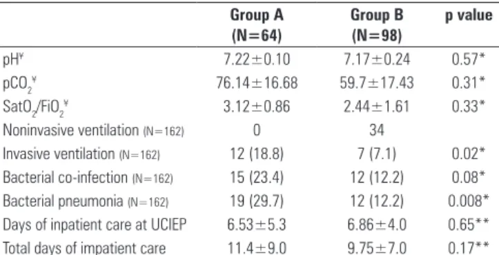
At the moment of intubation, the diferences in the blood gas analysis and SatO2/FiO2 values between groups A and B were not statistically signiicant (Table 2).
In group B, the number of children who required invasive ventilation was signiicantly lower than the
number in group A (group A: 12/64 versus group B:
7/98; p=0.02) (Table 2).
Regarding the response to treatment, the number of co-infections was similar in the two groups (group A: 15/46 versus group B: 12/98; p=0.08). However, in group B, there were fewer cases of bacterial pneumonia (group A: 19/64 versus group B: 12/98; p=0.008) (Table 2).
he average number of days of inpatient care at the UCIEP was 6.73±4.5 days (minimum: 1; maximum: 35). here were no statistically signiicant diferences in the durations of inpatient care between the two groups (Table 2).
here was no record of mortality in either of the groups.
DISCUSSION
Since Akingbola et al.’s 1993 publication of the irst paper about pediatric applications of NIV,(8) many case reports and case series have been published, and there have been many positive results for the use of NIV in several situations of respiratory failure.(6,13)
Several of these studies have been conducted in adults with chronic obstructive lung disease (COPD),
Table 2 - Clinical progression and average blood gas analysis and SatO2/FiO2
values before intubation
Group A (N=64)
Group B (N=98)
p value
pH¥ 7.22±0.10 7.17±0.24 0.57*
pCO2¥ 76.14±16.68 59.7±17.43 0.31*
SatO2/FiO2¥ 3.12±0.86 2.44±1.61 0.33*
Noninvasive ventilation (N=162) 0 34
Invasive ventilation (N=162) 12 (18.8) 7 (7.1) 0.02*
Bacterial co-infection (N=162) 15 (23.4) 12 (12.2) 0.08* Bacterial pneumonia (N=162) 19 (29.7) 12 (12.2) 0.008* Days of inpatient care at UCIEP 6.53±5.3 6.86±4.0 0.65** Total days of impatient care 11.4±9.0 9.75±7.0 0.17**
SatO2/FiO2 - oxygen saturation/fraction of inspired oxygen; UCIEP – pediatric intensive and special care unit. The results are expressed as frequencies (%) or means ± standard deviations. ¥Values for the 19 patients who were intubated, including12 in cohort A and 7 in
and they have demonstrated that NIV can reduce the rate of bacterial lung infection and improve survival.(18,19) Additionally, several studies have shown that NIV reduces the need for endotracheal intubation and the incidence of nosocomial infection.(20-22) Intubation and invasive ventilation are associated with high morbidity, as they are associated with local tissue lesions, pneumonia and extensions of hospital stay. By avoiding intubation, NIV has the potential to avoid these problems. Contrary to invasive ventilation, NIV leaves the respiratory tract intact, which preserves defense mechanisms and allows the patient to eat, speak and eliminate secretions.(23)
In the present study, two groups of children with the same disease and causal agent were compared. he only diference between the groups was that they had diferential access to NIV. When NIV was used, the need for intubation and the incidence of infectious complications decreased.
he present results are not explained by the inclusion of more severe cases in group A. In fact, the risk factors for severe RSV infection are pre-existing factors;(24,25) in the present sample, the distributions of age and the presence of preterm birth, chronic lung disease, a congenital heart defect and cerebral palsy were similar in the two groups. Additionally, there were no statistically signiicant diferences between the groups in the severity criteria, including the blood gas analysis at admission, the presence of a bacterial co-infection or the progression to ARDS or hypotension.
he co-infection rates were consistent with previous studies (group A: 23.4% versus group B: 12.2%).(26,27) he deinitions of co-infection and bacterial pneumonia were based on clinical, laboratory and radiographic criteria, rather than microbiological criteria. However, the same criteria were used for both groups, and the absence of microbiological criteria is justiied by the fact that identical criteria cannot be obtained for children who are invasively or noninvasively ventilated or who are not ventilated. Because the rate of bacterial co-infection was similar between the two groups at admission, the reduction of the number of children who were noninvasively ventilated most likely led to the occurrence of fewer infections related to the endotracheal tube.
he average duration of inpatient care at the UCIEP was similar to what has been described in other studies,(26) and there was no signiicant reduction in the number of days of inpatient care in group B. he length of hospital stay in children with bronchiolitis can be prolonged for several reasons, such as problems with feeding, social
problems and variation in discharge criteria.
he main limitations of this study were its retrospective design with comparisons between historical cohorts, thus limiting data collection and the losses of medical records.
CONCLUSION
In cases of severe RSV bronchiolitis, NIV was efective and avoided the need for conventional mechanical ventilation in a signiicant number of patients. NIV reduced the number of patients with bacterial pneumonia. hus, small infants with severe RSV bronchiolitis might beneit from the timely introduction of NIV.
RESUMO
Objetivos: Analisar se a ventilação não invasiva diminui a necessidade de intubação endotraqueal e se alterou a evolução clínica, relativamente a complicações infecciosas, da bronquioli-te por vírus sincicial respiratório com insuiciência respiratória.
Metodos: Estudo retrospectivo de coortes: cohorte A, de crianças internadas na unidade de cuidados intensivos e espe-ciais pediátrica antes da introdução da ventilação não invasiva (2003-2005); cohorte B, de crianças internadas após a intro-dução de ventilação não invasiva (2006-2008). Excluindo a ventilação não invasiva, a terapêutica de suporte foi igual nos dois grupos. Foram incluídas crianças com o diagnóstico de bronquiolite por vírus sincicial respiratório e insuiciência res-piratória entre novembro 2003 e março 2008. Analisaram-se variáveis demográicas, clínicas e gasimétricas.
Resultados: Incluídas 162 crianças, 75% com idade <3 meses. Grupo A: 64 crianças; Grupo B: 98 (34 necessitaram de ventilação não invasiva). Ambos os grupos apresentaram distribuição semelhante relativamente à idade, antecedentes de prematuridade, cardiopatia congénita, paralisia cerebral e doença pulmonar crónica. Na admissão, os valores da ga-simetria e o número de apneias não apresentaram diferenças
estaticamente signiicativas nos dois grupos. No Grupo B, o
número de crianças que necessitou de ventilação invasiva foi menor (Grupo A: 12 versus Grupo B: 7; p=0,02), veriicando--se uma diminuição do número de casos de pneumonia
bacte-riana (Grupo A:19/64 versus Grupo B:12/98; p=0,008). Não
se registou mortalidade.
Conclusão: Neste trabalho, comparando crianças com a mesma patologia, antes e depois da introdução de ventilação não invasiva como apoio ventilatório inicial, veriicou-se di-minuição das complicações infecciosas e da necessidade de entubação.
REFERENCES
1. American Academy of Pediatrics. Subcommittee on Diagnosis and Management of Bronchiolitis. Diagnosis and management of bronchiolitis. Pediatrics.2006;118(4):1774-93.
2. Martinón-Torres F, Rodríguez Núñez A, Martinón Sánchez JM. Bronquiolitis aguda: evaluación del tratamiento basada en la evidencia. An Pediatr (Barc). 2001;55(4):345-54.
3. Lopéz Guinea A, Casado Flores J, Martín Sobrino MA, Espínola Docio B, la Calle Cabrera T, Serrano A, et al. Bronquiolitis grave. Epidemiología y evolución de 284 pacientes. An Pediatr (Barc). 2007;67(2):116-22. 4. Mayordomo-Colunga J, Medina A, Rey C, Los Arcos M, Concha A,
Menéndez S. Predictores de éxito y de fracaso en la ventilación no invasiva en la bronquiolitis aguda. An Pediatr (Barc). 2009;70(1):34-9.
5. Mejías A, Chávez-Bueno S, Jafri HS, Ramilo O. Respiratory syncytial virus infections: old challenges and new opportunities. Pediatr Infect Dis J. 2005;24(11 Suppl):S189-96, discussion S196-7.
6. Davison C, Ventre KM, Luchetti M, Randolph AG. Efficacy of interventions for bronchiolitis in critically ill infants: a systematic review and meta-analysis. Pediatr Crit Care Med. 2004;5(5):482-9.
7. Corneli HM, Zorc JJ, Majahan P, Shaw KN, Holubkov R, Reeves SD, Ruddy RM, Malik B, Nelson KA, Bregstein JS, Brown KM, Denenberg MN, Lillis KA, Cimpello LB, Tsung JW, Borgialli DA, Baskin MN, Teshome G, Goldstein MA, Monroe D, Dean JM, Kuppermann N; Bronchiolitis Study Group of the Pediatric Emergency Care Applied Research Network (PECARN). A multicenter, randomized, controlled trial of dexamethasone for bronchiolitis. N Engl J Med. 2007;357(4):331-9. Erratum in N Engl J Med. 2008;359(18):1972. Majahan, Prashant [corrected to Mahajan, Prashant]. 8. Akingbola OA, Servant GM, Custer JR, Palmisano JM. Noninvasive
bi-level positive pressure ventilation: management of two pediatric patients. Respir Care. 1993;38:1092-8.
9. Thill PJ, McGuire JK, Baden HP, Green TP, Checchia PA. Noninvasive positive-pressure ventilation in children with lower airway obstruction. Pediatr Crit Care Med. 2002;5(4):337-42. Erratum in Pediatr Crit Care Med. 2004;5(6):590.
10. Díaz Lobato S, Mayoralas Alises S. Ventilación no invasiva. Arch Bronconeumol 2003;39(12):566-79.
11. Rimensberger PC. Noninvasive pressure support ventilation for acute respiratory failure in children. Schweiz Med Wochenschr. 2000;130(49):1880-6.
12. McNamara F, Sullivan CE. Nasal CPAP treatment in an infant with respiratory syncytial virus-associated apnea. Pediatr Pulmonol. 1997;24(3):218-21. 13. Nunes P, Abadesso C, Almeida E, Silvestre C, Loureiro H, Almeida H.
Ventilação não invasiva numa unidade de cuidados intensivos pediátricos. Acta Med Port. 2010;23(3):399-404.
14. Campion A, Huvenne H, Leteurtre S, Noizet O, Binoche A, Diependaele JF, et al. [Non-invasive ventilation in infants with severe infection presumably due to respiratory syncytial virus: feasibility and failure criteria]. Arch Pediatr. 2006;13(11):1404-9. French.
15. Larrar S, Essouri S, Durand P, Chevret L, Haas V, Chabernaud JL, et al. [Effects of nasal continuous positive airway pressure ventilation in infants with severe acute bronchiolitis]. Arch Pediatr. 2006;13(11):1397-403. 16. Khemani RG, Patel NR, Bart RD 3rd, Newth CJ. Comparison of the pulse
oximetric saturation/fraction of inspired oxygen ratio and the PaO2/fraction of inspired oxygen ratio in children. Chest. 2009;135(3):662-8.
17. Medina A, Pons M, Esquinas A. Ventilación no invasiva en pediatria. Barcelona: Ergón; 2004. Disponible en http://secip.blogspot.com/search/ label/Libros%20de%20Interes.
18. Girou E, Schortgen F, Delclaux C, Brun-Buisson C, Blot F, Lefort Y, et al. Association of noninvasive ventilation with nosocomial infections and survival in critically ill patients. JAMA. 2000;284(18):2361-7.
19. Antonelli M, Conti G, Rocco M, Bufi M, De Blasi RA, Vivino G, et al. A comparison of noninvasive positive-pressure ventilation and conventional mechanical ventilation in patients with acute respiratory failure. N Engl J Med. 1998;339(7):429-35.
20. Nourdine K, Combes P, Carton MJ, Beuret P, Cannamela A, Ducreaux JC. Does noinvasive ventilation reduce the ICU nosocomial infection risk? A prospective clinical survey. Intensive Care Med. 1999;25(6):567-73. 21. Peter JV, Moran JL, Phillips-Hughes J, Warn D. Noninvasive ventilation
in acute respiratory failure--a meta-analysis update. Crit Care Med. 2002;30(3):555-62.
22. Lightowler JV, Wedzicha JA, Elliot MW, Ram FS. Non-invasive positive pressure ventilation to treat respiratory failure resulting from exacerbations of chronic obstructive pulmonary disease: Cochrane systematic review and meta-analysis. BMJ. 2003;326(7382):185.
23. Mehta S, Hill NS. Noninvasive ventilation. Am J Respir Crit Care Med. 2001;163(2):540-77. Review.
24. Wang EE, Law BJ, Stephens D. Pediatric Investigators Collaborative Network on Infections in Canada (PICNIC) prospective study of risk factors and outcomes in patients hospitalized with respiratory syncytial viral lower respiratory tract infection. J Pediatr.1995;126(2):212-9.
25. Hernando Puente A, López-Herce Cid A, Bellón Cano JM, Villaescusa JU, Santiago Lozano MJ, Sánchez Galindo A. Factores pronósticos de evolución complicada en la bronquiolitis que requiere ingreso en cuidados intensivos pediátricos. An Pediatr (Barc). 2009;70(1):27-33.
26. Duttweiler L, Nadal D, Frey B. Pulmonary and systemic bacterial co-infections in severe RSV bronchiolitis. Arch Dis Child. 2004;89(12):1155-7. 27. Thorburn K, Harigopal S, Reddy V, Taylor N, van Saene HK. High incidence