ISSN: 2167-7964
OMICS Journal of Radiology
The International Open Access
OMICS Journal of Radiology
Executive Editors
Abass Alavi
University of Pennsylvania, USA
Donghoon Lee
University of Washington, USA
Chun Lam James CHOW
University of Toronto, Canada
Rashid A. Fawwaz
Columbia University, USA
Bensheng Qiu
University of Washington, USA
T
his article was originally published in a journal by OMICS
Publishing Group, and the attached copy is provided by OMICS
Publishing Group for the author’s beneit and for the beneit of
the author’s institution, for commercial/research/educational use
including without limitation use in instruction at your institution,
sending it to speciic colleagues that you know, and providing a copy
to your institution’s administrator.
All other uses, reproduction and distribution, including without
limitation commercial reprints, selling or licensing copies or access,
or posting on open internet sites, your personal or institution’s
website or repository, are requested to cite properly.
Review Article Open Access
Dulce Antunes1* and Teresa Margarida Cunha2
1Centro Hospitalar Lisboa Norte–Hospital St Maria, Lisbon, Portugal 2Instituto Português de Oncologia de Lisboa Francisco Gentil, Lisbon, Portugal
Keywords:
Uterine cervical carcinoma; Radiation therapy; CT scans; MR imagingIntroduction
Cervical carcinoma still represents a signiicant public health problem. Around 11,069 cases of invasive cervical cancer were diagnosed in the United States in 2008 and 3869 women died from the disease. Worldwide, cervical cancer has an even greater impact, with nearly 529,828 new cases and 275,128 deaths reported annually [1] .
Patients with early stage cervical cancer treated with surgery or radiotherapy are likely to have a good prognosis [2]. However, substantial treatment failure still occurs and about 30% of the patients treated for cervical carcinoma develop progressive or recurrent tumors. Patients who sufer recurrence have a dismal prognosis with a 1-year survival rate of between 15 and 20% [2].
Recurrent cervical cancer is deined as local tumor re-growth or the development of nodal or distant metastases at least 6 months ater the lesion has regressed. Residual disease is that which is evident within 6 months of primary treatment. he average rate of recurrence is that approximately two-thirds of cases recur within the irst 2 years following initial treatment, and 90% of cases recur by 5 years [3].
he risk of treatment failure depends on the initial staging and, consequently, on the chosen therapeutics. Risk factors for recurrence of cervical carcinoma include the histological type, size, depth of stromal invasion, and the nodal status at presentation.
Pelvic recurrence can locate centrally or laterally in the pelvis. Extra-pelvic recurrence most commonly involves para-aortic lymph nodes, lungs, liver and bone [4].
Imaging Surveillance
he early detection of recurrent disease in the pelvis may pose a diagnostic challenge, because symptoms are nonspeciic, physical examination of the irradiated pelvis is oten limited and cytological examination of the irradiated cervix has a poor sensitivity [5]. For these reasons and given that the therapeutic options for recurrence of cervical carcinoma depend on the location and extent of the recurrence, imaging can be crucial for early diagnosis and adequate management [4].
Abstract
Uterine cervical carcinoma is one of the most common malignancies occurring in females and although the longer survival provided by the advances in early diagnosis and effective treatment, 30% of the patients develop persistent or
recurrent disease. Recurrent disease is deined by local tumor re-growth or development of nodal or distant metastases
at least 6 months after the lesion has regressed. Recurrent disease can occur centrally in the uterus or vaginal vault,
laterally in the pelvic wall, can present as pelvic and extra-pelvic lymph node disease and as distant metastases.
Although CT can be useful for surveillance MR is the most accurate imaging tool for characterization of pelvic
recurrence. MR indings depend on previous therapeutics so the knowledge of imaging features of surgery and of the irradiated pelvis are crucial. Dynamic contrast-enhanced subtraction MR and diffusion weighted images give a valuable contribute in differential diagnosis of pelvic recurrence and inlammatory effects of radiation therapy but biopsy and serial imaging may be warranted.
The authors review the spectrum of imaging indings of recurrent cervical carcinoma.
*Corresponding author: Dulce Antunes, Centro Hospitalar Lisboa Norte–Hospital
St Maria, Lisbon, Portugal, E-mail:[email protected]
Received May 06, 2013; Accepted August 12, 2013; Published August 19, 2013
Copyright: © 2013 Antunes D, et al. This is an open-access article distributed
under the terms of the Creative Commons Attribution License, which permits
unrestricted use, distribution, and reproduction in any medium, provided the original author and source are credited.
Recurrent Cervical Cancer: How Can Radiology be Helpfull
Citation: Antunes D, Cunha TM (2013) Recurrent Cervical Cancer: How Can Radiology be Helpfull. OMICS J Radiology 2: 138 doi:10.4172/2167-7964.1000138
here are no formal recommendations based on a good level of evidence for the follow up of the patients treated for cervical carcinoma [6].
Mabuchi et al. showed that routine physical history, physical examination, and CT scans were the most common irst abnormal ttests leading to 86% of the recurrent disease diagnosis [7]. CT is an efective diagnostic tool for detection of recurrent tumor [8], however has little use in diferentiating recurrent tumor from post-therapeutic changes [9]. here is no consensus in the reviewed literature regarding the indication of pelvic MR for the follow-up of patients with medically treated cervical carcinoma and MR is usually performed when there is a clinical suspicion of recurrence. Ater surgery there is also no consensus for a systematic MR follow up with an exception for trachelectomy. Patients submitted to trachelectomy should be evaluated by MR at 6 months and 1 year given the higher recurrence rate comparing to patients who undergo brachytherapy or chemotherapy [10].
Normal Post-herapeutical Imaging Findings
Imaging evaluation of the treated cervical carcinoma depends on the knowledge of the normal post-therapeutical aspects ater surgery and chemo-radiation therapy. Usually follow up MR is not indicated during the irst 6 months ater completion of radiation therapy due to limitations of MR in diferentiating recurrent tumor from post-operative and post-radiation changes [9].
Page 2 of 6
Citation:Antunes D, Cunha TM (2013) Recurrent Cervical Cancer: How Can Radiology be Helpfull. OMICS J Radiology 2: 138 doi: 10.4172/2167-7964.1000138
upper third of the vagina, excision of parametrial and paravaginal tissue, including the sacro-uterine ligaments, and dissection of pelvic and para-aortic lymph nodes.
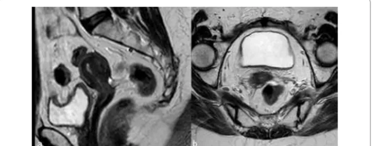
he vaginal fornix is represented, both on CT and MR, by a sot-tissue linear structure [11] (Figure 1). MR has the advantages of multiplanar evaluation of the fornix and better deinition of the vaginal wall. Normal vaginal vault has a strongly hypointense muscular wall in T2WI, with well-deined and regular contour. Frequently there is ibrotic scar tissue whose T2WI signal is slightly higher than the normal muscular tissue.
Laparoscopic vaginal radical trachelectomy is a fertility-preserving alternative to radical hysterectomy or chemoradiation in young women with stage IA2 to IB1 cervical cancers [12]. he technique involves a proximal vaginotomy, cervical resection, and paracervical and paravaginal dissection. he cervix is resected, with preservation of the corpus uteri and an end-to-end anastomosis with the remaining vagina. Bilateral pelvic lymphadenectomy is also performed [13].
he imaging indings of the uterus ater trachelectomy include the end-to-end anastomosis of the uterine corpus to the vaginal vault (Figure 2), frequently with a posterior neofornix that can present with nodular coniguration. hese MR features should be known to avoid false positive diagnosis of recurrent tumor.
he para-vaginal dissection may cause vaginal wall thickening that is more prominent in the irst 6 months and slowly diminishes till 1 year. Sometimes there is also loss of the low signal intensity on T2WI and this difuse thickening can be mistaken for recurrent
disease iniltrating the vaginal wall [13].
In the evaluation of patients submitted to radiation therapy MR is more efective than CT [8]. In postmenopausal patients, radiation therapy does not result in any signiicant changes of the uterus at MR imaging. In premenopausal patients the junctional zone and outer myometrium become indistinct and, ater 6 months, the endometrium becomes thin and hypointense [14].
Tumors treated with RT respond with a decrease in size and signal intensity on MR imaging (Figure 3). he response may be immediate (3-6 months) or, in larger tumors, delayed (6-9 months) [15]. Hricak et al. reviewed the MR images of 69 patients submitted to radiotherapy to cervical cancer and reported that the reconstitution of the normal zonal anatomy of the cervix and homogeneous cervical low signal have high negative predictive value (97%) for recurrence [14]. An early (2–3 months) and signiicant decrease in the signal intensity and volume of the tumor indicates a positive response to radiation therapy and a high probability of complete remission [16].
Tumor Recurrence
he accuracy of MR for the diagnosis of recurrence ater radiotherapy depends on the elapsed time since the ending of the therapeutics. In the irst 6 months necrosis, inlammation and edema contribute to the higher signal intensity on T2WI making diferential diagnosis with tumoral disease harder [17].
It is consensual that dynamic contrast-enhanced subtraction MR imaging for the diferentiation of benign from malignant lesions is advisable for recurrence detection.
Difusion-weighted MR also gives a valuable contribute for the evaluation of the irradiated cervix [18]. Hyperintense signal on high b-value images associated with lower ADC values are suggestive of recurrent tumor.
Multiple studies found signiicant increases in lesion ADC values in patients treated with combined chemo-radiation therapy, however the post-therapeutical values might be inferior to normal cervical tissue (due to the presence of edema, hyaline degeneration and granulation tissue). Chen et al. found that the diference of ADC values between normal cervical tissue and cervical area ater therapy was statistically signiicant (P < 0.01) and reported that the optimal ADC threshold values for distinguishing between cervical area before and ater therapy is 1.255×10⁻³ mm2/s (sensitivity of 95.5% and
speciicity of 100%) and between normal cervical tissue and cervical area ater therapy is 1.525×10⁻³ mm2/s (sensitivity and speciicity of
70% and 81.8%, respectively) [19].
Weber et al. found that the most helpful inding for diferentiating recurrent cervical tumor from radiation ibrosis was the presence of
Figure 1: Sagittal (a) and axial T2WI (b): the vaginal vault is visualized as a
linear soft tissue structure.
Figure 2: Twenty six year old woman with FIGO IB1 cervical cancer, submitted
to trachelectomy. Two years later sagittal T2WI (a and b) depict the utero-vaginal anastomosis and the regularity and low signal intensity of the utero-vaginal wall, better characterized after distension with ultrasound gel (b).
Figure 3: First MR evaluation after inishing chemoradiation, Sagittal (a) and
Page 3 of 6
a sot-tissue mass with higher signal intensity than that of adjacent pelvic sidewall muscle on T2WI [20] and Hricak et al. documented that these indings carry a recurrence probability of 86% [14]. However the recurrent tumor may show difuse growth making diferential with post-therapeutics scar more diicult [9].
Recurrence exhibits an earlier and more pronounced enhancement compared with scar tissue, on condition that MR is not performed earlier than 6 months ater the end of therapy [9].
Findings like widened endocervical canal and cervical difuse high signal intensity are nonspeciic and MR might not be conclusive (Figure 4).
he parametrium sot tissues may also undergo ibrotic changes and appear hypointense. his post-radiation imaging appearance may mimic parametrial invasion [21] thus becoming indistinguishable from the tumor. he intravenous administration of contrast material and DWI images can also help distinguishing between radiation-induced parametrial ibrosis and residual or recurrent disease [19].
When morphological and functional MR evaluation is inconclusive biopsies may be warranted (Figure 5). Roy et al. [22] showed transrectal ultrasound guided biopsy to be a feasible, safe and accurate method for establishing a histopathological diagnosis of recurrence and CT guided biopsy of lymph nodes and metastases is a current procedure (Figure 6).
In addition, serial or follow-up MR imaging is useful for distinguishing recurrent disease from radiation-induced ibrosis, as the latter is expected to remain stable or diminish over time [8].
Most cervical tumors are 18F-luorodeoxyglucose (FDG) avid, with exception to adenocarcinomas, which may reveal low FDG uptake. Revision of studies using FDG-PET/CT in the evaluation of
recurrence/metastases in patients with uterine cancer revealed high diagnostic accuracy with 90-93% sensitivity, 81-100% speciicity and 87-96% accuracy [23]. It is especially useful for the assessment of peritoneal dissemination, lymph node metastases, local recurrence, and bone/muscle metastases and may have the greatest utility in situations in which tumor marker levels are rising and conventional imaging studies show negative or equivocal indings [3,23].
Central Tumor Recurrence
About 30-45% of recurrences ater radical hysterectomy are central-pelvic-type, generally situated above the vaginal cuf, between the bladder and the rectum (Figure 7). Five-year survival rates reported by literature for these central recurrences vary from 6% to 77% . Patients with a central recurrence seem to have a better prognosis compared to those with recurrence of the pelvic wall [24].
Central recurrence can afect the cervix, vagina, parametria and ovaries and grow to involve the bladder, rectum, or side wall. Ater radical hysterectomy most tumors show supra-vaginal recurrence (20%) at the vaginal stump (Figure 8) and at the recto-vaginal space [9]. Skip lesions in the vagina may be seen in recurrent cervical cancer, in which case lesions may be seen in the distal vagina (Figure 9). Patterns of involvement of the vagina follow the growth pattern of the primary cervical cancer (eg, as a mass- like lesion or as an iniltration along the vaginal wall) [25].
Figure 4: First MR evaluation after inishing chemoradiation. Sagittal (a)
and axial T2WI (b) reveal a small cervix, predominantly hypointense. A slight central hyperintensity is noticed but attributed to inlammatory effects of radiation. These indings were stable in subsequent evaluations.
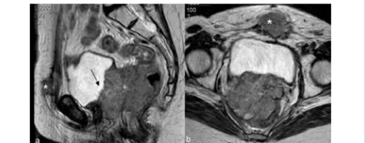
Figure 5: Uterine cervical carcinoma FIGO IIB that inished chemoradiation
4 months before. In the sagittal (a) and axial (b) T2WI a central area of high signal intensity is identiied in the cervix (arrow). This area could be related to post-radiation inlammatory effects (note the prominence of peri-rectal adipose tissue and low signal intensity striation of the parametria). Although it presents slight high signal intensity in b-1000 DWI (c) and signs of water restriction in the ADC map (d) (dashed arrows). Biopsies conirmed tumoral
recurrence.
Figure 6: Cervical cancer FIGO IIIB submitted do chemoradiation. Twelve
months after inishing therapy surveillance CT (a, b - axial contrast enhanced CT) revealed para-aortic lymph node enlargement (a) and a solid nodule in the vaginal vault (arrow in b). The vaginal vault nodule was biopsied under trans-rectal ultrasound guidance (c) and the para-aortic lymph node submitted to CT-guided biopsy (d). Both revealed neoplastic squamous cells according
to central and lymph node recurrence.
Figure 7: Uterine cervical tumor FIGO IIB (a-sagittal T2 WI) treated with
chemoradiation. A year after inishing therapy MR (b and c-sagittal and axial T2WI) show central cervical recurrence without signs of parametrial invasion.
Figure 8: Vaginal vault recurrence. Axial (a) and sagittal (b) T2WI images
show a small low signal intensity solid tumor in the vaginal vault (arrow). This lesion is high signal intensity on DWI with b-1000 (c) and has low signal
Page 4 of 6
Citation:
Antunes D,
Cunha TM
(2013) Recurrent Cervical Cancer: How Can Radiology be Helpfull. OMICS J Radiology 2: 138 doi: 10.4172/2167- 7964.1000138
Citation:Antunes D, Cunha TM (2013) Recurrent Cervical Cancer: How Can Radiology be Helpfull. OMICS J Radiology 2: 138 doi: 10.4172/2167-7964.1000138
Kim et al. reported a higher incidence of central recurrence among patients treated by radiation therapy than by surgery [26].
he most common CT and MR imaging feature of recurrent tumor of the primary site is an irregular mass averaging 4-7 cm in diameter between the bladder and rectum [26]. Recurrent tumor may be recognized on CT scans obtained with intravenous contrast material, on which it may appear as a sot-tissue mass with diminished enhancement compared with that of normal cervical tissue [8]. On MR recurrence has high signal intensity on T2WI, similar to the corresponding primary tumor [8] (Figure 10).
Hydrometra, depicted by CT and MR as a symmetrically enlarged uterine corpus containing non-enhanced luid [9], can be associated with central recurrence due to the obstruction of the internal os by the tumor. However it can also be due to radiogenic ibrosis and its appreciation depends on the deinition of a tumoral mass (Figure 11).
Central pelvic recurrence with anterior extension may lead to ureteral obstruction by direct encasement of the ureter or by tumor iniltration of the bladder wall, which results in obstruction at the ureteral oriice [9].
Although the loss of the adipose cleavage plan of the tumoral mass with bladder and rectum rise the suspicion of visceral invasion the imaging diagnosis depends on the identiication of luminal tumoral mass. he MR imaging indings of bladder invasion include nodularity and irregularity of the bladder wall, masses protruding into the bladder lumen, high signal intensity of the anterior aspect of the posterior bladder wall, and abnormal sot-tissue strands in the utero-vesical space [27]. Invasion of the rectum usually occurs at the recto-sigmoid junction and is detected by mass efect, spiculation, and luminal narrowing (Figure 12) [28].
Pelvic Side Wall Recurrence
Pelvic side wall recurrence is characterized either by direct invasion of the mass to the pelvic side-wall by means of a linear strand of sot tissue and involving loss of the fat planes, or by conluence of the solid mass and the muscle of the pelvic wall [27]. At times pelvic recurrences are identiied as pelvic side wall masses that are not associated with a centrally located pelvic mass [28].
Recurrent tumor of the pelvic sidewall may manifest with pain due to nerve iniltration or leg edema due to obliteration of the lymphatics [9].
he imaging criteria are the same used for the initial staging: the distance tumor–side wall is less than 3 mm; the piriformis and obturator internus muscles can be enlarged and demonstrate an enhancing sot-tissue mass; the iliac vessels can be encased and narrowed by tumor; direct extension and destruction of the pelvic bones may occur.
Kim et al. reported that there is no signiicant diference in the incidence of pelvic side-wall extension between patients who undergo surgery and those treated by radiation therapy [26].
Lymph Node Recurrence
In uterine cervical carcinoma lymphatic involvement has traditionally been separated into primary and secondary nodal groups. he irst of these consists of the paracervical, parametrial, internal and external iliac, and obturator nodes, while those comprised by the second include the sacral, common iliac, inguinal and para-aortic [29]. In general, paracervical and parametrial lymph nodes are involved irst, followed by the obturator nodes, the remaining external iliac nodes, and the internal iliac nodes. Extra-pelvic lymph nodes, including the supra-clavicular, para-aortic (Figure 13), and
Figure 9: Distal vaginal recurrence. Patient submitted to chemoradiation for
cervical carcinoma. Nodularity of distal vagina was noticed. Sagittal (a) and axial (b) T2WI reveal parietal thickening of the lower third of the vagina with high signal intensity on the T2WI. Recurrence of the cervical carcinoma was conirmed by vaginal biopsy. The cervix had the usual post-therapy aspects (small, diffuse low signal intensity) and no signs of cervical recurrence were identiied.
Figure 10: Cervical uterine carcinoma FIGO IIA1 treated with chemoradiation.
Sagittal T2WI shows the primary tumor (a) with high signal intensity suggestive of hyper-cellularity. Fourteen months later sagittal T2WI (b) reveals cervical recurrence and patient was submitted to hysterectomy. Sixteen months later (c-sagittal and d-axial T2WI) pelvic RM depicts a vaginal vault recurrence with
the same signal characteristics of the initial tumor.
Figure 11: About 2 years after chemoradiation for cervical cancer, surveillance
CT found hydrometra and MR was made for better characterization. Sagittal T2WI (a) depicts a high cervical recurrence causing obstruction of the cervix, with hydrometra. Axial T2WI (b and c) reveal recurrent tumor in the cervix with obstructing the internal os and hydrometra. The tumor doesn’t invade the parametria but the left parametrium is strongly hypointense (arrow in b) and there is left ureteral dilatation (arrowhead in c). These indings, as also the rectum and sigmoidal parietal thickening, were assigned to inlammatory
effects of radiation.
Figure 12: Central recurrence of uterine cervical carcinoma after hysterectomy
Page 5 of 6
inguinal, are common sites of distant metastases. he prevalence of recurrence at extra-pelvic lymph nodes is higher in patients treated with radiation therapy than in those who undergo surgery; this is probably because this tumor stage is generally more advanced. Multiple extra-pelvic and extra-abdominal nodal sites of recurrence, including the para-bronchial, supra-clavicular, and axillary nodes, have been reported but are less frequently involved than the primary and secondary groups [8].
Yang et al. compared the results of CT, MR and pathological evaluation of 949 lymph nodes in 43 women with biopsy proven cervical cancer and concluded that helical CT and MRI imaging showed similar accuracy in lymph node evaluation (89.5% and 85.5%, respectively) [18].
he indings of nodal metastases range from scattered, minimally enlarged lymph nodes to large, conglomerate nodal masses at CT and MR imaging [8]. Although enlarged lymph nodes may be hyperplastic and lymph nodes smaller than 1 cm may contain metastatic disease, nodes with a transverse diameter greater than 10 mm are usually considered malignant. Central necrosis within a lymph node is a strong predictor of metastatic disease (positive predictive value of
100%) [18,30]. Recent studies showed the potential of DW imaging to diferentiate benign from malignant pelvic lymph nodes based on their underlying mean ADC value [31].
Distant Recurrent Disease
Ater the pelvis and lymph nodes, the solid organs of the abdomen are the most frequent sites of involvement of recurrent cervical carcinoma [28].
Liver metastases are present in one-third of patients and appear as solid masses with variable enhancement. Adrenal metastases are usually from cervical adenocarcinomas and are present in approximately 15% of patients. Peritoneal metastases appear as sot-tissue masses and ascites and are present in 5%–27% of cases in autopsy series (Figure 14) [15,28].
Imachi et al. documented pulmonary metastases in 6% of 817 patients with uterine cervical carcinoma and 81% of those had local recurrence or other distant metastatic lesions [29]. Lymphangitic carcinomatosis is seen in less than 5% of patients and appears as difuse interstitial lung disease [25]. Mediastinal or hilar adenopathy and pleural lesions or efusions are present in approximately one-third of patients with metastatic disease to the chest [15].
he prevalence of osseous metastases (Figure 15) in the setting of recurrent cervical carcinoma ranges from 15% to 29%. he vertebral bodies are the most frequently involved bones, followed by the pelvis, ribs, and extremities. he most common mechanism of bone involvement is by direct extension of neoplasm from para-aortic nodes into the adjacent vertebral bodies (Figure 13). It can also occur by lymphatic or hematogenous spread or from direct extension of pelvic recurrence. Bone metastases may appear as destructive lesions associated with sot-tissue masses of variable size. he absence of a sot tissue mass, slow progression, blastic elements, and sharply deined borders on CT suggest radiation necrosis [30]. In some lesions gadolinium-enhanced fat-suppressed T1WI is useful for the diagnosis by the deinition of foci of enhancement within the marrow space [28].
Conclusion
Uterine cervical cancer recurrence is common (about two thirds within the irst 2 years) and carries a dismal prognosis.
CT and MRI play key roles in identifying recurrent disease. Although both have reasonable sensitivities [3] it is generally accepted that MR is superior in distinction between tumor and post-radiotherapy inlammatory and ibrotic changes. Dynamic contrast-enhanced subtraction and difusion weighted MR imaging are advisable. PET/CT has been considered a highly accurate imaging technique for restaging [23].
Knowledge of the wide spectrum of imaging indings ater surgery and radiotherapy is crucial for evaluation ater cervical cancer therapy. Reconstitution of the normal zonal anatomy of the cervix and homogeneous cervical low signal have high negative predictive value (97%) for recurrence. he most speciic sign of central recurrence is a hyperintense sot-tissue mass (T2WI) with earlier and pronounced enhancement. Imaging criteria of pelvic wall recurrence are the same used for the initial staging (distance tumor–side wall is less than 3 mm). Published studies show a certain degree of overlap between benign and malignant pelvic lymph nodes, similar accuracy for CT and MR and have not been able to deine the negative predictive value of DW imaging [31]. Lung, bone and liver are the most common locations of distance recurrence [32].
Figure 13: Lymph node recurrence. Non-enhanced CT depicting left
para-aortic lymph node conglomerate with invasion of the psoas muscle and
erosion of the adjacent vertebral body.
Figure 14: Uterine cervical cancer FIGO IIB treated with chemoradiation. Surveillance CT (axial non-enhanced CT) reveals hydrometra (a), ascites (a)
and peritoneal (b) and pulmonary (c) metastases.
Figure 15: Uterine cervical carcinoma. Sagittal T2WI (a) shows the primary
Page 6 of 6
References
1. Ferlay J, Shin HR, Bray F, Forman D, Mathers C, et al. (2008) GLOBOCAN
cancer incidence and mortality Worldwide: IARC CancerBase No. 10.
Available from International Agency for Research on Cancer, Lyon, France . 2. Long HJ 3rd (2007) Management of metastatic cervical cancer: review of the
literature. J Clin Oncol 25: 2966-2974.
3. Liyanage SH, Roberts CA, Rockall AG (2010) MRI and PET scans for primary
staging and detection of cervical cancer recurrence. Womens Health (Lond Engl) 6: 251-267.
4. Gadducci A, Tana R, Cosio S, Cionini L (2010) Treatment options in recurrent
cervical cancer (Review). Oncol Lett 1: 3-11.
5. Nishie A, Stolpen AH, Obuchi M, Kuehn DM, Dagit A, et al. (2008) Evaluation
of locally recurrent pelvic malignancy: performance of T2- and diffusion-weighted MRI with image fusion. J Magn Reson Imaging 28: 705-713.
6. Kew FM, Roberts AP, Cruickshank DJ (2005) The role of routine follow-up
after gynecological malignancy. Int J Gynecol Cancer 15: 413-419.
7. Mabuchi S, Isohashi F, Maruoka S, Hisamatsu T, Takiuchi T, et al. (2012)
Post-treatment follow-up procedures in cervical cancer patients previously treated with radiotherapy. Arch Gynecol Obstet .
8. Jeong YY, Kang HK, Chung TW, Seo JJ, Park JG (2003) Uterine cervical carcinoma after therapy: CT and MR imaging indings. Radiographics 23: 969-981.
9. Hamm B, Forstner R, Baert A, Knauth M, Sartor K(2007) MRI and CT of the female pelvis. Medical Radiology: Diagnostic imaging. Springer Verlag, New York: 166-172.
10. Balleyguier C, Sala E, Da Cunha T, Bergman A, Brkljacic B, et al. (2011)
Staging of uterine cervical cancer with MRI: guidelines of the European Society of Urogenital Radiology. Eur Radiol 21: 1102-1110.
11. Sugimura K, Okizuka H (2002) Postsurgical pelvis: treatment follow-up.
Radiol Clin North Am 40: 659-680, viii.
12. Chen Y, Xu H, Zhang Q, Li Y, Wang D, et al. (2008) A fertility-preserving
option in early cervical carcinoma: laparoscopy-assisted vaginal radical trachelectomy and pelvic lymphadenectomy. Eur J Obstet Gynecol Reprod Biol 136: 90-93.
13. Sahdev A, Jones J, Shepherd JH, Reznek RH (2005) MR imaging
appearances of the female pelvis after trachelectomy. Radiographics 25:
41-52.
14. Hricak H, Swift PS, Campos Z, Quivey JM, Gildengorin V, et al. (1993)
Irradiation of the cervix uteri: value of unenhanced and contrast-enhanced MR imaging. Radiology 189: 381-388.
15. Pannu HK, Corl FM, Fishman EK (2001) CT evaluation of cervical cancer:
spectrum of disease. Radiographics 21: 1155-1168.
16. Flueckiger F, Ebner F, Poschauko H, Tamussino K, Einspieler R, et al.
(1992) Cervical cancer: serial MR imaging before and after primary radiation therapy-a 2-years follow-up study. Radiology 184: 89-93.
17. Kinkel K, Ariche M, Tardivon AA, Spatz A, Castaigne D, et al. (1997)
Differentiation between recurrent tumor and benign conditions after treatment of gynecologic pelvic carcinoma: value of dynamic contrast-enhanced subtraction MR imaging. Radiology 204: 55-63.
18. Yang WT, Lam WW, Yu MY, Cheung TH, Metreweli C (2000) Comparison of
dynamic helical CT and dynamic MR imaging in the evaluation of pelvic lymph
nodes in cervical carcinoma. AJR Am J Roentgenol 175: 759-766.
19. Chen J, Zhang Y, Liang B, Yang Z (2010) The utility of diffusion-weighted MR
imaging in cervical cancer. Eur J Radiol 74: e101-106.
20. Weber TM, Sostman HD, Spritzer CE, Ballard RL, Meyer GA, et al. (1995)
Cervical carcinoma: determination of recurrent tumor extent versus radiation changes with MR imaging. Radiology 194: 135-139.
21. Addley HC, Vargas HA, Moyle PL, Crawford R, Sala E (2010) Pelvic imaging
following chemotherapy and radiation therapy for gynecologic malignancies. Radiographics 30: 1843-1856.
22. Roy D, Kulkarni A, Kulkarni S, Thakur MH, Maheshwari A, et al. (2008)
Transrectal ultrasound-guided biopsy of recurrent cervical carcinoma. Br J Radiol 81: 902-906.
23. Kitajima K, Murakami K, Kaji Y, Sakamoto S, Sugimura K (2011) Established,
emerging and future applications of FDG-PET/CT in the uterine cancer. Clin Radiol 66: 297-307.
24. Peiretti M, Zapardiel I, Zanagnolo V, Landoni F, Morrow CP, et al. (2012) Management of recurrent cervical cancer: a review of the literature. Surg Oncol 21: e59-66.
25. Parikh JH, Barton DP, Ind TE, Sohaib SA (2008) MR imaging features of
vaginal malignancies. Radiographics 28: 49-63.
26. Kim JE, Park HA, Kim KH, Lim D, Chin SY (1988). Patterns of recurrent cervical carcinoma on CT. J Korean Radiol Soc 24:1130-1134.
27. Choi JI, Kim SH, Seong CK, Sim JS, Lee HJ, et al. (2000) Recurrent uterine
cervical carcinoma: spectrum of imaging indings. Korean J Radiol 1:
198-207.
28. Fulcher AS, O’Sullivan SG, Segreti EM, Kavanagh BD (1999) Recurrent cervical carcinoma: typical and atypical manifestations. Radiographics 19 Spec No: S103-116.
29. Imachi M, Tsukamoto N, Matsuyama T, Nakano H (1989) Pulmonary
metastasis from carcinoma of the uterine cervix. Gynecol Oncol 33: 189-192.
30. Walsh JW, Amendola MA, Hall DJ, Tisnado J, Goplerud DR (1981) Recurrent carcinoma of the cervix: CT diagnosis. AJR Am J Roentgenol 136: 117-122.
31. Thoeny HC, Forstner R, De Keyzer F (2012) Genitourinary applications of
diffusion-weighted MR imaging in the pelvis. Radiology 263: 326-342.
32. Engin G (2006) Cervical cancer: MR imaging indings before, during, and
after radiation therapy. Eur Radiol 16: 313-324.
Submit your next manuscript and get advantages of OMICS Group submissions
Unique features:
• User friendly/feasible website-translation of your paper to 50 world’s leading languages • Audio Version of published paper
• Digital articles to share and explore
Special features:
• 250 Open Access Journals • 20,000 editorial team • 21 days rapid review process
• Quality and quick editorial, review and publication processing
• Indexing at PubMed (partial), Scopus, EBSCO, Index Copernicus and Google Scholar etc • Sharing Option: Social Networking Enabled
• Authors, Reviewers and Editors rewarded with online Scientiic Credits
• Better discount for your subsequent articles
Submit your manuscript at: http://www.omicsonline.org/submission
Citation:Antunes D, Cunha TM (2013) Recurrent Cervical Cancer: How Can Radiology be Helpfull. OMICS J Radiology 2: 138 doi: 10.4172/2167-7964.1000138
Citation: Antunes D, Cunha TM (2013) Recurrent Cervical Cancer: How Can Radiology be Helpfull. OMICS J Radiology 2: 138 doi: