Review article
Malformações Müllerianas: revisão da abordagem atual
Sérgio Conti Ribeiro
1, Renata Assef Tormena
2, Thais Villela Peterson
2, Marina de Oliveira Gonzáles
3,
Priscila Gonçalves Serrano
3, José Alcione Macedo de Almeida
4, Edmund Chada Baracat
5Department of Gynecology and Obstetrics, Faculdade de Medicina da Universidade de São Paulo (FMUSP), São Paulo, Brazil
1MD, PhD. Head of Laparoscopic Surgery Group, Department of Gynecology and Obstetrics, Faculdade de Medicina da Universidade de São Paulo (FMUSP), São Paulo, Brazil. 2MD. Attending physician, Laparoscopic Surgery Group, Department of Gynecology and Obstetrics, Faculdade de Medicina da Universidade de São Paulo (FMUSP), São Paulo, Brazil. 3Medical student, Faculdade de Medicina da Universidade de São Paulo (FMUSP), São Paulo, Brazil.
4MD, PhD. Head of Pediatric and Adolescent Group, Department of Gynecology and Obstetrics, Faculdade de Medicina da Universidade de São Paulo (FMUSP), São Paulo, Brazil. 5MD, PhD. Chief professor, Department of Gynecology and Obstetrics, Faculdade de Medicina da Universidade de São Paulo (FMUSP), São Paulo, Brazil.
ABSTRACT
The aim of this paper was to discuss the embryological aspects of Müllerian duct anomalies and to analyze the current diagnostic methods and therapy. Müllerian anomalies are congenital defects of the female reproductive tract resulting from failure in the development of the Müllerian ducts and their
associated structures. Their cause has yet to be fully clariied, and it is currently believed to be multifactorial. Symptoms appear principally during adolescence or early adulthood, and affect the reproductive capacity of these women. When clinically suspected, investigations leading to diagnosis
include imaging methods such as hysterosalpingography, ultrasonography and magnetic resonance. The classiication of these malformations relates to their embryogenesis, and deines the therapy and prognosis. Müllerian anomalies consist of a wide range of defects that may vary from patient to patient.
Therefore, their management must also be individual, taking anatomical and clinical characteristics into consideration, as well as the patient’s wishes.
RESUMO
O objetivo deste trabalho foi discutir as malformações müllerianas desde seus aspectos embriológicos, analisando os atuais métodos diagnóstico e
terapêuticos. As malformações müllerianas são anomalias congênitas do trato reprodutivo feminino decorrentes de falha do desenvolvimento dos ductos de Müller e estruturas associadas. Sua causa não foi completamente elucidada, acreditando-se, atualmente, que seja multifatorial. Os sintomas se
manifestam, principalmente, durante a adolescência e início da vida adulta, e afetam a capacidade reprodutiva dessas mulheres. A partir da suspeita clínica, a investigação diagnóstica inclui métodos de imagem, como a histerosalpingograia, ultrassonograia e ressonância magnética. A classiicação
das malformações está relacionada à sua embriogênese e direciona a terapêutica e prognóstico. As malformações müllerianas são um grupo amplo de anomalias que variam de paciente para paciente. Portanto, sua abordagem também é individual, devendo-se considerar os aspectos anatômicos,
clínicos e o desejo da paciente. KEY WORDS:
Urogenital abnormalities.
Müllerian ducts. Embryonic development.
Laparoscopy. Infertility.
Gynecology.
PALAVRAS-CHAVE: Anormalidades urogenitais.
Ductos de Müller. Desenvolvimento embrionário.
Laparoscopia. Infertilidade.
INTRODUCTION
Müllerian duct anomalies consist of a set of structural malforma-tions resulting from abnormal development of the paramesonephric or Müllerian ducts. The prevalence of these anomalies ranges from 0.001 to 10% in the general population and from 8-10% in women with an adverse reproductive history.1,2 The embryological development of the
female reproductive system is closely related to the development of the urinary system, and anomalies in both systems may occur in up to 25% of these patients.3 Other associated malformations may affect the
gas-trointestinal tract (12%) or musculoskeletal system (10-12%).3,4
Gonad formation begins between the fifth and sixth weeks of preg-nancy, with the appearance of the urogenital ridge developing from the intermediate mesoderm and the migration of the germinative cells orig-inating in the coelomic epithelium. Female development is determined by the absence of the Y chromosome (and consequent absence of the factor that determines testicle development) and by the presence of two X chromosomes.3,4
At around the ninth week, the ovaries are formed and the Wolff-ian (mesonephric) and MüllerWolff-ian ducts coexist. Absence of testosterone leads to involution of the Wolffian duct, whereas absence of anti-Mül-lerian hormone allows differentiation of the Mülanti-Mül-lerian duct. The cau-dal portions of these ducts merge to form the uterovaginal canal, which later gives rise to the cervix and uterus, as well as to the upper third of the vagina. Complete development of the vagina occurs through fusion of the structures of the urogenital sinus and the Müllerian tubercle. The cranial portion of the duct, i.e. the part that does not fuse, opens into the peritoneal cavity, giving rise to the Fallopian tubes.3,4
The causes of Müllerian anomalies have yet to be fully clarified. The karyotypes are normal (46 XX) in 92% of the women with Mül-lerian anomalies and abnormal (sex chromosome mosaicism) in 8% of these women. The majority of these developmental abnormalities are infrequent and sporadic, and are thus attributed to polygenic and multifactorial causes.5 A recent study attributed persistence of the
in-trauterine septum to a deficiency in the antiapoptotic protein Bcl 2, which is responsible for the process of apoptosis and absorption of the septum.5
Events such as hypoxia that occur during pregnancy, the use of medications such as methotrexate or diethylstilbestrol (DES), ionizing radiation and viral infections may also contribute towards the occur-rence of Müllerian malformations.6,7 Among the drugs that induce
Mül-lerian malformations, thalidomide and DES lead to malformations of the uterine cavity. DES, a nonsteroidal estrogen, was widely used in the 1950s, principally in the United States, for treating various obstetri-cal conditions, particularly miscarriage and preeclampsia. Consequent-ly, anomalies consisting notably of vaginal adenocarcinoma and defor-mations of the uterine cavity (specifically, T-shaped uterus) were iden-tified in the daughters of women who had taken this medication.8 A
study carried out on rats exposed intrauterinely to DES reported modi-fications to Dkk2, Nkd2, sFRP1, hox, Wnt and Eph gene expression. These have been correlated with changes induced by this drug.9
In a study on cases of congenital uterine and vaginal agenesis (May-er-Rokitansky-Küster-Hauser syndrome), possible mutations and
poly-morphisms were evaluated in the anti-Müllerian hormone gene and in the gene for its receptor. Analysis of the hox genes (the genes relating to embryogenesis) and the N314D gene (the gene that codifies the en-zyme galactose-1-phosphate uridyl transferase [GALT] for galactose me-tabolism) failed to find any positive correlation with the presence of malformations.10,11
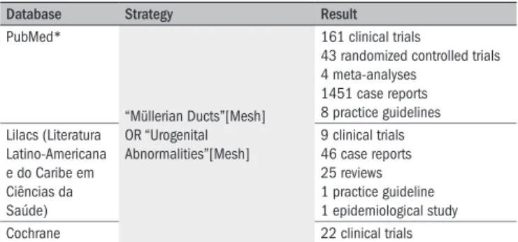
The aim of this study was to discuss embryology, diagnostic methods and therapy in cases of Müllerian duct anomalies. We searched PubMed (National Library of Medicine, Bethesda, Maryland, United States) by using the following terms: “Müllerian Ducts”[Mesh] OR “Urogenital Abnormalities”[Mesh]. The search strategy is described in Table 1.
PHYSIOPATHOLOGY
Uterine malformations result from failure in organogenesis or from fusion or reabsorption of the Müllerian ducts. Failures in organogen-esis are related to incomplete development of one or both Müllerian ducts, thereby leading to agenesis, uterine hypoplasia or a unicornu-ate uterus.3
Fusion defects result from incomplete merging of the caudal por-tion with the Müllerian ducts (lateral fusion) or incomplete merging of the structures of the urogenital sinus with the Müllerian tubercle (verti-cal fusion). Failures in lateral fusion may result in uterus didelphys, bi-cornuate uterus or arcuate uterus. When the defect occurs in vertical fu-sion, anomalies such as imperforate hymen, transverse vaginal septum, oblique vaginal septum or absence of the cervix may result. Following caudal fusion of the ducts, the remaining portion of the central septum is reabsorbed. Reabsorption failure results in a uterus with a partial or complete septum. Certain malformations of the uterine cavity also lead to the formation of hypoplastic uterus, infantile uterus, agenesis of the cervix and T-shaped uterus.3
CLASSIFICATION
The definition of uterine anomalies proposed by the American Fer-tility Society12 classifies uterine malformations into seven separate
cat-egories:
Class I: Hypoplasia/uterine agenesis. Class II: Unicornuate uterus:
a) Has a functioning endometrium and communication with the main uterine cavity.
Database Strategy Result
PubMed*
“Müllerian Ducts”[Mesh] OR “Urogenital Abnormalities”[Mesh]
161 clinical trials
43 randomized controlled trials 4 meta-analyses
1451 case reports 8 practice guidelines
Lilacs (Literatura Latino-Americana e do Caribe em Ciências da Saúde)
9 clinical trials 46 case reports 25 reviews 1 practice guideline 1 epidemiological study
Cochrane 22 clinical trials
*Limits: added to PubMed in the last 10 years; Female; English; Portuguese.
b) Also has an endometrial structure that responds to hormonal stim-ulus; however, there is no communication with the external genital tract.
c) Has a rudimentary structure with no activity, attached to a more fully developed uterine horn.
d) Results from the development of only one Müllerian duct, with complete agenesis of the contralateral duct.
Class III: Uterus didelphys. Class IV: Bicornuate uterus:
a) Complete: when the indentation produced in the fundic region is deep, thus indicating that fusion failed from the level of the cervical region.
b) Incomplete: when the division is higher, not extending to the level of the cervix, indicated by the shallower indentation in the contour of the region of the uterine fundus.
Class V: Septate uterus:
a) When the septum extends into the internal cervical ostium, possi-bly including the cervical canal, and divides the cervix into two tun-neled cavities. A vaginal septum is often also present.
b) When the septum does not divide the uterine cavity along its entire length, and circulation exists between the two chambers.
Class VI: Arcuate uterus:
This is a rather insignificant anomaly of the uterine cavity in which, generally, no abnormalities in the external contour of the uterus are visi-ble. The small fundal cleft or impression with a protruding uterine horn becomes more noticeable during pregnancy.
Class VII: T-shaped uterus resulting from the use of DES.
CLINICAL IMPLICATIONS
Müllerian anomalies are frequently asymptomatic and are often missed in routine gynecological examinations. Nevertheless, a history of pelvic pain following the menarche, dysmenorrhea and an increase in abdominal volume are complaints suggestive of uterine anomalies. In addition, primary amenorrhea and changes to menstrual flows may be present.13
Among the ductal differentiation malformations, vaginal agenesis presents with primary amenorrhea and dyspareunia. In cases of uteri with a functional endometrium, hematometra and hematocolpos are frequent findings.
A unicornuate uterus is seldom symptomatic unless associated with other malformations. If a rudimentary, noncommunicating uterine horn is present together with a functional endometrium, hematometra and sometimes hematosalpinges may be found.14
The clinical presentation of anomalies associated with defects in fu-sion and reabsorption of the septum varies clinically according to the
Miscarriage (%)
Premature delivery (%)
Fetal survival (%)
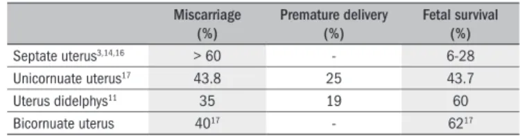
Septate uterus3,14,16 > 60 - 6-28
Unicornuate uterus17 43.8 25 43.7
Uterus didelphys11 35 19 60
Bicornuate uterus 4017 - 6217
Table 2. Reproductive prognosis
duct segment affected. The presence of a vaginal septum is perceived by patients as an obstacle to sexual relations and may be confirmed by speculum examination. When the menstrual flow is obstructed, patients may report pain.12
Uterine septum is generally an asymptomatic condition and is often only diagnosed when couples with a history of repeated miscarriage or infertility are undergoing investigation. Likewise, lateral fusion defects, which are responsible for uterus didelphys and bicornuate uterus, are of-ten detected only when women undergo imaging tests.12
Anomalies resulting from failure in vertical fusion, such as cervical agenesis, transverse vaginal septum and imperforated hymen, are associ-ated with primary amenorrhea, hematocolpos and hematometra.15
The reproductive prognosis for patients with Müllerian malforma-tions is shown in Table 2.3,12,16-19 The hypotheses developed to explain
the poor obstetrical prognosis for women with the diverse Müllerian malformations include decreased intraluminal volume; inadequate vas-cularization of regions such as the septum; presence of a medial wall or an unfused uterine horn; and greater uterine contractility and irritabil-ity, thereby leading to miscarriages and premature deliveries.12
DIAGNOSIS
Due to the complexity of presentations, diagnosing of Müllerian malformations requires the use of more than one imaging method in 62% of the cases.20,21 Hysterosalpingography (HSG) is the method
tra-ditionally used to evaluate the cervical canal, uterine cavity and Fallo-pian tubes. Its efficacy in diagnosing anomalies is debatable and varies according to the specific type of malformation.20 The specificity values
range from 6 to 60%, depending on the malformation investigated and the technician’s skill.1,22 HSG enables accurate evaluation of tube
per-meability and can detect the presence of uterine septa, intrauterine syn-echiae, submucous fibroids and endometrial polyps. However, it cannot be performed on patients who are virgins and it does not allow the ex-ternal uterine anatomy to be viewed, which hampers the differential di-agnosis between uterus didelphys and septate uterus.3,22,23 Moreover, the
method exposes the patient to ionizing radiation; the injection of con-trast may cause allergies and discomfort; and there is also a risk of uter-ine perforation and infection.1
Ultrasonography has a sensitivity of around 44%, varying accord-ing to the specific type of malformation under evaluation, the patient’s body composition, the radiologist’s experience and the type of transduc-er used. Transvaginal ultrasonography allows a more detailed analysis of the endometrium, uterine cavity and cervix. The specificity of this ex-amination ranges from 85 to 92%.24-26 Recently, three-dimensional
ul-trasonography has shown high specificity and sensitivity in evaluations on all uterine anomalies, including Müllerian malformation.27
The specificity of magnetic resonance imaging (MRI) ranges from 96 to 100% for diagnosing Müllerian malformations.1 In cases of
uter-ine hypoplasia, the images show a small endometrial cavity and a short-er distance between the utshort-erine horns.20 In these patients, the presence
if there is no communication with the cervical canal, the accumulated retrograde menstruation will be easily seen.20
Uterus didelphys is visible in axial sections, which show two sepa-rate chambers. However, the anatomy of the wall is intact. A vaginal septum may or may not be present.20
The MRI criteria for diagnosing a bicornuate uterus consist of the presence of divergent uterine horns and concavity at the contour of the uterine fundus. This type of anomaly appears as a heart-shaped uterus. On the other hand, in cases of septate uterus, the external con-tour of the uterus is normal and the septum is seen as a difference in signal intensity, according to its composition. Fibrous septa are seen as low-intensity signals on T2-weighted images and muscular septa as in-termediate-intensity T2 signals.28 Nowadays, cytogenetic analysis on
pa-tients with developmental anomalies of the Müllerian ducts may only be useful for family counseling.29
THERAPEUTIC MANAGEMENT
The treatment for Müllerian anomalies varies according to the spe-cific type of malformation found in each patient. A systematic search (descriptor: Müllerian anomalies or uterine anomalies, default tag: Ti-tle/Abstract) was conducted in the Lilacs, PubMed and Cochrane data-bases. Two randomized controlled trials were identified.30,31 Most of the
studies on therapeutic issues (827 studies) were restricted to reports on single cases or small series.
Anomalies of the vaginal septum should be resected at the time of diagnosis, thereby resolving problems of dyspareunia and permitting adequate drainage of menstrual flow.32
Patients with a confirmed diagnosis of cervical agenesis should be referred for hysterectomy, preferentially performed laparoscopically. Several surgical attempts to create a cervix have resulted in tragic out-comes, often associated with fatal complications.19 The prospects for
pregnancy using in vitro fertilization techniques should be evaluated
in the light of the obstetrical complications, and possible alternatives should be offered to these women. The use of a surrogate womb may be the best option in such cases. In cases of uterine septum, the resec-tion should be performed hysteroscopically, in order to improve the re-productive prognosis for these patients by decreasing the incidence of miscarriage, premature delivery and infertility.17 The advantages of
hys-teroscopy include the shorter duration of surgery, smaller blood loss, lower costs, reduced morbidity and shorter hospital stay, compared with abdominal surgery.30,31
In cases of complete uterine septum, resection of the cervical sep-tum may be related to cervical incompetence and secondary infertility. A randomized controlled trial performed to evaluate the safety and ef-ficacy of resection of the cervical septum during hysteroscopic metro-plasty showed that this procedure was safer and easier with resection than with preservation of the cervical septum.32
The use of estrogen therapy or an intrauterine device are post-surgical alternatives for minimizing formation of uterine adherences (synechiae).32,33 The follow-up in these cases includes hysteroscopy, one
to three months after the initial surgery.31 Laparoscopy should be used
to excise obstructed, rudimentary uterine horns and adjacent tubes in
patients with a unicornuate uterus. It should also be used for hysterec-tomy in cases of cervical agenesis and in neovaginoplasty procedures in cases of vaginal agenesis.19 In many women, the malformation results
in obstructed and retrograde menstruation, thereby facilitating the de-velopment of endometriosis. During laparoscopy, this diagnosis may be confirmed and the endometrial foci may be resected.
CONCLUSIONS
Müllerian anomalies consist of a wide range of defects that may vary from patient to patient. Therefore, their management must also be indi-vidual, taking anatomical and clinical characteristics into consideration, as well as the patient’s wishes.
REFERENCES
1. Pui MH. Imaging diagnosis of congenital uterine malformation. Comput Med Imaging Graph. 2004;28(7):425-33.
2. Propst AM, Hill JA 3rd. Anatomic factors associated with recurrent pregnancy loss. Semin Reprod Med. 2000;18(4):341-50.
3. Golan A, Langer R, Bukovsky I, Caspi E. Congenital anomalies of the müllerian system. Fertil Steril. 1989:51(5):747-55.
4. Li S, Qayyum A, Coakley FV, Hricak H. Association of renal agenesis and mullerian duct anomalies. J Comput Assist Tomogr. 2000;24(6):829-34.
5. Rackow BW, Arici A. Reproductive performance of women with müllerian anomalies. Curr Opin Obstet Gynecol. 2007;19(3):229-37.
6. Homer HA, Li TC, Cooke ID. The septate uterus: a review of management and reproductive outcome. Fertil Steril. 2000;73(1):1-14.
7. Sharara FI. Complete uterine septum with cervical duplication, longitudinal vagi-nal septum and duplication of a revagi-nal collecting system. A case report. J Reprod Med. 1998;43(12):1055-9.
8. Milhan D. DES exposure: implications for childbearing. Int J Childbirth Educ. 1992;7(4):21-8.
9. Suzuki A, Urushitani H, Sato T, et al. Gene expression change in the Müllerian duct of the mou-se fetus expomou-sed to diethylstilbestrol in utero. Exp Biol Med (Maywood). 2007;232(4):503-14.
10. Burel A, Mouchel T, Odent S, et al. Role of HOXA7 to HOXA13 and PBX1 genes in various for-ms of MRKH syndrome (congenital absence of uterus and vagina). J Negat Results Biomed. 2006;5:4.
11. Klipstein S, Bhagavath B, Topipat C, Sasur L, Reindollar RH, Gray MR. The N314D polymor-phism of the GALT gene is not associated with congenital absence of the uterus and vagina. Mol Hum Reprod. 2003;9(3):171-4.
12. The American Fertility Society classiications of adnexal adhesions, distal tubal occlusion, tubal occlusion secondary to tubal ligation, tubal pregnancies, müllerian anomalies and intrauterine adhesions. Fertil Steril. 1988;49(6):944-55.
13. Fedele L, Bianchi S, Frontino G. Septums and synechiae: approaches to surgical correction. Clin Obstet Gynecol. 2006;49(4)767-88.
14. Johansen K. Pregnancy in a rudimentary horn. Two case reports. Obstet Gynecol. 1969;34(6):805-8.
15. Nawroth F, Schmidt T, Freise C, Foth D, Römer T. Is it possible to recommend an “optimal“ postoperative management after hysteroscopic metroplasty? A retrospective study with 52 infertile patients showing a septate uterus. Acta Obstet Gynecol Scand. 2002;81(1):55-7. 16. Green LK, Harris RE. Uterine anomalies. Frequency of diagnosis and associated obstetric
complications. Obstet Gynecol. 1976;47(4):427-9.
17. Heinonen PK. Reproductive performance of women with uterine anomalies after abdomi-nal or hysteroscopic metroplasty or no surgical treatment. J Am Assoc Gynecol Laparosc. 1997;4(3):311-7.
18. Raga F, Bauset C, Remohi J, Bonilla-Musoles F, Simón C, Pellicer A. Reproductive impact of congenital Müllerian anomalies. Hum Reprod. 1997;12(10):2277-81.
19. Acién P. Reproductive performance of women with uterine malformations. Hum Reprod. 1993;8(1):122-6.
20. Scarsbrook AF, Moore NR. MRI appearances of müllerian duct abnormalities. Clin Radiol. 2003;58(10):747-54.
anomalies in adults: evaluation of practice. Fertil Steril. 2008;89(1):219-22.
22. Braun P, Grau FV, Pons RM, Enguix DP. Is hysterosalpingography able to diagnose all uterine malformations correctly? A retrospective study. Eur J Radiol. 2005;53(2):274-9. 23. Sørensen SS. Hysteroscopic evaluation and endocrinological aspects of women with
mülle-rian anomalies and oligomenorrhea. Int J Fertil. 1987;32(6):445-52.
24. Doyle MB. Magnetic resonance imaging in müllerian fusion defects. J Reprod Med. 1992;37(1):33-8.
25. Pellerito JS, McCarthy SM, Doyle MB, Glickman MG, DeCherney AH. Diagnosis of uterine anomalies: relative accuracy of MR imaging, endovaginal sonography, and hysterosalpingo-graphy. Radiology. 1992;183(3):795-800.
26. Ozsarlak O, De Schepper AM, Valkenburg M, Delbeke L. Septate uterus: hysterosalpingogra-phy and magnetic resonance imaging indings. Eur J Radiol. 1995;21(2)122-5. 27. Ferreira AC, Mauad Filho F, Nicolau LG, Gallarreta FMP, Paula WM, Gomes DC.
Ultra-sonogra-ia tridimensional em ginecologUltra-sonogra-ia: malformações uterinas. [Three-dimensional ultrasound in gynecology: uterine malformations]. Radiol Bras. 2007;40(2):131-6.
28. Imaoka I, Wada A, Matsuo M, Yoshida M, Kitagaki H, Sugimura K. MR imaging of disorders associated with female infertility: use in diagnosis, treatment, and management. Radiogra-phics. 2003;23(6):1401-21.
29. Regev M, Kirk R, Mashevich M, Bistritzer Z, Reish O. Vertical transmission of a mutation in exon 1 of the WT1 gene: lessons for genetic counseling. Am J Med Genet A. 2008;146A(18):2332-6. 30. Fayez JA. Comparison between abdominal and hysteroscopic metroplasty. Obstet Gynecol.
1986;68(3):399-403.
31. Colacurci N, De Franciscis P, Mollo A, et al. Small-diameter hysteroscopy with Versapoint versus resectoscopy with a unipolar knife for the treatment of septate uterus: a prospective randomized study. J Minim Invasive Gynecol. 2007;14(5):622-7.
32. Parsanezhad ME, Alborzi S, Zarei A, et al. Hysteroscopic metroplasty of the complete uterine septum, duplicate cervix, and vaginal septum. Fertil Steril. 2006;85(5):1473-7. 33. Vercellini P, Fedele L, Arcaini L, Rognoni MT, Candiani GB. Value of intrauterine device
insertion and estrogen administration after hysteroscopic metroplasty. J Reprod Med. 1989;34(7):447-50.
Sources of funding: None Conlict of interest: None
Date of irst submission: June 2, 2008 Last received: March 20, 2009
Accepted: March 23, 2009
Address for correspondence:
Sérgio Conti Ribeiro
Rua Joaquim Floriano, 466 — Conjunto 708 — Itaim Bibi São Paulo (SP) — Brasil — CEP 04534-002 Tel/Fax. (+55 11) 3079-5050