ABSTRACT
ORIGINAL AR
INTRODUCTION Adequate postoperative analgesia is consi-dered to be a major key for reducing pul monary morbidity and improving the outcome.l-3 Thoracic epidural anesthesia (TEA) has been proposed as a reliable analgesic method follo-wing thoracic surgery.3 Most of these benefi ts, however, relate to the postoperative or to the so-called perioperative period. Some case reports have suggested that sympathetic block secon dary to thoracic or spinal anesthesia might cause impaired intraoperative ventilation by increasing airway resistance and bronchial reactivity.4,5
A similar controversy surrounds some aspects of respiratory mechanics in anesthe-tized humans. Although previous studies have demonstrated that systemic local anesthetic secondary to TEA does not alter airway resis-tance in humans, and even attenuates bron-chial hyperreactivity,6,7 the effects of thoracic sympathetic block on respiratory mechanics, particularly on its compliance, have been poorly explained.
Pressure-volume curves are a feasible method for studying respiratory system mechanics, and the use of a low-fl ow infl a-tion technique has been established as a reliable and quick method for obtaining these curves.8,9
The remaining doubts are whether the ad-vantageous postoperative effects of TEA begin intraoperatively, and whether these benefi ts outweigh the undesirable intraoperative effects of thoracic sympathetic block.
OBJECTIVE To evaluate the effects of intraoperative thoracic sympathetic block on the elastic, viscoelastic and resistive properties of the respiratory system and its components (chest wall and pulmonary parenchyma), through analysis of pressure-volume curves obtained under quasi-static conditions.
METHODS
Patient selection
Patient selection
Patients requiring pulmonary segmental resection, pulmonary lobectomy, pulmonary biopsy or mediastinal nodular resection were eligible for the study. Those classifi ed on the American Society of Anesthesiologists (ASA) scale as having a physical status score of 3 or higher were excluded, as were those presenting with moderate or severe obstruc-tive respiratory disease, or any degree of restrictive respiratory disease, as diagnosed by spirometry and/or clinical signs. Written informed consent was obtained from each patient, and the study was approved by the hospital’s Ethics Committee.
Patients were randomly assigned to one of two anesthetic solutions that were injected into the epidural space: 15 ml of 0.5% bupivacaine plus epinephrine 1:200,000 and 2 mg mor-phine chlorohydrate (bupivacaine group), or 15 ml 0.9% NaCl plus epinephrine 1:200,000 and 2 mg morphine chlorohydrate (placebo group). Randomization and blindness were achieved by having an anesthesiologist who was not involved in the study make the draw for the two solutions by means of a randomiza-tion table that identifi ed the patients.
Anesthesia
Anesthesia
All patients were administered epidural and general anesthesia. They were premedi-cated with 10 mg of diazepam orally the night before surgery and 0.1 mg/kg of midazolam intramuscularly 45 minutes before surgery. Once in the operating room, the electrocardio-gram (CB-5), arterial pressure, pulse oximetry and temperature were monitored.
Epidural anesthesia was performed in the T7-T8 interspace by the loss-of-resistance technique, with patients in the sitting position. If a loss of resistance could not be achieved in
Anesthesia Department, Hospital das Clínicas, Faculdade de Medicina da
Universidade de São Paulo, São Paulo, Brazil
CONTEXT AND OBJECTIVE: Thoracic epidural an-esthesia (TEA) following thoracic surgery presents known analgesic and respiratory benefi ts. How-ever, intraoperative thoracic sympathetic block may trigger airway hyperreactivity. This study weighed up these benefi cial and undesirable effects on intraoperative respiratory mechanics.
DESIGN AND SETTING: Randomized, double-blind clinical study at a tertiary public hospital.
METHODS: Nineteen patients scheduled for partial lung resection were distributed using a random number table into groups receiving active TEA (15 ml 0.5% bupivacaine, n = 9) or placebo (15 ml 0.9% saline, n = 10) solutions that also contained 1:200,000 epinephrine and 2 mg morphine. Under general anesthesia, fl ows and airway and esophageal pressures were recorded. Pressure-volume curves, lower infl ection points (LIP), resistance and compli-ance at 10 ml/kg tidal volume were established for respiratory system, chest wall and lungs. Student’s t test was performed, including confi -dence intervals (CI).
RESULTS: Bupivacaine rose 5 ± 1 dermatomes upwards and 6 ± 1 downwards. LIP was higher in the bupivacaine group (6.2 ± 2.3 versus 3.6 ± 0.6 cmH2O, p = 0.016, CI = -3.4 to -1.8). Respiratory system and lung compliance were higher in the placebo group (respectively 73.3 ± 10.6 versus 51.9 ± 15.5, p = 0.003, CI = 19.1 to 23.7; 127.2 ± 31.7 versus 70.2 ± 23.1 ml/cmH2O, p < 0.001, CI = 61 to 53). Resistance and chest wall compliance showed no difference.
CONCLUSION: TEA decreased respiratory system compliance by reducing its lung component. Resistance was unaffected. Under TEA, positive end-expiratory pressure and recruitment maneu-vers are advisable.
this interspace, attempts were made to use the T8-T9 and T9-T10 interspaces. Patients for whom locating the epidural space continued to be impossible after these three approaches were excluded from the study, in order to avoid a different spread of local anesthetic within the epidural space. Once the epidural space was located, the assigned solution was injected and an epidural catheter positioned in order to allow for postoperative analgesia. The patient was then maintained in the supine position for 30 mi nutes. Following this, an anesthesiologist who was not involved in the study and was unaware which solution had been injected tested the level of the block by applying a thermal bilateral stimulus to the mid-axillary and mid-clavicular lines, from the cervical region to the pubis. If there was a difference between the right and left sides, the lower anesthetic spread was recorded.
After the extent of TEA was assessed, the patient received 100% oxygen by means of a facemask for five minutes. Anesthesia was induced using 2 mg/kg of propofol and 0.5µg/kg of sufentanil citrate, and oral tracheal intubation was performed by using a 37 left endobronchial tube (Smith Industries Medical Systems Inc./Portex, Keene, New Hampshire, United States), facilitated by 0.1 mg/kg of ve-curonium bromide. Correct tube positioning was checked by means of fi beroptic bronchos-copy. Anesthesia was maintained using con-tinuous infusion pumps (ANNETM Anesthesia Infuser, Abbott Laboratories, Chicago, Illinois, United States) for propofol, vecuronium and sufentanil and, when necessary, small boluses of these same agents.
After induction, radial and right atrial pressures were invasively measured via intra-vascular catheters.
During these and the subsequent pro-cedures, the patients were ventilated using a circulating system with CO2 absorber con-nected to the anesthesia machine (Intermed Linea anesthesia apparatus, São Paulo, Brazil) in volume controlled mode, square-wave (constant) fl ow of 30 l/min, respiratory rate of 10 breaths per minute, tidal volume of 8 ml per kilo and positive end-expiratory pressure (PEEP) level of 5 cmH2O. The fresh gas fl ow composition was a mixture of air and oxygen in equal parts. Inspired and expired gas analy-sis was performed with a Capnomac Ultima respiratory monitor (Datex Instrumentarium, Helsinki, Finland).
Respiratory mechanics data
Respiratory mechanics data acquisition
acquisition
Data acquisition on respiratory mechanics was performed prior to the start of surgery.
Im-mediately before each measurement, the airways were cleaned in order to remove accumulated mucus. Thus, ventilation was stopped, the fresh gas manifolds were closed, and the ventilator was adjusted as follows: volume-controlled mode, square-waveform (constant) flow of 6 l/min, tidal volume of 1000 ml, respiratory rate of three breaths per minute, and zero posi-tive end-expiratory pressure (ZEEP). An inspi-ratory pause of fi ve seconds was applied after this tidal volume was reached, in order to obtain a plateau and determine the resistance.
The resistance relative to the tracheal can-nula was measured by connecting the proximal end of the cannula to the anesthesia machine Y-piece, with a pneumotachograph inserted between them and the distal end of the can-nula left open, as described previously.10 This value was removed from the peak airway pressure before analysis.
Total resistance (Rmax), minimum resis-tance (Rmin) and additional resisresis-tance (DR) were determined for the respiratory system, chest wall and lungs using previously described methods.7,11-13 Rmin refl ects the opposition to air fl ow through the airways in the respiratory system(Rmin, rs), chest wall (Rmin, w) and lung parenchyma (Rmin, L). DR represents the additional resistance secondary to volume redistribution and/or tissue relaxation fol-lowing airway fl ow cessation in the respiratory system (DR, rs), chest wall (DR, w) and lung parenchyma (DR, L).12
Airway pressure (Paw) and inspiratory and expiratory fl ows were measured using a vari-able-area pneumotachograph (Bicore CP-100 respiratory monitor, Irvine, California, United States). The sensor (Var-Flex Flow Trans-ducer, Allied Healthcare, California, United States) was inserted between the proximal tip of the endobronchial tube and the Y-piece. For each patient, the anesthesia apparatus fl ow controls were calibrated by means of a Timeter RT-200 (Allied Healthcare, California, United States) to ensure that the set fl ows were abso-lutely correct during measurements.
Esophageal pressure was measured using an air-fi lled catheter (SmartCath Esophageal Catheter, BEAR Medical Systems, California, United States) inserted orally and connected to the Bicore CP-100 monitor. Catheter posi-tioning in the lower third of the esophagus was confi rmed by means of the occlusion test.14
Tidal volumes were obtained by integra-tion of the fl ow curve.
Data formatting and analysis
Data formatting and analysis
The analog Bicore signals were recorded in ASCII format on a PC (IBM Computers,
São Paulo, Brazil) by using an analog-to-digital converter (CAD 12 bit/32 channels, Lynx, São Paulo, Brazil) for one minute at 200 Hz. The fi les were converted to Excel for Windows 2000 format (Microsoft, São Paulo, Brazil) before analysis. Analysis of the fl ow curve allowed determination of the beginnings of the inspiratory and expiratory phases, as well as the beginning and end of the inspiratory pause, in accordance with a previous study.10 The fl ow values were double-checked by ob-serving the inspiratory time on the pressure curve (the 1000 ml tidal volume had to be reached in exactly 10 seconds to assure a fl ow equal to 100 ml/s).
Intrinsic PEEP (PEEPi), which was con-sidered to be any pressure measured at zero fl ow, was subtracted when detected during an expiratory pause of fi ve seconds. After the pres-sure-volume curves were built, a polynomial trend line was obtained for each curve, to re-move artifacts from the cardiac rhythm. These trend lines and the equations originating from them were used for the data analysis.
Quasi-static compliance for the respi-ratory system (Crs), chest wall (Cw), and lung parenchyma (CL) were calculated by dividing the tidal volume at end-inspira-tion by airway pressure (Paw), esophageal pressure (Pes) and the difference between them (Paw-Pes). The tidal volume used for statistical analysis was 10 ml/kg, as proposed by Gattinoni et al.15
The lower inflection point (LIP) was obtained by fi nding the intersect between the starting compliance (the ratio between the fi rst 100 ml infl ation and the corresponding pres-sure) and the infl ation compliance (the slope of the pressure-volume curve in its most linear segment), also in accordance with the method proposed by Gattinoni et al.15
Statistical analysis
Statistical analysis
RESULTS A total of 29 patients were initially re-cruited. Of these, four were excluded because of uncorrected fl ow settings that were noticed during the analysis; one because epidural ca-theter placement was impossible; two because esophageal catheter location in accordance with the established reference method14 was impossible; two because the plateau interval was shorter than fi ve seconds; and one that was considered to be an epidural block failure since no spread of sensory block could be detected even though this patient received the bupiva-caine solution. Consequently, the compliance and resistance of 19 patients (9 in the bupiva-caine group and 10 in the placebo group) were analyzed. The demographic characteristics and spreads of the sensory epidural block for these 19 patients are presented in Table 1.
The patients underwent spirometry evaluation one day before surgery, in order to apply the exclusion criteria. Those scheduled for the bupivacaine group presented mean forced vital capacity of 97.8% ± 7.9% of the predicted, forced expiratory volume in one second (FEV1) of 96.9% ± 7.3% of the predicted and a mean forced expiratory fl ow rate measured over the middle portion of the forced vital capacity (FEF25-75) of 87.8% ± 9.1% of the predicted. The patients from the placebo group presented mean forced vital capacity of 98.1% ± 17.8%, FEV1 of 88.1% ± 20.2% and mean FEF25-75 of 85.1% ± 23.7%. The measured spirometric values in both groups were within normal ranges for the Brazilian population.16 Although the smoking habit was not an exclusion criterion, only fi ve patients (three in thebupivacaine group and two in the placebo group) had smoked over the last two years before this study. Among these, there was one active smoker in each group at the time of the study.
The lower infl ection points were higher in the bupivacaine group than in the placebo group (6.2 ± 2.3 and 3.6 ± 0.6 cmH2O respec-tively, p = 0.016, CI = -3.4 to -1.8).
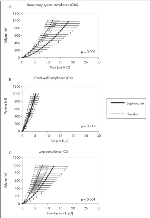
Higher respiratory system compliance was observed among patients assigned to the placebo than among those assigned to the bupi-vacaine solution, for a tidal volume of 10 ml/kg (73.3 ± 10.6 and 51.9 ± 15.5 ml/cmH2O respectively for placebo and bupivacaine solu-tions, p = 0.003, CI = 19.1 to 23.7). Lung com-pliance was also higher in the placebo group (127.2 ± 31.7 and 70.2 ± 23.1 ml/cmH2O, p < 0.001, CI = 61 to 53). No difference was found between the groups concerning chest wall compliance (186.3 ± 52.7 and 179.1 ± 30.7 ml/cmH2O for the bupivacaine and
Table 1. Characteristics and ventilation settings of the 19 patients studied for
respira-tory mechanics, presented as mean ± standard deviation
0.9% saline 0.5% bupivacaine p [CI]
Age (years) 46.4 ± 15.4 43.1 ± 13.4 0.628 Gender (male/female) 5/5 1/8 0.141 Weight (kg) 64.6 ± 7.7 68.8 ± 9.3 0.298 Height (m) 1.64 ± 0.1 1.61 ± 0.1 0.324 Body mass index (kg/m2) 24.2 ± 3.5 26.6 ± 3.5 0.147
Puncture level* 7 8 0.485
Cranial spread* 0 5 ± 1 < 0.001 Caudal spread* 0 6 ± 1 < 0.001 Tidal volume (TD, ml) 591.4 ± 63.6 598.3 ± 77.3 0.849 FIO2 (mmHg) 0.68 ± 0.1 0.68 ± 0.2 0.981 SpO2 (mmHg) 99 ± 1 99 ± 1 0.412 EtCO2 (mmHg) 41 ± 4 36 ± 4 0.022 [4.5 to 5] PaO2 (mmHg) 327.9 ± 104 264.8 ± 138 0.273 [47.2 to 79.1] PaCO2 (mmHg) 42.2 ± 5.3 40.8 ± 3.7 0.494 [2.2 to 0.8] *Height of block presented as number of dermatomes.
Table 2. Airway pressures and resistance in the 19 patients studied
0.9% saline 0.5% bupivacaine p [CI]
P’max, aw 12.3 ± 2.1 18.9 ± 5.6 0.003 [-8.3 to -5] P2, aw 10.9 ± 2.4 17.1 ± 5.2 0.003 [-7.5 to -4.9] P1, aw 11.9 ± 2.2 18.3 ± 5.3 0.009 [-7.5 to -4.9] Pmax, es 5.3 ± 1.1 4.9 ± 2.1 0.565 [0 to 0] P2, es 4.5 ± 1.3 4.2 ± 1.9 0.696 [0 to 1] Rmax, rs 13.8 ± 5 18.1 ± 8.4 0.192 [-5.9 to -2.7] Rmax, w 8.1 ± 4.7 6.6 ± 3.7 0.443 [2 to 1.1] Rmax, L 5.7 ± 2.7 11.5 ± 8.7 0.059 [-8.6 to -3] Rmin, rs 3.8 ± 2 6.1 ± 5.6 0.223 [-4 to 0] Rmin, L 3.8 ± 2 6.1 ± 5.6 0.223 [-4 to -0.7] DR, rs 10.1 ± 3.9 12 ± 4.6 0.349 [-2.2 to -1.6] DR, w 8.1 ± 4.7 6.6 ± 3.7 0.443 [2 to 1.1] DR, L 2 ± 1.5 5.5 ± 5.4 0.061 [-5.4 to -1.7] Calculated resistance presented as mean ± standard deviation. P’max, aw = maximumtracheal pressure minus pressure gener-ated by the tracheal cannula (cmH2O); P2, aw = pressure at end of slow decay to a inspiratory airway plateau (cmH2O); P1,
aw = pressure at the end of fast drop during the airway inspiratory pause (cmH2O); Pmax, es = maximum esophageal pressure
(cmH2O); P2, es = esophageal pressure at end of inspiratory pause (cmH2O); Rmax = total resistance (cmH2O.s.L-1) generated
by airway (Rmax, rs), chest wall (Rmax, w) and lung parenchyma (Rmax, L); Rmin = true airway (Rmin, rs) and lung (Rmin, L) resistance (cmH2O.s.L-1); and DR = residual resistance (cmH2O.s.L-1) of respiratory system (DR, rs), chest wall (DR, w) and lung
parenchyma (DR, L). CI = Confi dence Index.
and its lung component, in patients under-going mechanical ventilation. The chest wall component and the resistance of the respiratory system did not present any infl uence from the epidural block.
Several mechanisms can lower lung compliance, including atelectasis, increased smooth muscle tone and stimulation of other contractile elements in the airways or lung parenchyma and small airway closure.
Computed tomography has shown that pulmonary atelectasis is a common fi nding fol-lowing the induction of anesthesia, occurring in almost 90% of all anesthetized patients.17,18 placebo groups, respectively, p = 0.719, CI =
-17.5 to 3.1). Respiratory system, chest wall and lung compliance curves are presented respectively in Figures 1A, B and C.
There was no difference between the two groups regarding the resistance of the respiratory system or its lung and chest wall components. The calculated resistance is presented in Table 2.
Atelectasis during anesthesia can be formed by reduced transmural alveolar distending pres-sure (compression atelectasis), gas absorption when using high-oxygen air mixtures (absorp-tion atelectasis) or reduced surfactant produc-tion or acproduc-tion.19,20 The formation of atelectasis right after induction and the use of similar air mixtures in both groups suggest compression atelectasis as the probable etiology. There is no previous information suggesting a
syner-Figure 1. Comparative results between bupivacaine and placebo groups showing mean ± standard deviation (SD) compliance curves for respiratory system (A), chest wall (B) and lung parenchyma (C), and also the statistical differences found (P). Paw = airway pressure; Pes = esophageal pressure.
Respiratory system compliance (CRS)
0 200 400 600 800 1000 1200
0 5 10 15 20 25 30
Paw (cm H2O)
Vo
lu
m
e
(m
l)
Chest wall compliance (Cw)
0 200 400 600 800 1000 1200
0 5 10 15 20 25 30
Pes (cm H2O)
Vo
lu
m
e
(m
l)
Lung compliance (CL)
0 200 400 600 800 1000 1200
0 5 10 15 20 25 30
Paw-Pes (cm H2O)
Vo
lu
m
e
(m
l)
A
B
C
p = 0.003
p = 0.719
p < 0.001
Bupivacaine
Placebo
gistic effect of general and thoracic epidural anesthesia on atelectasis formation, but there is a possibility that atelectasis after muscle paralysis, as demonstrated by Tokics et al.,21 may be further increased under epidural an-esthesia. As the risks imposed by pulmonary artery catheterization were not justifiable in most of the patients studied, respiratory shunting was not calculated. Nevertheless, the fi nding of similar PaCO2 values in both groups
p = 0.719
p < 0.001
and of reduced EtCO2 in the bupivacaine group suggests a ventilation-to-perfusion mis-match, probably secondary to the respiratory component, since there was no documented shift in the distribution of intrathoracic blood volume or pulmonary blood volume during epidural anesthesia.22
Increased smooth muscle tone or stimu-lation of other contractile elements in the airways induced by TEA should be associated with increased airway resistance. Although the values of respiratory system resistance (R, rs and its components) and interrupter lung re-sistance (Rmin, L) found in both groups were signifi cantly higher than the corresponding values previously reported in normal anes-thetically paralyzed humans,23 no signifi cantly higher values were found in the bupivacaine group. Previous reports on the effect of TEA on respiratory system resistance in patients with documented bronchial hyperreacti vity showed increased acetylcholine threshold concentration, but this was correlated with local anesthetic blood serum concentration rather than with any effects from epidural sympathetic blockade.6 These results suggest that pulmonary sympathetic innervation ef-fects on airway resistance are not relevant for clinical practice.
Small airway closure, either as a result of higher tonus in small airways or as a result of reduced FRC, would be a possible mecha-nism accounting for the diminished CL and the signifi cantly higher LIP in the bupivacaine group. The slightly, although not statistically signifi cant, increased residual resistance in the bupivacaine group, either from the lung tissue or from the small airways, may have contributed towards a difference that could be noticed as reduced compliance rather than as enhanced resistance of the respiratory system. The contracted peripheral airway may stretch the lung tissue, thus decreasing its compliance, as showed by the rightward shift of the pres-sure-volume curve in the bupivacaine group (Figure 1). It is also possible that sympathetic blockage may trigger isotonic contraction of lung tissue. Peripheral lung tissue has been identifi ed as having the ability to respond directly to contractile stimulation, thus sug-gesting that lung parenchyma might play a role in obstructive diseases.24 It seems reasonable to consider the small airways as a possible site for sympathetic direct action.
This study was not designed to investigate the intraoperative effects of TEA, but those relating to the sympathetic blockade that is se-condary to it. Thoracic epidural anesthesia was the tool that made thoracic sympathectomy
p = 0.003
Volume (ml)
Volume (ml)
1. Zwissler B. Editorial: Thoracic anesthesia. In: Barash PG, Van Aken H, editors. Current opinion in anaesthesiology. Philadel-phia: Lippincott Williams & Wilkins; 2001. p. 47-9. 2. Rodgers A, Walker N, Schug S, et al. Reduction of
post-operative mortality and morbidity with epidural or spinal anaesthesia: results from overview of randomised trials. BMJ. 2000;321(7275):1493.
3. Ballantyne JC, Carr DB, deFerranti S, et al. The comparative effects of postoperative analgesic therapies on pulmonary outcome: cumulative meta-analyses of randomized, controlled trials. Anesth Analg. 1998;86(3):598-612.
4. Wang CY, Ong GS. Severe bronchospasm during epidural anaesthesia. Anaesthesia. 1993;48(6):514-5.
5. McGough EK, Cohen JA. Unexpected bronchospasm during spinal anesthesia. J Clin Anesth. 1990;2(1):35-6. 6. Groeben H, Schwalen A, Irsfeld S, Tarnow J, Lipfert P, Hopf HB.
High thoracic epidural anesthesia does not alter airway resistance and attenuates the response to an inhalational provocation test in patients with bronchial hyperreactivity. Anesthesiology. 1994;81(4):868-74.
7. Groeben H. Effects of high thoracic epidural anesthesia and local anesthetics on bronchial hyperreactivity. J Clin Monit Comput. 2000;16(5-6):457-63.
8. Rodriguez L, Marquer B, Mardrus P, et al. A new simple method to perform pressure-volume curves obtained under quasi-static conditions during mechanical ventilation. Intensive Care Med. 1999;25(2):173-9.
9. Lu Q, Vieira SR, Richecoeur J, et al. A simple automated method for measuring pressure-volume curves during mechanical venti-lation. Am J Respir Crit Care Med. 1999;159(1):275-82.
10. Benseñor FE, Vieira JE, Auler JO Jr. Guidelines for inspiratory fl ow setting when measuring the pressure-volume relationship. Anesth Analg. 2003;97(1):145-50.
11. D’Angelo E, Robatto FM, Calderini E, et al. Pulmonary and chest wall mechanics in anesthetized paralyzed humans. J Appl Physiol. 1991;70(6):2602-10.
12. Reta GS, Riva JA, Piriz H, Medeiros AS, Rocco PR, Zin WA. Effects of halothane on respiratory mechanics and lung histo-pathology in normal rats. Br J Anaesth. 2000;84(3):372-7. 13. Auler JO Jr, Miyoshi E, Fernandes CR, Benseñor FE, Elias
L, Bonassa J. The effects of abdominal opening on respira-tory mechanics during general anesthesia in normal and morbidly obese patients: a comparative study. Anesth Analg. 2002;94(3):741-8.
14. Baydur A, Behrakis PK, Zin WA, Jaeger M, Milic-Emily J. A simple method for assessing the validity of the esophageal bal-loon technique. Am Rev Respir Dis. 1982;126(5):788-91. 15. Gattinoni L, Pesenti A, Avalli L, Rossi F, Bombino M.
Pressure-volume curve of total respiratory system in acute respiratory failure. Computed tomographic scan study. Am Rev Respir Dis. 1987;136(3):730-6.
16. Neder JA, Andreoni S, Castelo-Filho A, Nery LE. Reference values for lung function tests. I. Static volumes. Braz J Med Biol Res. 1999;32(6):703-17.
17. Hedenstierna G, Rothen HU. Atelectasis formation during anesthesia: causes and measures to prevent it. J Clin Monit Comput. 2000;16(5-6):329-35.
18. Oczenski W, Schwarz S, Fitzgerald RD. Vital capacity manoeu-vre in general anaesthesia: useful or useless? Eur J Anaesthesiol. 2004;21(4):253-9.
19. Hedenstierna G. Alveolar collapse and closure of airways: regular effects of anaesthesia. Clin Physiol Funct Imaging. 2003;23(3):123-9.
20. Magnusson L, Spahn DR. New concepts of atelectasis during general anaesthesia. Br J Anaesth. 2003;91(1):61-72. 21. Tokics L, Hedenstierna G, Strandberg A, Brismar B, Lundquist
H. Lung collapse and gas exchange during general anesthesia: effects of spontaneous breathing, muscle paralysis, and positive end-expiratory pressure. Anesthesiology. 1987;66(2):157-67. 22. Hachenberg T, Holst D, Ebel C, et al. Effect of thoracic
epidural anaesthesia on ventilation-perfusion distribution and intrathoracic blood volume before and after induction of general anaesthesia. Acta Anaesthesiol Scand. 1997;41(9):1142-8. 23. D’Angelo E, Prandi E, Tavola M, Calderini E, Milic-Emily
J. Chest wall interrupter resistance in anesthetized paralyzed humans. J Appl Physiol. 1994;77(2):883-7.
24. Dolhnikoff M, Morin J, Ludwig MS. Human lung parenchyma responds to contractile stimulation. Am J Respir Crit Care Med. 1998;158(5 Pt 1):1607-12.
25. Rothen HU, Sporre B, Engberg G, Wegenius G, Hedenstierna G. Airway closure, atelectasis and gas exchange during general anaesthesia. Br J Anaesth. 1998;81(5):681-6.
Sources of funding: Fapesp (Fundação de Amparo à Pesquisa do Estado de São Paulo). Grant number 00/10847-0.
Confl ict of interest:Not declared
Date of fi rst submission:November 11, 2005
Last received:November 30, 2006
Accepted:November 30, 2006
REFERENCES possible. This is the reason why epinephrine
was added to both solutions. We did not in-tend to evaluate whether the fi ndings detected might be related to the sympathetic blockade or to the action of bupivacaine itself.
It would be diffi cult to ascribe the results to the gender composition of the bupivacaine and placebo groups, since the medical literature does not establish differences for respiratory mecha nics between male or female subjects, to the best of our knowledge. It is also important to emphasize that we studied patients with preserved respira-tory function, as confi rmed by the preoperative spirometric and laboratory analyses.
An advantageous correlation between postoperative analgesia, particularly with TEA, and better postoperative respiratory function has already been established. Ease of chest
expansion, ability to cough and cooperation with physiotherapy work assure less atelectasis, pulmonary infection and respiratory failure. This study intended to evaluate whether such respiratory improvement begins intraoperatively or whether the postoperative benefi ts can hide intraoperative drawbacks. On reducing lung compliance, probably byincreasing atelectasis, it may be assumed that intraoperative thoracic sympathetic block increases the postoperative work of breathing and consequently makes weaning more difficult.Besides atelectasis, another possibility is that decreased lung compli-ance is a consequence of small airway closure and reopening during tidal breathing, thus implying a risk of low lung volume injury. Both possibili-ties must be considered separately, since they are unrelated.25 Tomographic studies may be useful
for clarifying thisquestion. In both hypotheses, however, undesirable effects could be minimized by applying adequate PEEP levels.
AUTHOR INFORMATION
Fábio Ely Martins Benseñor, MD, PhD. Attending physician, Anesthesia Department, Hospital das Clínicas, Faculda-de Faculda-de Medicina da UniversidaFaculda-de Faculda-de São Paulo, São Paulo, Brazil.
Joaquim Edson Vieira, MD, PhD. Attending physician, Anesthesia Department, Hospital das Clínicas, Facul-dade de Medicina da UniversiFacul-dade de São Paulo, São Paulo, Brazil.
Jose Otávio Costa Auler Júnior, MD, PhD. Full professor, Anesthesia Department, Hospital das Clínicas, Facul-dade de Medicina da UniversiFacul-dade de São Paulo, São Paulo, Brazil.
Address for correspondence: Fabio Ely Martins Benseñor
Rua Mauá, 934/936
São Paulo (SP) — Brasil — CEP 01028-000 Tel./Fax (+55 11) 4617-3723
E-mail: [email protected]
Copyright © 2007, Associação Paulista de Medicina
RESUMO
Bloqueio simpático torácico reduz a complacência do sistema respiratório
CONTEXTO E OBJETIVO: Os benefícios pós-operatórios da anestesia peridural torácica (APT) na analgesia e respiração após toracotomias são conhecidos. Contudo, bloqueio simpático torácico pode desencadear hiperreatividade das vias aéreas. Este estudo pesou tais efeitos benéfi cos e indesejáveis na mecânica respiratória intra-operatória.
TIPO DE ESTUDO E LOCAL:Estudo clínico, randomizado, duplo-cego realizado em hospital público terciário.
MÉTODOS: Uma tabela de números aleatórios dividiu 19 pacientes submetidos a ressecção pulmonar parcial entre duas soluções administradas na APT: ativa (15 ml 0,5% bupivacaína, n = 9) ou placebo (15 ml 0,9% NaCl, n = 10). Ambas continham epinefrina 1:200,000 e morfi na 2 mg. Sob anestesia geral, pressões esofágicas e de vias aéreas foram registradas. Curvas de pressão versus volume, pontos de infl exão inferior (PII), resistências e complacências sob volume corrente de 10 ml.kg-1 foram aferidos para sistema respiratório, parede torácica e pulmões. O teste t de Student foi realizado (p < 0,005), incluindo intervalos de confi ança (IC).
RESULTADOS: A dispersão cefálica e caudal da bupivacaína foi, respectivamente, de 5 ± 1 e de 6 ± 1 dermátomos. A curva PII foi maior no Grupo Bupivacaína (6,2 ± 2,3 versus 3,6 ± 0,6 cm H2O, p = 0,016, IC = -3,4 a -1,8). Complacências do sistema respiratório e pulmões foram maiores no Grupo Placebo (respectivamente 73.3 ± 10.6 versus 51.9 ± 15.5, p = 0,003, IC = 19,1 a 23,7, e 127,2 ± 31,7 versus
70,2 ± 23,1 ml.cm H2O-1, p < 0,001, IC = 61 a 53). Resistências e complacências da parede torácica não mostraram diferenças.
CONCLUSÃO: APT diminui a complacência do sistema respiratório por reduzir seu componente pulmonar. Resistências não são afetadas. Sob APT, pressão positiva expiratória fi nal e manobras de recrutamento são recomendáveis.