Major Article
Corresponding Authors: Dr. Saif Hameed.
e-mail: [email protected]
Received 2 May 2017
Accepted 10 August 2017
Insights into the intracellular mechanisms of citronellal
in Candida albicans: implications for reactive
oxygen species-mediated necrosis, mitochondrial
dysfunction, and DNA damage
Venkata Saibabu
[1], Shweta Singh
[1], Moiz A. Ansari
[1],
Zeeshan Fatima
[1]and Saif Hameed
[1][1]. Amity Institute of Biotechnology, Amity University Haryana, Gurugram (Manesar), India.
Abstract
Introduction: Citronellal (Cit) possesses antifungal activity and has possible implications for reactive oxygen species (ROS) generation in Candida albicans. In this study, the effects of Cit on ROS generation and the mechanisms by which Cit exerts anti-Candida effects were examined. Methods: A 2′,7′-dichlorodihydroluorescein diacetate assay was used to assess oxidative
damage. Cell necrosis was determined by low cytometry after FITC-Annexin V staining. Mitochondrial function was studied based on mitochondrial potential, metabolic activity (MTT assay), and phenotypic susceptibility on a non-fermentable carbon
source. Membrane intactness and DNA damage were estimated by a propidium iodide (PI) uptake assay and 4',6-diamidino-2-phenylindole (DAPI) staining. Results:ROS generation was enhanced in response to Cit, leading to necrosis (2%). Additional hallmarks of cell death in response to Cit, such as mitochondrial membrane depolarization and DNA damage, were also observed. Cit treatment resulted in dysfunctional mitochondria, as evidenced by poor labeling with the mitochondrial membrane potential-sensitive probe rhodamine B, reduced metabolic activity (61.5%), and inhibited growth on a non-fermentable carbon
source. Furthermore, Cit induced DNA damage based on DAPI staining. These phenotypes were reinforced by RT-PCR showing differences in gene expression (30-60%) between control and Cit-treated cells. Finally, PI uptake in the presence of sodium azide conirmed non-intact membranes and suggested that Cit activity is independent of the energy status of the cell. Conclusions:
Cit possesses dual anticandidal mechanisms, including membrane-disruptive and oxidative damage. Taken together, our data
demonstrated that cit could be used as a prominent antifungal drug.
Keywords: Reactive oxygen species. Necrosis. Mitochondria. DNA. Propidium iodide. Candida albicans.
INTRODUCTION
The opportunistic pathogen Candida albicans is normally a commensal organism in humans, but in immunocompromised conditions, such as in patients with AIDS, diabetes, organ transplantation, and cancer, it causes mucosal, cutaneous, or invasive mycosis1. Prolonged usage of antifungals has recently,
in some instances, led to the emergence of multidrug-resistant strains of Candida2.In addition, high costs and severe toxicity
along with stagnation in the development of new drugs have become major impediments to effective antifungal therapy.
Thus, phytochemical research has steadily increased, owing to their safety proiles and low side effects, with promising results3.
Citronellal (Cit) is a monoterpenoid
(3,7-dimethyloct-6-en-1-al) found in Cymbopogon plants. It gives the characteristic
lemon-scented aroma to citronella oil. The anticandidal potential
of Cit has been demonstrated; it primarily targets fungal cell membranes and inhibits virulence traits, such as morphological
shifts and bioilm formation, and it may promote the generation
of ROS4. This study aimed to add to the existing literature about
ROS generation in Cit-treated cells and elucidate other potential targets of Cit in C. albicans. The results conirmed that Cit increases ROS levels and elicits cell necrosis, mitochondrial dysfunction, and deoxyribonucleic acid (DNA) damage in C. albicans. We also showed that the energy status of the cell is not important for the antifungal mechanisms of Cit.
METHODS
All media chemicals, Yeast Extract Peptone Dextrose (YPD), and propidium iodide (PI) were purchased from Himedia (Mumbai, India).Citronellal, DCFDA (2,7-dichloroluorescein diacetate), DAPI (4',6-diamidino-2-phenylindole), the Annexin
Saibabu V et al - Anticandidal mechanismsof citronellal
primerswere obtained from Sigma Chemicals (St. Louis, MO,
USA) and IDT (San Diego, CA, USA). Rhodamine B was
purchased from CDH (Delhi, India) and sodium azide (NaN3)
was purchased from Fischer Scientiic (Waltham, MA, USA).
Growth media and strains
The laboratory Candida strain SC5314 was used; its whole genome was sequenced in 2004 and it is considered the most widely used laboratory strain. Itwas cultured in YPD broth with yeast extract 1% (w/v), peptone 2% (w/v), and dextrose 2%
(w/v). For agar plates, 2% (w/v) agar was added to the media. Candida strains were stored in 30% (v/v) glycerol stock at
-80°C. The cells were freshly revived twice on YPD broth and transferred to agar plates. The cells were grown overnight at
30°C on agar plates before each analysis to ensure the revival of the strains.
Phenotypic susceptibility assay
Phenotypic susceptibility assays were performed as described elsewhere4. Briely, for the spot assay, 5μL of ivefold
serially diluted yeast cultures (cells suspended in normal saline to an OD600 of 0.1 (1 × 10⁶ cells mL-1) were spotted on YPD
plates in the absence (control) and presence of Cit (250µg mL-1).
Growth was not affected by the presence of a solvent (dimethyl sulfoxide) (data not shown). Growth differences were measured after incubation for 48h at 30°C.
RT-PCR
Ribonucleic acid (RNA) was isolated and reverse
transcriptase-polymerase chain reaction (RT-PCR) was
performed as described earlier5. In brief, cells were diluted in
50mL of fresh YPD broth at OD600 of 0.1 (106 cells mL-1) in
the absence (control) and presence of Cit (250µg mL-1) and
grown at 30°C until reaching OD600 of 1.0. RNA isolation was
performed by the TRIzol method and reverse-transcriptase (RT) PCR was performed using the RevertAid H Minus Kit
(Invitrogen, Carlsbad, CA, USA), following the manufacturer’s
instructions. Briely, 5µg of isolated RNA was DNase-treated
at 37°C for 30 min and the reaction was terminated by adding
1µL of 25mM EDTA and incubated at 65°C for 60 min. RNA was subsequently primed with oligo (dT)18 for cDNA synthesis
at 42°C for 60 min. Reverse transcription was terminated by
heating at 70°C for 5 min. The synthesized cDNA product (2µL) was directly used for PCR ampliication (50µL) using gene-speciic forward and reverse primers The ampliied products
were separated by electrophoresis and the densities of bands (for genes of interest) were measured and normalized to that of the constitutively expressed actin gene (ACT1).
Analysis of apoptotic markers
Candida albicans spheroplasts were prepared as described earlier5. Briely, Cit-treated (MIC
80) cells were incubated with Zymolyase (100U/g wet weight) at 37°C for 3h and vortexed to
remove the cell wall. Spheroplast preparation was monitored by adding 0.2% sodium dodecyl sulfate to lyse the enzyme-digested cells and visualized under a microscope (Olympus, Mumbai, India). Cell necrosis was determined by the externalization
of phosphatidyl serine (PS), an apoptotic marker, using a
Fluorescein Isothiocyanate (FITC) Annexin V Apoptosis Detection Kit. (Sigma Chemicals). Cells were analyzed by low
cytometry (Beckman Coulter, Brea, CA, USA) using a 488-nm
excitation wavelength and a 515-nm band pass ilter for FITC detection and a ilter of >560nm for PI detection. A total of 10,000 events were counted at the low rate. Data analysis was
performed using Summit v4.3 (Beckman Coulter).
Fluorescence microscopy
Experimental cultures were grown in the presence of Cit at 30°C for 3h with shaking. Cells were harvested and washed three times with phosphate-buffered saline (PBS) stained with PI (1µg mL-1), an indicator of membrane damage, DCFDA
(1µg mL-1), an indicator of ROS generation, Rhodamine B
(0.5µg mL-1), an indicator of mitochondrial membrane potential (∆Ψm), and DAPI (2µg mL-1), an indicator of DNA damage, for
20 min in the dark6-8. For PI experiments, Candida cells were
pre-incubated with 5mM NaN3 for 60 min. Subsequently, the unbound dye was removed by washing with the same buffer.
Specimens were then examined under a Cos Lab luorescence
microscope (Ambala Cantt, India).
Hemolytic activity assay
The hemolytic activity of Cit was assayed as described
elsewhere9,with modiications. In brief, fresh human red blood
cells (hRBCs) collected in the presence of an anti-coagulant from a healthy volunteer (author donated voluntarily) were
washed three times in PBS. The drug was added to the suspension of red blood cells (4%, v/v) in PBS to a inal volume of 1mL and incubated at 37°C for 35 min. The samples
were then centrifuged for 2 min at 2000rpm, and the release of hemoglobin was monitored by measuring the absorbance (Asample) of the supernatant at 540nm. For negative and positive controls, hRBCs in PBS (Ablank) and in 1% (inal concentration
v/v) Triton X-100 were used, respectively. The percentage of
hemolysis was calculated according to the following equation: Hemolysis (%) = [(Asample - Ablank)/(ATriton - Ablank)]*100.
RESULTS
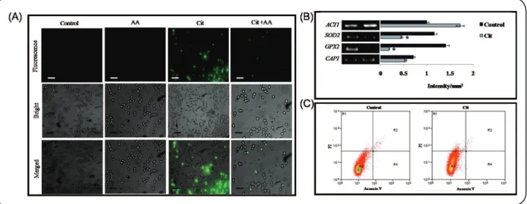
Cit induces ROS generation, leading to necrosis in Candida albicans
We irst conirmed the effect of Cit on ROS production in C. albicans. This was achieved using DCFDA, an oxidant-sensitive
probe, to assess ROS generation; in this analysis, increased fluorescence within cells indicates enhanced ROS levels6. As expected, the luorescence in Cit-treated cells was considerably higher than that in untreated cells. The ampliied luorescence intensity was
reverted when the treated cells were incubated with an antioxidant, such as ascorbic acid (Figure 1A). Differential expression of genes associated with oxidative stress, i.e., SOD2 (Superoxide dismutase 2), CAP1 (Adenylate Cyclase Associated Protein 1), and GPX2 (Glutathione Peroxidase 1), was quantiied by RT-PCR and these
FIGURE 1 - Effect of Citronellal on ROS generation and necrosis. (A) Fluorescent microscopy of DCFDA for the detection of increased ROS in the presence
of Cit, and reversion to normal levels after treatment with AA. Scale bar depicts 20µm. (B) RT-PCR of differentially regulated genes in response to Cit. The
left panels show transcript levels of SOD2, CAP1, and GPX2 in Control cells (Lane 1) and Cit-treated cells (250μg/mL) (Lane 2). The right panel shows the
quantitation (density expressed as intensity/mm2) of the respective transcripts normalized against constitutively expressed ACT1 transcripts. (C) Measurement of
apoptosis or necrosis by tracking FITC and PI in the absence (control) or presence of Cit (1mgmL-1). Cit treatment showed necrosis in 2% of cells. AA:ascorbic
acid; Cit: Citronellal; ACT1: Actin 1; SOD2: superoxide dismutase 2; GPX2: glutathione peroxidase 1; CAP1: adenylate cyclase associated protein 1; ROS: Reactive Oxygen Species; DCFDA: 2’,7’ –dichloroluorescin diacetate; RT-PCR: Reverse Transcriptase Polymerase Chain Reaction; FITC: Fluorescein
isothiocyanate ; PI: Propidium Iodide.
FITC labeling indicates the presence of late apoptotic cells, while PI labeling conirms the presence of necrotic cells7. Our low cytometry analysis revealed that almost 2% cells underwent
necrosis following Cit treatment, with no apoptosis (Figure 1C).
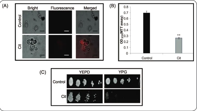
Cit leads to dysfunctional mitochondria
We subsequently evaluated the role of mitochondria in Cit-induced cell death. We examined mitochondrial membrane
potential (∆Ψm) by employing a Rhodamine B probe8. Exposure
to Cit resulted in substantial hyperpolarization of mitochondria in C. albicans, confirming mitochondrial dysfunction (Figure 2A). Next, we studied the activity of mitochondrial dehydrogenases, which are indicators of the metabolic activity of functional mitochondria10. Our results showed that
Cit had inhibitory effects on the activity of mitochondrial dehydrogenases in C. albicans (Figure 2B). Finally, even at a
non-lethal Cit concentration (250µg mL-1), cells were unable to
grow when non-fermentable glycerol was provided as the sole carbon source (Figure 2C).
Anticandidal activity of Cit is independent of energy status
NaN3 blocks intracellular ATP by inhibiting cytochrome oxidase11. The effect of the cellular energy status was evaluated
by estimating PI uptake, which binds to nucleic acids only in injured membranes, as in Cit-treated cells. We observed that cell viability was substantially reduced in the presence of Cit, regardless of the presence of NaN3 (Figure 3A). Moreover, the downregulation of NPC2 (NPC Intracellular Cholesterol
Transporter 2), a gene involved in sterol transport, further
conirmed the loss of membrane function (Figure 3B).
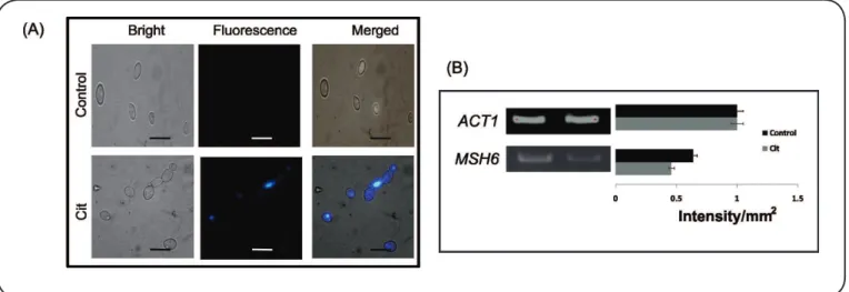
Cit induces DNA damage
To conirm DNA damage, we utilized the DAPI staining method. DAPI binds to AT sites within the minor groove of DNA, where its luorescence can be assessed as a direct indicator
of DNA damage. During ROS formation, the permeability of the
dye increases, resulting in deep blue nuclear luorescence. C. albicans cells exposed to Cit exhibited enhanced luorescence,
indicating DNA damage (Figure 4A). We further explored whether DNA damage in the presence of Cit was related to
defects in DNA repair mechanisms. Based on RT-PCR, the
downregulation of MSH6 (MutS protein homolog 2) was involved in DNA repair (Figure 4B).
Hemolytic activity of Cit
The hemolytic activity of Cit against human erythrocytes
is a major parameter related to the antifungal toxicity against human cells. Accordingly, hRBCs were isolated and hemolysis was evaluated in the presence of Cit. We observed only 15%
hemolysis at the MIC of Cit in comparison to Triton X (control),
showing 100% hemolysis (Figure 5).
DISCUSSION
The results of this study elucidate the oxidative stress mechanism induced by Cit. The induction of oxidative stress
contributes to the anticandidal activity of Cit, possibly via the augmentation of excessive ROS production4. Our luorescence microscopy-mediated DCFDA assay confirmed that ROS
FIGURE 2 - Effect of Citronellal on mitochondrial functioning. (A) Fluorescent microscopy of Rhodamine B for the analysis of mtΨm in the presence of Cit.
Control shows the quenching of luorescence in comparison to Cit-treated cells, which show clear luorescence, suggesting a change in the mitochondrial membrane
potential. Scale bar depicts 10 µm. (B) Effect of Cit (250μgmL-1) on mitochondrial activity in Candida. albicans based on an MTT assay. Mean OD
570 of three
independent sets of experiments is depicted on the Y-axis. (C) Phenotypic susceptibility assays in the absence (control) and presence of Cit (250μgmL-1) in YPD
(fermentable carbon source) and YPG (Yeast Extract-Peptone-Glycerol) media (non-fermentable carbon source). YEPD: Yeast extract Peptone Dextrose; YPG:
Yeast Extract Peptone Glycerol; Cit: Citronellal; MTT: 3-(4,5-dimethylthiazol-2-yl)-2,5-diphenyltetrazolium bromide; PI: Propidium Iodide.
FIGURE 3 - Effect of energy depletion on anticandidal activity of Citronellal. (A) Fluorescent microscopy of PI for the detection of membrane damage in
the presence of Cit. Scale bar depicts 25µm. Cells were pre-incubated with 5mM NaN3 at 30°C for 60 min. Fluorescence in NaN3+Cit-treated cells suggests
that antifungal activity of Cit is energy-independent. (B) RT- PCR of NPC2 in response to Cit. The left panels show transcript levels of NPC2 in Control cells (Lane 1) and Cit-treated cells (250μg/mL) (Lane 2). The right panel shows the quantitation (density expressed as intensity/mm2) of the respective transcripts
normalized against constitutively expressed ACT1. NaN3: Sodium Azide; Cit: Citronellal; ACT1: Actin 1;NPC2: NPC Intracellular Cholesterol Transporter 2; RT-PCR: Reverse Transcriptase Polymerase Chain Reaction.
FIGURE 5 - Hemolytic activity of Citronellal against human erythrocytes.
Hemolysis was determined by recording absorbance at 450nm in Cit-treated
cells and cells treated with 1% Triton X-100 (reference for 100% hemolysis).
Data are presented as the means of triplicate experiments. Cit: Citronellal.
FIGURE 4 - Effect of Citronellal on DNA damage. (A) Fluorescent microscopy of DAPI for the detection of DNA damage in the presence of Cit. Scale bar
depicts 10µm. (B) RT-PCR of MSH6 in response to Cit. The left panel shows transcript levels of MSH6 in Control cells (Lane 1) and Cit-treated cells (250μg/mL) (Lane 2). The right panel shows the quantitation (density expressed as intensity/mm2) of the respective transcripts normalized against constitutively expressed
ACT1 transcripts. ACT1: Actin 1; MSH6: MutS protein homolog 2; DNA: deoxyribonucleic acid; DAPI: 4',6-diamidino-2-phenylindole; Cit: Citronellal; RT-PCR: reverse transcriptase polymerase chain reaction.
effects of Cit against C. albicans involve ROS production. Sod2p (superoxide dismutase 2 protein) is a manganese-dependent mitochondrial protein that plays an essential role in protection against oxidative damage12. Further, Cap1p, which is
homologous to Saccharomyces cerevisiae Yap1p, is responsible for the activation of genes related to oxidative stress. When ROS accumulates, Cap1p oxidizes and accumulates in the nucleus, further activating genes carrying Cap1 response elements in their promoters13. However, Yap1p is not directly oxidized, but
needs a glutathione peroxidase (Gpx)-like protein to transduce
Yap1p signaling by creating a disulide bond between Gpx3p and
Cys36 (evolutionarily conserved redox-sensitive residues) and Yap1p with Cys59814. It is well-known that ROS accretion plays
a pivotal role in the necrosis of yeast cells6. For instance, natural
compounds, such as melittin, silymarin, and silibinin, induce apoptotic pathways via ROS generation15-17.Thus, we further
checked whether Cit-mediated ROS accumulation also leads to apoptosis or necrosis in Candida cells. A low cytometry analysis
conirmed that Cit treatment leads to necrosis in approximately
2% cells (Figure 1C).
A hallmark of yeast cells involved in ROS generation and apoptosis is dysfunctional mitochondria18. Since mitochondria
are key organelles governing apoptotic events, we evaluated
their role in Cit-induced cell death. To access the functionality of mitochondria, we irst studied the loss of ∆Ψm, a distinctive
feature of cells undergoing apoptosis. Moreover, it plays a role
in ATP synthesis via mitochondrial oxidative phosphorylation. Thus, ∆Ψm is a sensitive indicator of the energy status of
mitochondria, which is determined by an electrochemical gradient maintained by the electron transport chain19. Our results conirmed that exposure to Cit resulted in signiicant
hyperpolarization of mitochondria in C. albicans, providing evidence for mitochondrial dysfunction (Figure 2A). Cell
death in yeast also correlates with altered ∆Ψm stemming from
oxidative damage caused by ROS accumulation20. Next, using an MTT assay, we found that the metabolic activity of functional
mitochondria was inhibited (Figure 2B) in the presence of Cit10. The impaired functionality of mitochondria was further
apparent when we compared the growth of Cit-treated cells on non-fermentative and fermentative carbon sources, indicating hypersensitivity in non-fermentable medium (Figure 2C). These
results conirmed that Cit leads to dysfunctional mitochondria. The observation of dysfunctional mitochondria prompted us to further examine the effect of ATP depletion on the activity of Cit. Enhanced PI uptake, as evidenced by increased luorescence
(Figure 3A), and the downregulation of NPC2, a gene involved in sterol transport (Figure 3B),in the presence of Cit indicate a
Cit leads to membrane disruption, but also revealed that Cit activity was unaffected by NaN3, suggesting that its antifungal effects are mediated by cellular functions, irrespective of energy consumption.
DNA damage is another key feature of cells involved in ROS generation and apoptosis21. Moreover, Cit induces
hypersensitivity in the presence of a DNA damaging agent4.
Based on DAPI staining, in the presence of Cit, cells showed
enhanced luorescence indicating DNA damage (Figure 4A) as well as the downregulation of MSH6, a gene involved in DNA repair (Figure 4B). These results conirm that Cit induces
DNA damage in C. albicans; however, whether DNA damage mediated by Cit occurs via the generation of ROS remains to
be validated. Finally, we assessed the toxicity of Cit against
mammalian cells, and we observed negligible toxicity in comparison to the positive control (Figure 5).
Taken together, in addition to membrane disruption,
the primary mode of anticandidal action for Cit, our results
conirmed other effects of Cit, including ROS generation,
necrosis, mitochondrial dysfunction, and DNA damage. Although the link between these mechanisms of disruption is
unclear, these indings suggest that Cit could trigger these events
after membrane damage against C. albicans. Further studies are warranted to effectively employ phytotherapeutics, such as Cit, for the treatment of Candida infections.
Acknowledgments
We are grateful to Joseph Heitman for providing the Candida SC5314 reference strain as a generous gift. We thank Smita Sundaram, Advanced
Instrumentation Research Facility (AIRF) at Jawaharlal Nehru University, New Delhi for helping in the low cytometry experiment. We thank Rajendra Prasad, Dean, Faculty of Science, Engineering and Technology for
encouragement.
Conlict of interest
The authors declare that there is no conlict of interest.
Financial support
The work was supported by the Science and Engineering Research Board (SERB), New Delhi (grant number SR/FT/LS-12/2012).
REFERENCES
1. Singh S, Fatima Z, Hameed S. Predisposing factors endorsing
Candida infections. Infez Med. 2015;23(3):211-23.
2. Tanwar J, Das S, Fatima Z, Hameed S. Multidrug resistance: an emerging crisis. Interdiscip Perspect Infect Dis. 2014; doi: 10.1155/2014/541340.
3. Patwardhan B, Vaidya AD. Natural products drug discovery: accelerating the clinical candidate development using reverse pharmacology approaches. Indian J Exp Biol. 2010;48(3):220-7.
4. Singh S, Fatima Z, Hameed S. Citronellal-induced disruption of membrane homeostasis in Candida albicans and attenuation of its
virulence attributes. Rev Soc Bras Med Trop. 2016 49(4):465-72.
5. Ansari MA, Fatima Z, Hameed S. Anticandidal effect and mechanisms of monoterpenoid, perillyl alcohol against Candida
albicans. PLoS One. 2016;11(9):e0162465.
6. Menezes RA, Amaral C, Batista-Nascimento L, Santos C, Ferreira
RB, Devaux F, et al. Contribution of Yap1 towards Saccharomyces
cerevisiae adaptation to arsenic-mediated oxidative stress. Biochem
J. 2008;414(2):301-11.
7. Eisenberg T, Carmona-Gutierrez D, Büttner S, Tavernarakis N,
Madeo F. Necrosis in yeast. Apoptosis. 2010;15(3):257-68.
8. Reungpatthanaphong P, Dechsupa S, Meesungnoen J, Loetchutinat C, Mankhetkorn S. Rhodamine B as a mitochondrial probe for measurement and monitoring of mitochondrial membrane potential in drug-sensitive and resistant cells. J Biochem Biophys Methods. 2003;57(1):1-16.
9. Asthana N, Yadav SP, Ghosh JK. Dissection of antibacterial and toxic activity of melittin: a leucine zipper motif plays a crucial role in determining its hemolytic activity but not antibacterial activity. J Biol Chem. 2004;279(53):55042-50.
10. Adam-Vizi V, Tretter L. The role of mitochondrial dehydrogenases in the generation of oxidative stress. Neurochem Int. 2013;62(5):757-63.
11. Wilson DF, Chance B. Azide inhibition of mitochondrial electron
transport. I. The aerobic steady state of succinate oxidation.
Biochim Biophys Acta. 1967;131(3):421-30.
12. Luk E, Yang M, Jensen LT, Bourbonnais Y, Culotta VC. Manganese activation of superoxide dismutase 2 in the mitochondria of
Saccharomyces cerevisiae. J Biol Chem. 2005;280(24):22715-20.
13. Komalapriya C, Kaloriti D, Tillmann AT, Yin Z, Herrero-de-Dios C, Jacobsen MD, et al. Integrative model of oxidative stress adaptation in the fungal pathogen Candida albicans. PLoS One. 2015;10(9):e0137750.
14. Rodrigues-Pousada CA, Nevitt T, Menezes R, Azevedo D, Pereira J, Amaral C. Yeast activator proteins and stress response: an overview.
FEBS Lett. 2004;567(1):80-5.
15. Lee J, Lee DG. Melittin triggers in Candida albicans through the reactive oxygen species-mediated mitochondria/caspase-dependent
pathway. FEMS Microbiol Lett. 2014;355(1):36-42.
16. Yun DG, Lee DG. Silibinin triggers yeast apoptosis related to
mitochondrial Ca2+ inlux in Candida albicans. Int J Biochem Cell Biol. 2016; 80:1-9.
17. Yun DG, Lee DG. Silymarin exerts antifungal effects via membrane-targeted mode of action by increasing permeability and inducing oxidative stress. Biochim Biophys Acta. 2017;1859(3):467-74.
18. Bhat AH, Dar KB, Anees S, Zargar MA, Masood A, Soi MA, et al. Oxidative stress, mitochondrial dysfunction and neurodegenerative diseases; a mechanistic insight. Biomed Pharmacother. 2015;74:101-10.
19. Brand MD, Chien LF, Ainscow EK, Rolfe DF, Porter RK. The causes and functions of mitochondrial proton leak. Biochim Biophys Acta. 1994; 1187(2):132-9.
20. Xu C, Wang J, Gao Y, Lin H, Du L, Yang S, et al. The anthracenedione compound bostrycin induces mitochondria mediated apoptosis in
the yeast. FEMS Yeast Res. 2010;10(3):297-308.
21. Rowe LA, Degtyareva N, Doetsch PW. DNA damage-induced reactive oxygen species (ROS) stress response in Saccharomyces
cerevisiae. Free Radic Biol Med. 2008;45(8):1167-77.