539
Rev Soc Bras Med Trop 50(4):539-542, July-August, 2017 doi: 10.1590/0037-8682-0485-2016
Short Communication
Corresponding author: Mario Luis Garcia de Figueiredo. e-mail: [email protected]
Received 23 November 2016 Accepted 28 March 2017
Cacipacore virus as an emergent
mosquito-borne Flavivirus
Mario Luis Garcia de Figueiredo
[1],[2], Alberto Anastacio Amarilla
[1],
Glauciane Garcia de Figueiredo
[1], Helda Liz Alfonso
[1], Veronica Lippi
[1],
Felipe Gonçalves Motta Maia
[3], Felipe Alves Morais
[4], Cristóvão Alves da Costa
[5],
Dyana Alves Henriques
[2], Edison Luis Durigon
[2], Luiz Tadeu Moraes Figueiredo
[3]and Victor Hugo Aquino
[1][1]. Laboratório de Virologia, Departamento de Análises Clínicas, Toxicológicas e Bromatológicas, Faculdade de Ciências Farmacêuticas de Ribeirão Preto, Universidade de São Paulo, Ribeirão Preto, SP, Brasil. [2]. Departamento de Microbiologia, Instituto de Ciências Biomédicas, Universidade de São Paulo, São Paulo, SP, Brasil. [3]. Centro de Pesquisa em Virologia, Faculdade de Medicina de Ribeirão Preto, Universidade de São Paulo, Ribeirão Preto, SP, Brasil.
[4]. Faculdade de Zootecnia e Engenharia de Alimentos, Universidade de São Paulo, Campus de Pirassununga, Pirassununga, SP, Brasil. [5]. Instituto Nacional de Pesquisa de Manaus, Manaus, AM, Brasil.
Abstract
Introduction: Cacipacore virus (CPCV), a possible bird-associated lavivirus, has yet to be detected in mosquitoes. Our purpose
is examining CPCV in mosquitoes from the Amazon region of Brazil. Methods: Approximately 3,253 Culicidae (grouped into 264 pools) were collected from the Amazon region during 2002-2006 and analyzed using a Flavivirus genus-speciic reverse
transcription- polymerase chain reaction followed by nested polymerase chain reaction assay and by nucleotide sequencing of amplicons. Results: Nucleotide sequences from ive mosquito samples showed high similarity to the those of CPCV originally
isolated in the Amazon region. Conclusions: This is the irst report of CPCV-infected mosquitoes which has implications on the
arbovirus maintenance in nature and transmission to man.
Keywords: Cacipacore virus. Mosquitoes. Emerging Flavivirus.
Cacipacore virus (CPCV) is a member of the Japanese
encephalitis virus (JEV) complex belonging to the genus
Flavivirus of the family Flaviviridae1. It was originally isolated
in 1977, from the blood of a bird (Percnostola ruifrons) in the Amazon region of Brazil2. Recently, antibodies against CPCV
have been reported in equines, non-human primates, and water buffalo in the central region of Brazil, suggesting virus circulation3-5. In addition, one patient from the Amazon region
exhibiting an acute febrile illness was found to be infected by CPCV in 20116. Here, we present the irst report of
CPCV-infected mosquitoes collected from the environment.
As part of the arbovirus surveillance programs (2002 to 2006), approximately 3,253 mosquitoes (Culicidae) were collected from urban and rural areas in Manaus (Amazonas State) and Monte Negro County (Rondonia State), using
attraction traps including the CO2, the cluster of differentiation (CD4) night model, and the Shannon. Captured specimens were
identiied based on morphological characteristics and grouped
into 264 pools (including 10-13 specimens per pool) and stored
at −80 °C7 .
Reverse transcription-polymerase chain reaction (RT-PCR) and nucleotide sequencing for arbovirus diagnosis
and identiication were performed using ribonucleic acid (RNA)
extracts from the mosquito pools while taking precautions to avoid contamination. The mosquito samples were triturated at
−4°C temperatures using plastic pestles and divided into two
aliquots: one for RNA extraction and the other for future virus
isolation. Aliquots were stored at −80°C until ready for use.
RNA was extracted from the mosquito pools, using the PureLink Viral ribonucleic acid/deoxyribonucleic acid (RNA/DNA) Kit (Invitrogen, USA) according to the manufacturer’s instructions. Subsequently, RT and Hemi-Nested-PCR Flavivirus assays that differentiate dengue, yellow fever, and other viruses based on amplicon size, were performed as previously reported8.
The PCR products were purified using the QIAquick Gel Extraction Kit (QIAGEN, Germany), following the manufacturer's protocol and subsequently sequenced with the ABI PRISM 3500 Genetic Analyzer (Applied Biosystems, Foster City, CA, USA). The sequences obtained in this study were submitted to GenBank under access numbers GU811223
540
de Figueiredo MLG et al - Cacipacore infecting mosquito in Amazon regions
Genus/species Number of adults
Male specimens
Female specimens
Number of pools
Anopheles sp. 81 - 81 7
Culex sp. 867 140 727 75
Coquillettidia sp. 252 50 202 19
Aedes aegypti 950 45 905 78
Aedes albopictus 244 10 234 18
Psorophora
albipes 97 - 97 8
Psorophora
albigenus 55 - 55 5
Psorophora ferox 60 - 60 6
Haemagogus
jantinomys 126 10 116 10
Haemagogus
leucocelaenus 271 1 270 20
Haemagogus
spegazzinii 250 241 9 18
Total 3,253 497 2,756 264
TABLE 1
Number of Culicidea (Diptera) collected for the study from 2002 to 2006.
Gender/species
Number of detected
pools Amplicon size (bp) Collection places/State Date of collection
Aedes aegypti 3 (33 females) ~ 232 Manaus City/Amazonas State 2005-2006
Anopheles sp. 1 (9 females) ~ 216 Montenegro County/Rondonia State 2002
Culex sp. 1 (8 females) ~ 275 Montenegro County/Rondonia State 2002 TABLE 2
Flavivirus amplicons obtained by reverse transcription- polymerase chain reaction followed by nested polymerase chain reaction assays for mosquitoes collected in the Brazilian Amazon region, 2002-2006.
- GU811223; CPCVBR/RO/Anopheles sp 43A5/2002 - GU811224; CPCVBR/AM/Aedes aegypti 17/2005 - GU811225; CPCVBR/AM/Aedes aegypti 46/2005 - GU811226; and CPCVBR/AM/Aedes aegypti 47/2005 - GU811227. CPCV
and others lavivirus sequences retrieved from GenBank were
aligned using the BioEdit v 7.09 program9. Phylogenetic trees
based on Neighbor-joining (NJ) methods were constructed using the Mega 5 software10.
Captured specimens were identiied based on morphological
characteristics as Aedes aegypti, Aedes albopictus, Anopheles
sp., Culex sp., Coquillettidia sp., Haemagogus janthinomys,
Haemagogus leucocelaenus, Haemagogus spegazzinii,
Psorophor albigenus, Psorophora albipes, and Psorophora
ferox7, as shown in Table 1.
For virus detection, we used a Hemi-Nested-PCR that did not include a specific primer for Cacipacore virus. Interestingly, CPCV amplicons were obtained using the primer
for dengue virus (DENV) type 2 and the CACV speciic origin
of the amplicons was only recognized following nucleotide sequencing8. Amplicons ~200-300bp in size, corresponding to
a section of the NS5 gene region of Flavivirus were obtained
from ive mosquito pools (1.89% positivity). Following further ampliication and puriication, it was possible to sequence the
amplicons. The pools infected by Flavivirus included Aedes aegypti, Anopheles sp., and Culex sp., as shown in Table 2.
The sequences, ranging from 216 to 275 nucleotides, were aligned with other sequences retrieved from GenBank. The
sequences exhibited 98-100% similarity with those of the
CPCV originally isolated in 1977 from a bird1. The Flavivirus
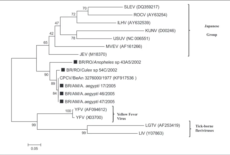
phylogenetic tree, shown in Figure 1, includes sequences of the viruses infecting our pools of mosquitoes, all of which
cluster within the same CPCV branch, conirming this virus is
the causative agent of the infections. The tree also shows that CPCV is related to Japanese encephalitis virus, corroborating a previous report by Kuno et al.1 also based on the NS5
gene sequence. Unfortunately, despite numerous attempts that exhausted our samples, we were unable to obtain larger nucleotide sequences from our mosquito pools.
Japanese encephalitis virus (JEV), Ilhéus virus (ILHV),
Rocio virus (ROCV), Saint Louis encephalitis virus (SLEV), and
West Nile virus (WNV) are all zoonotic avian viruses transmitted
by Culicidae mosquitoes. Thus, it is possible that CPCV, a
closely related virus, shares the same zoonotic characteristics because it was originally isolated from a bird.
Aedes aegypti, an urban anthropophilic mosquito that lives around human houses, is the main vector for DENV in Brazil and has been involved in huge outbreaks over the last 30 years,
with more than 10 million reported cases. Over the last three
years, Aedes aegypti has been involved in the transmission of
Chikungunya virus (CHIKV) and Zika virus (ZIKV), which
541
Rev Soc Bras Med Trop 50(4):539-542, July-August, 2017
Japanese
Group
Tick-borne flaviviruses SLEV (DQ359217)
ROCV (AY63254)
ILHV (AY632539)
KUNV (D00246)
USUV (NC 006551)
MVEV (AF161266)
JEV (M18370)
BR/RO/Anophelessp 43A5/2002 BR/RO/Culexsp 54C/2002
CPCV/BeAn 3276000/1977 (KF917536 )
BR/AM/A.aegypti17/2005 BR/AM/A.aegypti46/2005 BR/AM/A.aegypti47/2005
YFV (AF094612)
YFV (X03700)
LGTV (AF253419)
LIV (Y07863)
99 100
99
70
72
78 47
42
65
90
89
84
0.05
Yellow Fever Virus
FIGURE 1 - Phylogenetic tree based on NS5 partial gene sequences. The tree was constructed using the Neighbor-joining method with 1,000 bootstrap replications. Branch lengths are proportional to the percentage of divergence. The Tamura-Nei nucleotide substitution model was used with a gamma distribution (shape parameter = 1). GenBank accession numbers, species, country of origin, and year of isolation are detailed in the tree. CPCVs isolated from mosquito samples are indicated with a square (▄). SLEV: Japanese encephalitis; ROCV: Rocio virus; ILHV: lhéus virus; KUNV: Kunjin virus; USUV: Usutu virus; MVEV: Murray Valley encephalitis virus; JEV: Japanese encephalitis; BR/RO: Brazil/Rondonia; CPCV: Cacipacoré virus; BR/AM/A: Brazil/Amazonas/aedes; YFV: yellow fever virus;. LGTV: Langat virus; LIV: Louping ill virus .
serologic diagnostic tests for dengue could be positive for
lavivirus cross-reactivity in patients with CPCV.
Culex pipiens are found worldwide, breeding in water
contaminated with organic matter. The conditions in poorly sanitized urban areas of Brazil support the propagation of this mosquito. Culex pipiens feed at night and are zoophilic; they are particularly more ornithophilic. Alphaviruses such
as Western Equine Encephalitis (WEEV), Venezuelan Equine
Encephalitis (VEEV), and Eastern Equine Encephalitis
(EEEV); Orthobunyaviruses such as Caraparu virus and
Oropouche virus; and Flaviviruses, such as SLEV, have been
isolated from Culex species in different regions of Brazil and Argentina. SLEV has also been isolated from birds, including migratory species, as well as rodents2,11-13. Thus, because CPCV
has been found miles apart, it is conceivable that the virus has a natural cycle involving Culex species as vectors and birds, including those with migratory habits, as reservoirs.
Anopheles mosquitoes, especially Anopheles darlingi,
are important vectors of parasites of the genus Plasmodium
in Brazil, which cause malaria. These mosquitoes have also been shown to be infected with alphaviruses, (VEEV),
orthobunyaviruses (Guaroa virus and Tacaiuma virus), and
the lavivirus SLEV2,11-13. Thus, it is possible that Anopheles
mosquitoes could also transmit CPCV. As observed in the phylogenetic analysis, CPCV is closely related to JEV, which has also been reported in Anopheles and Aedes spp. However, further studies are necessary to check vector competence of these mosquitoes and the risk of CPCV emergence producing outbreaks14.
The report of one patient from the Amazon region with an acute febrile illness caused by CPCV could resemble reports warning of ZIKV infections (only 13 reported human cases in six decades)15. However, over the past few years, ZIKV has
emerged as an important pathogen causing worldwide epidemics associated with Aedes mosquitoes, especially in Brazil. Thus, CPCV may represent an important public health threat similar to ZIKV.
Cacipacore virus is a zoonotic lavivirus that infects different
species of mosquitoes, including the urban anthropophilic
Aedes aegypti, which possesses great potential to emerge in
urban Brazilian areas and cause human disease. Thus, further
542
vectors. This study, using molecular biology technique, shows Aedes aegypti, Anopheles sp and Culex sp mosquitoes from the Amazon region can be infected by Cacipacoré virus, a Flavivirus until recently obscure but recently reported as causative of
human acute febrile illness. These indings highlight a potential
vectorial condition for emergence of this Flavivirus as a public health problem in Brazil.
Acknowledgements
We wish to thank Dr. Luis Marcelo Aranha, coordinator of the Center for Advanced Instituto de Ciências Biomédicas/Universidade de São Paulo (ICB/ USP) in Rondônia (ICB5/USP) for assistance in capturing mosquitoes.
Conlict of interest
The authors declare that no competing interests exist.
Financial support
This work was supported by the São Paulo State Research Council (FAPESP) Grant No. 2015/04882-3 and the Brazilian Government Research Council [Conselho Nacional de Desenvolvimento Cientíico e Tecnológico (CNPq)] Grant No. 158828/2010-0
REFERENCES
1. Kuno G, Chang G, Tsuchiya K, Karabatsos N, Cropp C. Phylogeny of the genus Flavivirus. J Virol. 1998;72(1):73-83.
2. Rosa JFST, Rosa APAT, Vasconcelos PFC, Pinheiro FP, Rodrigues SG, Rosa EST, et al. Arboviruses isolated in the Evandro Chagas
Institute, including some described for the irst time in the Brazilian
Amazon region, their known hosts, and their pathology for man. In: Travassos da Rosa APA, Vasconcelos PFC, Rosa JFST, editors. An overview of arbovirology on Brazil and neighboring countries. Belem: Instituto Evandro Chagas; 1998. p. 19-31.
3. Pauvolid-Corrêa A, Campos Z, Juliano R, Velèz J, Nogueira RMR, Komar N. Serological evidence of widespread circulation of West
Nile virus and other laviviruses in equines of the Pantanal, Brazil.
PLoS Negl Trop Dis. 2014;8(2):e2706.
4. Batista PM, Andreotti R, Almeida PS, Marques AC, Rodrigues SG,
Chiang JO, et al. Detection of arboviruses of public health interest
in free-living New World primates (Sapajus spp.; Alouatta caraya) captured in Mato Grosso do Sul, Brazil. Rev Soc Bras Med Trop. 2013,46(6):684-90.
5. Casseb AR, Cruz AV, Jesus IS, Chiang JO, Martins LC, Silva SP,
et al. Seroprevalence of laviviruses antibodies in water buffaloes
(Bubalus bubalis) in Brazilian Amazon. J Venom Anim Toxins Incl Trop Dis. 2014;20:9.
6. Batista WC, Tavares GS, Vieira DS, Honda ER, Pereira SS, Tada
MS. Notiication of the irst isolation of Cacipacore virus in a
human in the State of Rondônia, Brazil. Rev Soc Bras Med Trop. 2011;44(4):528-30.
7. Forattini OP. Culicidologia Médica: Identiicação, Biologia, Epidemiologia. Vol. 2. São Paulo: Editora da Universidade de São Paulo; 2002. 864p.
8. Morais Bronzoni RV, Baleotti FG, Ribeiro Nogueira RM, Nunes M, Moraes Figueiredo LT. Duplex reverse transcription-PCR followed
by nested PCR assays for detection and identiication of Brazilian alphaviruses and laviviruses. J Clin Microbiol. 2005;43(2):
696-702.
9. Hall T. BioEdit: a user-friendly biological sequence alignment editor and analysis program for Windows 95/98/NT. Nucl Acids Symp Ser. 1999;41:95-8.
10. Tamura K, Peterson D, Peterson N, Stecher G, Nei M, Kumar S. MEGA5: Molecular evolutionary genetics analysis using maximum likelihood, evolutionary distance, and maximum parsimony methods. Mol Biol Evol. 2011;28(10):2731-9.
11. Coimbra TLM, Rocco IM, Suzuki A, Pereira L, Souza L, Nassar E, et al. Arthropod- and Rodent-borne viruses detected in São Paulo State, Brazil. In: Travassos da Rosa APA, Vasconcelos PFC, Travassos da Rosa JFST, editors. An overview of arbovirology on Brazil and neighboring countries. Belem: Instituto Evandro Chagas; 1998. p. 168-176.
12. Sabattini MS, Aviles G, Monath TO. Historical, epidemiological and ecological aspects of arboviruses in Argentina: Flaviviridae, Bunyaviridae and Rhabdoviridae. In: Travassos da Rosa A,
Vasconcelos PFC, Travassos da Rosa JFS, editors. An Overview of
Arbovirology on Brazil and Neighboring Countries. Belem, Pará, Brazil: Instituto Evandro Chagas; 1998. p. 113-34.
13. Vasconcelos P, Travassos da Rosa A, Pinheiro F, Shope RE, Travassos da Rosa J, Rodriges S, et al. Arboviruses pathogenic for man in Brazil. In: Travassos da Rosa A, Vasconcelos PFC,
Travassos da Rosa JFS, editors. An Overview of Arbovirology on
Brazil and Neighboring Countries. Belem, Pará, Brazil: Instituto Evandro Chagas; 1998. p. 72-99.
14. Huang YJ, Higgs S, Horne KM, Vanlandingham DL. Flavivirus-mosquito interactions. Viruses. 2014;6(11):4703-30.
15. Petersen LR, Jamieson DJ, Powers AM, Honein MA. Zika Virus. N Engl J Med. 2016;374(16):1552-63.