Morphometric analysis of swine carotid artery angioplasty
with or without cobalt-chromium stent implantation
Análise morfométrica da carótida de suínos submetidos a angioplastia com ou sem implante
de stent de cromo-cobalto
João Luiz de Lara Elesbão1, Adamastor Humberto Pereira2, Marco Aurélio Grüdtner3, Fabiola Meyer4
Abstract
Background: Intimal hyperplasia is the most common delayed response to angioplasty. he use of cobalt-chromium stents is well studied in the coronary circulation; however, there are few studies on their use in the carotid and peripheral circulation.
Objective: To analyze the intimal reaction in a swine carotid artery undergoing simple angioplasty and angioplasty followed by implantation of cobalt-chromium stent.
Materials and methods: We carried out angioplasty in the right common carotid artery and angioplasty with cobalt-chromium stent in the left common carotid artery in eight swine. Four weeks later, all animals were sacriiced for arterial tissue sampling and preparation of histological slides. Slide images were scanned and analyzed using a digital morphometry program. Statistical analysis was performed by mean values and standard deviations of the areas in each group, using the Student’s t test. A p value of < 0.05 was considered signiicant.
Results: Angioplasty with cobalt-chromium stent implantation resulted in a higher degree of hyperplasia compared with simple angioplasty. he diference was statistically signiicant when the lumen area, the internal elastic lamina area, and the external elastic lamina area were compared between the two groups. No statistically signiicant diference was found when the media layers of both groups were compared.
Conclusion: Cobalt-chromium stent implantation resulted in more intimal hyperplasia than simple angioplasty, however the stent was not enough to reduce the arterial lumen.
Keywords: Angioplasty, stents, swine.
Resumo
Contexto: A hiperplasia intimal é a reação tardia mais comum decorrente da angioplastia. O uso de stents de cromo-cobalto é bem estudado na circulação coronariana, porém não há muitos estudos que abordem o uso desses stents nas circulações carotídea e periférica.
Objetivo: Analisar mediante morfometria a reação intimal presente na artéria carótida de suínos submetidos a angioplastia isoladamente e a angioplastia seguida de implante de stent de cromo-cobalto.
Materiais e métodos: Em oito suínos, foi realizada angioplastia da artéria carótida comum direita e angioplastia seguida de implante de um stent de cromo-cobalto na artéria carótida comum esquerda. Após 4 semanas, os animais foram submetidos a eutanásia para a retirada de amostras de tecido arterial e preparo de lâminas histológicas. As imagens das lâminas foram digitalizadas e analisadas por programa de morfometria digital. A análise estatística foi realizada através da média e desvio padrão das áreas em cada grupo, utilizando-se o Teste t de Student. O valor de p < 0,05 foi considerado signiicativo.
Resultados: O implante do stentprovocou maior grau de hiperplasia comparado à angioplastia isolada. A diferença em resposta ao implante de stent foi estatisticamente signiicativa quando as áreas do lúmen, da lâmina elástica interna e da lâmina elástica externa foram comparadas entre os dois grupos. Não se observou diferença signiicativa quando se realizou a comparação entre as camadas médias dos dois grupos.
Conclusão: O implante de stent de cromo-cobalto gerou um espessamento intimal maior do que o produzido apenas pela angioplastia, porém ele não foi suiciente para reduzir o lúmen arterial.
Palavras-chave: Angioplastia, stents, suínos.
1 Mestre. Cirurgião vascular. Oicial Médico. Chefe, Centro Cirúrgico, Hospital Militar de Área, Porto Alegre, RS.
2 Doutor. Cirurgião vascular. Preceptor, Programa de Residência Médica em Cirurgia Vascular, Hospital de Clínicas de Porto Alegre (HCPA). Professor adjunto. Orientador, Programa de Pós Graduação em Medicina, Universidade Federal do Rio Grande do Sul (UFRGS), Porto Alegre, RS, Brazil.
3 Doutor. Cirurgião Vascular. Preceptor, Residência Médica em Cirurgia Vascular, HCPA, Porto Alegre, RS, Brazil. 4 Médica veterinária, Unidade de Experimentação Animal, UFRGS, Porto Alegre, RS, Brazil.
rosclerotic lesions, restenosis is still the main long-term limitation of endovascular therapy.1-3 he choice of stent alloy likely plays a major role in the intimal response to stent placement.3,4 Although some experimental studies have shown encouraging results with the use of biode-gradable alloys, these results have yet to be conirmed in human trials.5,6 Most commercially available stents are made from metallic alloys that difer not only in mecha-nical properties (biofunctionality), but also in compatibi-lity with the recipient’s body (biocompatibicompatibi-lity).3,7 hese two factors are of the utmost importance in analyzing in-lammation and cell proliferation in the arterial wall.3,7-9 At least four mechanisms are involved in long-term post-stenting intimal hyperplasia: vascular injury caused by the procedure itself; continuous presence of an intravascu-lar foreign body; chronic vessel wall strain; and delayed reendothelialization.10
Both intimal hyperplasia and increased local thrombo-genicity are determined by the characteristics of the metal alloy from which the stent is made and by its surface coa-ting.4 Among the main alloys used in stent manufacturing (stainless steel, cobalt-chromium, and nitinol), stainless steel is the least resistant to corrosion and should, theore-tically, be used only on a temporary basis. Titanium and cobalt-chromium alloys are subject to less corrosion in the body, but release metallic ions that are deposited in the tis-sues adjacent to the stent. here are no available data on the long-term complications of this metal deposition process.7
he present study seeks to conduct a comparative, di-gital morphology-based analysis of the intimal reaction oc-curring in the swine carotid artery ater simple angioplasty and angioplasty with cobalt-chromium stent placement.
Materials and methods
he study sample comprised eight Large White pigs, from diferent breeding stock, with a mean age of 8 weeks and a mean weight of 20 kg (range, 18–22 kg). he study itself was approved by the Ethics Committee of the Hospital de Clínicas de Porto Alegre Graduate Research Group, and followed humane experimentation principles set forth by the Brazilian College of Animal Experimentation (Colégio Brasileiro de Experimentação Animal, COBEA).
he balloon-expandable stents used in the study (deployed diameter, 4 mm; length, 16 mm) were kindly
nous general anesthesia followed by local anesthetic inil-tration of the incision site. Preoperative fasting, sedation, IV access, luid replacement, and postoperative analgesia were provided according to Animal Experimentation Unit protocols.
he criteria for exclusion from the study were throm-bosis or rupture of the angioplasty segment, reintervention for bleeding, death of the animal before the established date of tissue collection, and technical issues in tissue prepara-tion or processing.
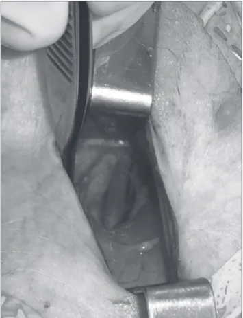
The procedure began with a left groin cutdown for exposure of the common femoral artery (Figure 1). After direct puncture to the common femoral artery was ob-tained with an 18G needle, a 0.035-inch hydrophilic gui-dewire was introduced, and a 6F introducer sheath was placed. Under fluoroscopic guidance, the guidewire was advanced to the aortic arch with a pigtail catheter inser-ted over it. After aortography and identification of the common carotid arteries, the left common carotid artery was selectively catheterized with a 5F vertebral catheter, and the 0.035-inch guidewire was replaced with a 0.014-inch guidewire. We then performed angioplasty and pla-ced a 4 x 16 mm balloon-expandable stent in the middle segment of the artery, maintaining an expansion pressu-re of 8 atm for 30 seconds. This was followed by selective catheterization of the right common carotid artery and balloon angioplasty, also with 30 seconds of 8-atm ex-pansion pressure, in the middle third of the artery, with a 4 x 16 mm balloon catheter. A catheter gauge 10–20% wider than the normal diameter of the CCA in 8-week-old swine was chosen to ensure balloon oversizing and subsequent circumferential strain and stretch. After the procedure, control arteriography was performed to con-firm artery patency.
Arteriography was performed through a 5F pigtail or vertebral catheter, with manual injection of iothalamate meglumine 1 mL/kg for contrast. Arteriograms were obtai-ned with a portable luoroscope (SK7-3) and recorded on DVD (Samsung).
animals were returned to the Animal Experimentation Unit for anesthesia and euthanasia according to Unit protocols.
Collected specimens were sent for digital morpho-metric analysis. Histology sections were obtained with a sliding microtome (Polycut S®, Leica AG, Germany), equi-pped with a type D, 16 cm-long, 5-micron tungsten carbide knife (LeicaAG, Germany), and stained according to the Verhoef–Van Gieson protocol (Figures 2, 3, and 4).
Morphometry was performed with the aid of a Quantimet 500 computerized image analysis system, un-der a Leica® microscope, at a magniication of ×2.5. he slides were examined in a blinded fashion, with no obser-ver interference. In each section, the area of the arterial lumen, the area within the internal elastic lamina (corres-ponding to the arterial lumen if no intimal proliferation were present) and external elastic lamina (external diame-ter of the vessel), and the approximate area bounded by the innermost points of any stent struts present in the section (that is, the approximate area bounded by the stent if no intimal proliferation were present) were measured. Based on these results, we calculated the area of the neointima and of the tunica media. he total area occupied by any
stent fragments present in the section was also measured, as was the extent of neointimal obstruction. Mean ove-rall wall thickness, thickness of the intima, and thickness of the media—at stent-containing points and the points between these—were calculated from the measurements of all stent segments present in each section. Mean mini-mum distance between the stent and the arterial lumen
Figure 1 – Approach to the common carotid artery
Figure 2 - Histological cross sections of the carotid artery. he elastic ibers that make up the internal and external elastic laminae (blue and red arrows respectively) are shown in black (Verhoef–Van Gieson stain, 2.5× magniication).
Figure 3 - Histological cross sections of the carotid artery showing stent fragments, some highlighted by green arrows (Verhoef–Van Gieson stain, 2.5× magniication).
(mean and standard deviation) for all parametric variables. Student’s t-test was used for comparison between groups.
The signiicance level (α) was set at 5%.
Results
All eight animals completed the study. Stent patency and patency of the angioplasty segment were assessed di-rectly during specimen collection. Gross (macroscopic) examination revealed a perivascular inlammatory reaction
intimal, and tunica media area in the analyzed specimens. Sections were obtained from the middle segment of the ca-rotid artery and the middle of the stent, sliced with a tun-gsten carbide knife, for intrastent assessment of the intimal hyperplasia process. Animals were divided into two groups, group 1 (angioplasty + stent) and group 2 (angioplasty only), as shown in Table 1.
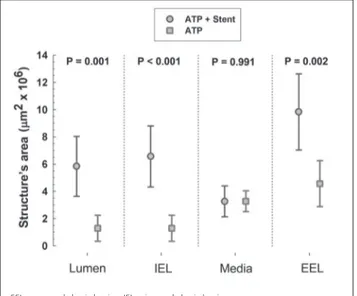
Intimal thickening (Figure 5) occurred to a greater ex-tent in the sex-tent placement group (group 1) than in the an-gioplasty-only group (group 2), but there was no diference in tissue reaction in the tunica media.
Discussion
Post-angioplasty restenosis is a multifactorial process that depends on two basic mechanisms: intimal hyperpla-sia and arterial remodeling.11,12 Experimental studies have shown that the response to vascular injury, regardless of mechanism, involves migration and proliferation of smoo-th muscle cells to and in smoo-the intima, wismoo-th synsmoo-thesis and de-position of extracellular matrix. hese events play a critical role in the pathophysiology of intimal hyperplasia, which leads to restenosis.13
Balloon angioplasty is currently accepted not only as a method for treatment of arterial occlusive disease, but also as a model for vascular injury.14,15 Angioplasty-induced cir-cumferential strain on the vessel wall leads to tearing of the internal elastic lamina and stretching of collagen and elas-tin ibers in the tunicae media and adventitia. Endothelial injury is followed by endothelial dysfunction, with sub-sequent platelet aggregation on the surface of the disrup-ted area and an inlammatory reaction that may extend to all layers of the vessel wall. Furthermore, hemodynamic
Variable Group Mean
(x 106 µm²)
SD (x 106 µm²)
SE
(x 106 µm²) p
Lúmen 1 5.841 2.200 0.777 p = 0.001
2 1.287 0.956 0.338
LEI 1 6.566 2.240 0.792 p < 0.001
2 1.287 0.956 0.338
LEE 1 9.832 2.787 0.985 p = 0.991
2 4.559 1.685 0.595
Média 1 3.266 1.134 0.401 p = 0.002
2 3.271 0.763 0.269
Table 1 – Morphometric data (N = 8)
EEL = external elastic lamina; IEL = internal elastic lamina.
Figure 5 – Comparison of lumen, IEL, media, and EEL measurements between the angioplasty only and angioplasty + stenting group. here is a signiicant diference in luminal area, IEL, and EEL measurements between the two groups. here is no statistically signiicant diference in tunica media measurement between the two groups.
changes secondary to vascular trauma may trigger cellular responses leading to arterial contraction, thus contributing to ibrocellular hyperplasia of the intima. Smooth muscle cells take on a “synthetic/proliferative” phenotype and mi-grate to the intima, becoming the dominant cell type in in-timal hyperplasia.12-15
Stents were developed with the purpose of mecha-nically supporting the arterial wall. As compared with balloon angioplasty, stent placement improves short- and midterm outcomes by reducing negative remodeling. he metallic alloys from which stents are manufactured seek a compromise between biofunctionality and biocompatibili-ty; however, the choice of material must focus primarily on biocompatibility. he main aspects to be taken into account in this respect are susceptibility of the alloy to corrosion and the efects of this corrosion on the recipient’s body.3,7
Experimental human models have irmly established that the extent of injury caused by angioplasty and stent pla-cement is directly associated with intimal hyperplasia.12,13,15 Large-animal models (mostly porcine) appear to relect the pathogenesis of restenosis better than small-animal models (mouse and rabbit) do, particularly with respect to preo-perative care and artery handling.16 Mouse studies suggest that bone marrow progenitor cells may also play a role in the restenosis process.17 he key limiting factor of animal models is the lack of preexisting atherosclerosis, which is present in humans. Dietary lipid supplementation of labo-ratory animals induces the formation of vulnerable athe-rosclerotic plaques,18 but subsequent intimal hyperplasia and restenosis are similar to those occurring in animals fed a normal diet.19 he use of an atherogenic diet thus appe-ars to provide no advantage in porcine animal models of restenosis.
A four-week interval between intervention and tissue sampling is enough in experimental studies of post-angio-plasty intimal hyperplasia. Past studies have reported deve-lopment of full-blown intimal hyperplasia, similar to that found in humans, within 28 days.12-14,20
In prior studies, Pasa et al.21 conducted morphometric analysis of intimal thickening in the swine carotid artery ater placement of 316L-grade stainless steel stents, without angioplasty, and concluded that intimal thickening indu-ced by the stent is greater than that produindu-ced by injury of the contralateral artery by the introducer sheath. he mere presence of a stent, even in the absence of rupture of the internal elastic lamina, is therefore enough to produce in-timal hyperplasia. Grudtner et al.22 and Dutra & Pereira23 investigated the process of neointimal formation in the swine aorta ater angioplasty and placement of e-PTFE and
Dacron-coated 316L-grade stainless steel as compared with results obtained ater placement of uncoated stents. he authors found no diference between the study groups, but conirmed that all stents, coated or otherwise, are associa-ted with signiicant formation of neointima. Castro et al.24 assessed the neointimal response to angioplasty with 316L stainless steel stenting in the swine iliac artery as compared with simple balloon angioplasty of the contralateral iliac ar-tery. In their study, stenting was associated with increased intimal hyperplasia, but no reduction of the arterial lumen, due to positive remodeling of the elastic lamina.
he aforementioned studies, all conducted in Brazil, only assessed intimal hyperplasia in the stent margins, as no cross-sections were obtained from the stented segments (Figures 2, 3, and 4). In the present study, microtome cross-sections of the middle segment of the stent were obtained with the aid of a tungsten carbide knife, which allowed as-sessment of the extent of intrastent injury. No study con-ducted thus far had gone beyond assessing alloy behavior in the coronary circulation, where device placement was associated with less intimal hyperplasia than other metallic alloys.25,26
In the early days of histology, ine sections meant for microscopic examinations were obtained manually with the aid of razors and knives. In the late 19th century, Chevalier and Purkinje separately developed the irst mechanical mi-crotomes.27 In the present study, we used a tungsten carbi-de knife-equipped microtome due to the resistance of the stent mesh and the need for precise sections. Unlike dia-mond blades, tungsten knives keep stent struts intact on cross-sectioning, minimizing the potential for stent remo-val artifacts.
Histological sections were obtained to assess intimal hyperplasia and luminal area preserved ater intervention. Luminal area was calculated by direct measurement of the area bounded by endothelium or by the stent itself. Intimal area was calculated by subtracting the luminal area from the area bounded by the internal elastic lamina, while the area of the tunica media was calculated by adding the va-lues obtained from luminal and intimal measurement and subtracting them from the value measured for the external elastic lamina. Intima and media measurements were ex-pressed as absolute igures, in square micrometers, by ave-raging the values of eight circumferential measurements.
Our luminal area indings highlight the importance of negative arterial remodeling ater simple angioplasty. Conversely, as it widens the arterial lumen (positive remo-deling), stent placement prevents intimal hyperplasia from signiicantly reducing luminal area. Stenting thus prevents negative arterial remodeling and supports the arterial wall.
Cobalt-chromium stents were developed for use in smaller and sinuous arteries, as cobalt-chromium alloys produce a more resistant and lexible mesh than stainless steel. heir struts are also thinner and provide greater ra-dial strength, allowing treatment of tortuous and calciied atherosclerotic lesions, particularly in the coronary circu-lation.26 heoretically, these stents’ thinner struts—which, of course, mean a lower amount of metal is present – could be associated with improved biotolerance. Another very useful feature of this alloy is that recipients of cobalt-chro-mium stents may undergo magnetic resonance imaging. Experimental studies in the swine coronary artery have sho-wn that polyphosphazene-coated cobalt-chromium stents may induce less neointimal formation than stainless steel stents coated with the same polymer.25 However, our review of the literature did not yield any experimental studies in the peripheral circulation to corroborate these indings.
he cobalt-chromium stents used in the present study did not appear to be associated with a reduction in intimal hyperplasia. he hyperplasia secondary to angioplasty and cobalt-chromium stent placement does not appear to di-fer from that found ater placement of devices made from 316L-grade stainless steel or nitinol in previous studies con-ducted in our experimental unit. Various studies concon-ducted by Pasa et al., Grudtner et al., Dutra et al. e Castro et al.21-24 have shown the role of angioplasty- and stent-induced vas-cular injury, regardless of alloy and coating, in the intimal hyperplasia process.
Long-term maintenance of arterial lumen patency is the ultimate purpose of stenting. Several studies have addressed the possibility of pharmacotherapy to suppress the intrastent restenosis process. he search for drugs that suppress myointimal proliferation and devices that induce a lesser degree of parietal reaction remains a challenge.
Conclusion
We conclude that, in swine, angioplasty of the common carotid artery with cobalt-chromium stent implantation
arterial lumen size due to positive remodeling.
References
1. Fattori R, Piva T. Drug-eluting stents in vascular intervention. Lancet. 2003;361:247-9.
2. Inoue S, Koyama H, Miyata T, Shigematsu H. Pathogenetic hetero-geneity of in-stent lesion formation in human peripheral arterial disease. J Vasc Surg. 2002;35:820-2.
3. Fischer A, Wienecke H, Brauer H, Erbel R. [Metallic biomaterials for coronary stents]. Z Kardiol. 2001;90:251-62.
4. Hansi C, Arab A, Rzany A, Ahrens I, Bode C, Hehrlein C. Diferences of platelet adhesion and thrombus activation on amorphous si-licon carbide, magnesium alloy, stainless steel, and cobalt chro-mium stent surfaces. Catheter Cardiovasc Interv. 2009;73:488-96.
5. Waksman R, Pakala R, Kuchulakanti PK, et al. Safety and eicacy of bioabsorbable magnesium stents in porcine coronary arteries. Catheter Cardiovasc Interv. 2006;68:607-17.
6. Messer RL, Wataha JC, Lewis JB, Lockwood PE, Caughman GB, Tseng WY. Efect of vascular stent alloys on expression of cellu-lar adhesion molecules by endothelial cells. J Long Term Ef Med Implants. 2005;15:39-47.
7. Gotman I. Characteristics of metals used in implants. J Endourol. 1997;11:383-9.
8. Koch W, Tiroch K, von Beckerath N, Schömig A, Kastrati A. Tumor necrosis factor-alpha, lymphotoxin-alpha, and interleukin-10 gene polymorphisms and restenosis after coronary artery stenting. Cytokine. 2003;24:161-71.
9. Versaci F, Gaspardone A. Prevention of restenosis after stenting: the emerging role of inlammation. Coron Artery Dis. 2004;15:307-11.
10. Welt FG, Rogers C. Inlammation and restenosis in the stent era. Arterioscler hromb Vasc Biol. 2002;22:1769-76.
11. Ouriel K. Peripheral arterial disease. Lancet. 2001;358:1257-64.
12. Toutouzas K, Colombo A, Stefanadis C. Inlammation and res-tenosis after percutaneous coronary interventions. Eur Heart J. 2004;25:1679-87.
13. Cwikiel W, Harnek J, Zoucas E, Stenram U. Proliferative respon-se in smooth muscle cells after angioplasty or inrespon-sertion of respon- self-expanding stents. An experimental study in pigs. Acta Radiol. 1997;38:124-8.
14. De Meyer GR, Bult H. Mechanisms of neointima formation: les-sons from experimental models. Vasc Med. 1997;2:179-89.
15. Wolf YG, Gertz SD, Banai S. Animal models in syndromes of accele-rated arteriosclerosis. Ann Vasc Surg. 1999;13:328-38.
17. Tsai S, Butler J, Raii S, Liu B, Kent KC. he role of progenitor cells in the development of intimal hyperplasia. J Vasc Surg. 2009;49: 502-10.
18. Shi ZS, Feng L, He X, et al. Vulnerable plaque in a Swine model of carotid atherosclerosis. AJNR Am J Neuroradiol. 2009;30:469-72.
19. França LH, Pereira AH, Perini SC. Self-expandable nitinol stent placement in homocysteinemic porcine aorta. Clinics. 2008;63:229-36.
20. Verheye S, Salame MY, Robinson KA, et al. Short- and long-term histopathologic evaluation of stenting using a self-expanding ni-tinol stent in pig carotid and iliac arteries. Catheter Cardiovasc Interv. 1999;48:488-96.
21. Pasa MB, Pereira AH, Castro Junior C. Morphometric analysis of intimal thickening secondary to stent placement in pig carotid ar-teries. Acta Cir Bras. 2008;23:165-72.
22. Grudtner MA, Pereira AH, Costa LF, Souza GG, Argenta R, Longhi JA. Efeitos a curto prazo de stents não recobertos e recobertos com politetraluoroetileno em aorta de suínos: um modelo expe-rimental. Acta Cir Bras. 2004;19:120-5.
23. Dutra CF, Pereira AH. Digital morphometric analysis of the aortic wall in pigs following implantation of dacron-covered stents ver-sus non-covered stents. Acta Cir Bras. 2004;19:210-219.
24. Castro Junior C, Pereira AH, Pasa MB. Morphometric analisis of in-timal reaction after stent implantation in iliac arteries submitted to angioplasty in pigs. Acta Cir Bras. 2006;21:139-43.
25. Stampl U, Sommer CM, hierjung H, et al. Reduction of late in-stent stenosis in a porcine coronary artery model by cobalt chro-mium stents with a nanocoat os polyphosphazene (Polyzene-F). Cardiovasc Intervent Radiol. 2008;31:1184-92.
26. Kereiakes DJ, Cox DA, Hermiller JB, et al. Usefulness of a cobalt chromium coronary stent alloy. Am J Cardiol. 2003;92:463-6.
27. Santos MB. Ergonomia, carga mental de trabalho, riscos e preven-ção de acidentes: o caso do trabalhador em histotécnica [disserta-ção]. Rio de Janeiro (RJ): Pontifícia Universidade Católica do Rio de Janeiro; 2007.
Correspondence: João Luiz Elesbão Rua Landel de Moura, 1379/1 – Tristeza CEP 91920-150 – Porto Alegre, RS, Brazil Tel.: +55 (51) 2111.9843 E-mail: [email protected]