Incidental detection of retroperitoneal
lymphangio-leiomyomatosis (LAM) - CT and MRI findings with
re-levance to the urologist
_______________________________________________
Chandan Phukan, Shailesh Prabhu, Vivek Venkatramani
Department of Urology, Christian Medical College, Vellore, India
_______________________________________________________________________________________
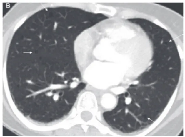
A 48 year old lady presented with blunt abdominal trauma following a road traffic acci-dent. She had no hematuria or other injuries. Vi-tals were normal. Examination was significant for left loin tenderness. Ultrasound was suspicious for retroperitoneal fluid. Computed tomography reve-aled normal abdominal viscera but multiple diffu-se low attenuating cystic retroperitoneal masdiffu-ses showing peripheral enhancement (Figure-1). Lung segments showed multiple thin walled parenchy-mal cysts bilaterally (Figure-2, arrows). T2-wei-ghted MRI revealed conglomerated cystic masses in the paraaortic and parailliac regions (Figure-3, arrows) suggesting multiple lymphangiomyomas.
574
RADIOLOGY PAGE
Figure 1 - Multiple diffuse low attenuating cystic masses with peripheral enhancement.
Figure 2 - Lung segments with multiple thin walled parenchymal cysts.
A diagnosis of lymphangioleiomyomatosis (LAM) was made. She was managed conservati-vely and was well at 6 months.
Lymphangioleiomyomatosis is a rare hamartomatous lesion affecting women of re-productive age who manifest with respiratory symptoms secondary to multiple pulmonary cysts (1). Abdominal manifestations include angiomyolipomas (20-54%), lymphangioleio-myoma (20%) and lymphadenopathy (40%) (1). The disease is slowly progressive and results in respiratory failure and death in 31-62% cases at 8-10 years (1). The importance of this entity to the urologist is its association with angio-myolipoma and tuberous sclerosis. Though cli-nical pulmonary manifestations are rare (2-3%)
Vol. 40 (4): 574-575, July . August, 2014
IBJU| INCIDENTAL DETECTION OF RETROPERITONEAL LYMPHANGIOLEIOMYOMATOSIS
575
ARTICLE INFO
Int Braz J Urol. 2014; 40: 574-5
_____________________
Submitted for publication: February 18, 2014 _____________________
Accepted after revision: March 18, 2014 Figure 3 - T2-weighted MRI with conglomerated cystic
masses in the paraaortic and parailliac regions.
in tuberous sclerosis, morphological changes similar to LAM are seen in a much larger per-centage (2).
REFERENCES
1. Pallisa E, Sanz P, Roman A, Majó J, Andreu J, Cáceres J: Lymphangioleiomyomatosis: pulmonary and abdominal findings with pathologic correlation. Radiographics. 2002; 22 Spec No: S185-98.
2. Abbott GF, Rosado-de-Christenson ML, Frazier AA, Franks TJ, Pugatch RD, Galvin JR: From the archives of the AFIP: lymphangioleiomyomatosis: radiologic-pathologic correlation. Radiographics. 2005; 25: 803-28.
_______________________ Correspondence address: