Laparoscopic Diagnosis and Treatment of Nonpalpable Testis
Francisco T. Denes, Fernando J. Saito, Frederico A. Silva, Amilcar M. Giron, Marcos Machado,
Miguel Srougi
Division of Urology, School of Medicine, University of Sao Paulo, USP, Sao Paulo, SP, Brazil
ABSTRACT
Introduction: Treatment of the cryptorchid testicle is justiied due to the increased risk of infertility and malignancy as well as the risk of testicular trauma and psychological stigma on patients and their parents. Approximately 20% of cryptorchid testicles are nonpalpable. In these cases, the videolaparoscopic technique is a useful alternative method for diagnosis and treatment.
Materials and Methods: We present data concerning 90 patients submitted to diagnostic laparoscopy for impalpable testicles. Forty-six patients (51.1%) had intra-abdominal gonads. In 25 testicles of 19 patients, we performed a two stage laparoscopic Fowler-Stephens orchiopexy. The other 27 patients underwent primary laparoscopic orchiopexy, in a total of 29 testicles.
Results: We obtained an overall 88% success rate with the 2 stage Fowler-Stephens approach and only 33% rate success using one stage Fowler-Stephens surgery with primary vascular ligature. There was no intraoperative complication in our group of patients. In the laparoscopic procedures, the cosmetic aspect is remarkably more favorable as compared to open surgeries. Hospital stay and convalescence were brief.
Conclusions: In pediatric age group, the laparoscopic approach is safe and feasible. Furthermore, the laparoscopic orchio-pexy presents excellent results in terms of diagnosis and therapy of the impalpable testis, which is why this technique has been routinely incorporated in our Department.
Key words: testis; cryptorchidism; laparoscopy
Int Braz J Urol. 2008; 34: 329-35
INTRODUCTION
Cryptorchidism is the most common geni-tourinary anomaly in male children. Its incidence can reach 3% in full term neonates, rising to 30% in premature boys (1). The treatment of the cryptorchid testicle is justiied by the increased risk of infertility and malignancy, as well as an associated inguinal hernia and the risk of trauma to the ectopic testicle against the pubis. Furthermore, the psychological stigma of a missing testis for the patient, as well as the parents’ anxiety are also factors that justiies this type of treatment (2,3).
About 20% of cryptorchid testicles are non-palpable. In these cases, the laparoscopic technique is a useful alternative method of diagnosis and treat-ment. We assessed our data and present our results, including a comparison between the laparoscopic and two stage Fowler-Stephens approaches.
MATERIALS AND METHODS
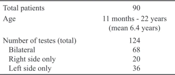
(37.8%) presented with bilateral, while 56 (62.2%) had unilateral impalpable cryptorchidism. The age and the laterality are presented in Table-1.
Preoperatively, all the patients were examined at least by two different examiners at different times, conirming the diagnosis. Another careful physical examination was performed in the operating room, with the patient under anesthesia. When the testicle was palpated on any one of these occasions, the patient was submitted to an open orchiopexy.
Although the preoperative ultrasound for lo-cation of the testicle was performed in some patients, with negative results in all, no patient underwent computerized tomography scan or magnetic nuclear resonance imaging for the same purpose.
There was no age limit for the laparoscopic procedure. The procedure was performed under
general anesthesia, with orotracheal ventilation and nasogastric and vesical tubes.
The laparoscopic technique (Figure-1) has previously been described (4-6).The laparoscopic indings were similar to those described by Castilho in 1990 (7), and are summarized in Table-2. Surgical
Table 1 – Age and laterality data.
Total patients 90
Age 11 months - 22 years
(mean 6.4 years) Number of testes (total)
Bilateral
Right side only
Left side only
124 68 20 36
management was performed based on the laparo-scopic indings (8,9). In cases of testicle absence , the procedure was interrupted, whereas in cases of intra-canalicular inguinal testis, open surgical explo-ration was performed. When intra-abdominal testes were found, immediate laparoscopic orchiectomy was performed for atrophic testicles, while patients with viable testicles underwent laparoscopic orchiopexy. The technique of this procedure has been previously described, stressing that in cases of low intra-abdomi-nal testicle (located less than 2 cm from the interintra-abdomi-nal inguinal ring) the procedure was straightforward, without transection of the spermatic vessels, while in those located higher (more than 2 cm from the internal inguinal ring) the vessels were sectioned to facilitate the appropriate descent of the testicle to the scrotum (6). When the vessels are transected, the testis is relo-cated into the scrotum either during the same surgical procedure (primary or one stage Fowler-Stephens) or the relocation is postponed for at least six months after vascular ligature (two stage Fowler-Stephens).
All the operated patients were followed-up for 6 to 100 months, and evaluated for the incidence of intra and post-operative complications, as well as for the inal location and morphology of the operated testes. These complications were classiied as normal (good size and consistence, in addition to appropriate position in the scrotum), atrophic (altered morphol-ogy, independent of the position) or malpositioned (normal morphology, but located above the scrotum).
In cases of unilateral disease, the evaluation of crypt-orchid testicle was based on the normal testicle. In cases of bilateral disease, this evaluation was based on clinical palpation as well as ultrasonography in some cases, comparing the obtained values with normal parameters in infancy and adulthood.
RESULTS
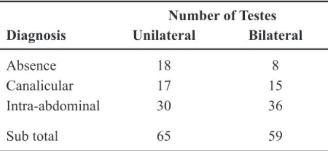
The initial laparoscopic indings are summa-rized in Table-3. One should note that ive patients had bilateral disease, in which we had different diagnostic indings in each affected side as emphasized in Table-3.
Eighteen patients (20%) presented absent testicles, four due to agenesis and 14 to vanishing
Table 2 – Laparoscopic indings classiication.
Absent testis Agenesis (absence of spermatic vessels and vas deferens). Vanishing testis (blind ending of spermatic vessels or vas)
Canalicular testis Penetration of vas and spermatic vessels into the internal inguinal ring with or without
directly seeing the testis
Abdominal testis Localized between the inferior renal pole and the ipsilateral internal inguinal ring. Can be a normal or an atrophic gonad.
Peeping testis Primarily in intra-abdominal position. The testis introduces itself into the inguinal canal due to the intra-abdominal pressure augmentation during the laparoscopic procedure. Usually associated with inguinal hernia and returns to its original position by pressing the inguinal region externally.
Table 3 – Laparoscopic results.
Number of Testes Diagnosis Unilateral Bilateral
Absence 18 8
Canalicular 17 15
Intra-abdominal 30 36
Sub total 65 59
testes. In these cases, the laparoscopic procedure was completed preventing the patients from any further unnecessary exploration. In 12 patients of this group, who were near pubertal age, a testicular prosthesis was inserted during the same surgical procedure.
Thirty-two patients (35.5%) had a diagnosis of intra-canalicular inguinal testicles. In this group, the dificulty in palpating the testis was due to re-gional obesity, the small size of the testis or general anesthesia, which facilitated the child’s examination. The majority of patients underwent a conventional inguinal exploration, and those with viable gonads (34.4%) had an orchiopexy during the same pro-cedure, while those with atrophic testicles (65.6%) underwent orchiectomy. Only one patient underwent a laparoscopic dissection of the inguinal testicle via the internal inguinal ring, where an atrophic testis was found, and removed. In this group, the older children also received testicular prosthesis at the same time. Interestingly, we observed that the presence of a hernia sac almost always indicates the presence of a canalicular testicle, particularly in peeping testis.
In 46 patients (51.1%), the testes were intra-abdominal, and treatment varied according to their morphology and position (Table-4). In three patients, four testicles were atrophic (one patient with bilateral disease). All were submitted to immediate scopic orchiectomy. In the other 43 patients laparo-scopic orchiopexy was performed. In 25 testicles of 19 patients, we performed the two stage laparoscopic Fowler-Stephens orchiopexy, with initial vascular transection. All were submitted to orchiopexy at least
six months later. In 21 testicles of 15 patients, the orchiopexy was also done by laparoscopic technique, while the remaining four testicles were positioned by conventional inguinal approach.
The other 27 patients underwent primary laparoscopic orchiopexy, in a total of 29 testicles. In 3 of these testicles (3 patients), vascular ligature and section were necessary, while the majority (26 tes-ticles) was relocated to the scrotum with preservation of the vascular pedicle.
Minimal follow-up period was approximately 6 months. Only 2 patients were lost to follow-up dur-ing a ifteen year protocol.
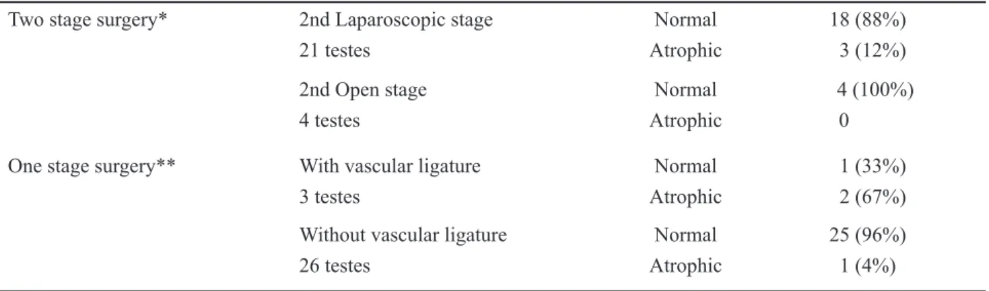
Of the 25 testicles advanced into the scrotum by the two step Fowler-Stephens technique, 18 (88%) presented good morphology and position in the scro-tum, while 3 testicles became atrophic. Considering this same group, an 85% success rate was achieved with the laparoscopic second stage, as compared to a 100% success rate with the open approach.
Of the 25 testicles submitted to the primary laparoscopic orchiopexy, without vascular transection, 96% were considered successful, with good position and normal morphology, with only one testis develop-ing atrophy. Among the testes submitted to primary orchiopexy with simultaneous vascular ligature, two presented atrophy, while one testis remained normal (success rate of 33%).
There was no intraoperative complication in our group of patients, and none required blood transfu-sion or convertransfu-sion to open procedure. All patients who underwent a laparoscopic diagnostic procedure alone
Table 4 – One stage vs. two stage Fowler-Stephens.
Two stage surgery* 2nd Laparoscopic stage Normal 18 (88%)
21 testes Atrophic 3 (12%)
2nd Open stage Normal 4 (100%)
4 testes Atrophic 0
One stage surgery** With vascular ligature Normal 1 (33%)
3 testes Atrophic 2 (67%)
Without vascular ligature Normal 25 (96%)
26 testes Atrophic 1 (4%)
could be fed on the same day and were discharged the following day. Those who underwent orchiopexy were discharged on the second post-operative day. Post-operative pain was minimal and treated with common analgesics or non-steroidal anti-inlammatory drugs, according to patients’ age.
In late follow up, we did not observe any post-operative complication in the abdominal or scrotal percutaneous ports, nor inguinal or incisional hernia.
COMMENTS
The treatment of non-descended testicles is mandatory due to the increased risk of infertility, pres-ent in up to 40% of the patipres-ents, as compared to 6% of control groups (10), including malignancy, which reaches 20 times that of normal adults (11).
Despite the recommendations for the treat -ment of the cryptorchid testis before 2 years of age, many of our patients were older, due to the socio-eco-nomic characteristics of the public health system in our country, the lack of parental information and dif-icult access to tertiary health care. Although fertility is already compromised in this age group, treatment is necessary not only for the risk of malignancy, but also for the satisfaction and improvement in the qual-ity of the patient’s life and parents´ concern for their children’s health (12).
In relation to diagnosis, some tests can be used for appropriate therapeutic planning. In the case of bi-lateral impalpable testes, the stimulation with human chorionic gonadotrophin has only a relative useful-ness, since a negative result, although suggestive of absent testes, cannot completely exclude the presence of a dysplasic gonad. Even in the case of a positive answer, it is not possible to establish the number, location and the laterality of the gonad (13). Despite a sensitivity of 70-90% in the diagnosis of inguinal testes, ultrasonography is not useful in intra-abdomi-nal cases(14). Although presenting a better quality, both computed tomography and nuclear magnetic resonance lack suficient sensitivity and speciicity to be considered as gold standard diagnostic tools (15). More recently, the magnetic angioresonance was introduced with sensibility of 96% and speciicity of
100%, but it is still a new method, with high costs, also requiring general anesthesia in children (16).
In relation to the treatment, the use of gonado-trophin for undescended testes presents a success rate of deinitive descent to the scrotum of 21 to 56%, with better results in bilateral cases (13,14). Surgical treat-ment via an inguinal incision is the main treattreat-ment option for palpable testicles, but can also be employed for the evaluation and treatment of impalpable testis. In this situation, however, surgical exploration can often require large incisions and extensive dissections, especially in bilateral cases. This can be avoided us-ing laparoscopic evaluation, with a sensitivity and speciicity reaching more than 90% (17,18).
In 20% of our patients with testicular agenesis or vanishing testis, laparoscopic surgery was the deci-sive diagnostic method and saved these patients from any further incision or unnecessary investigation. In patients with intra-canalicular testicles, laparoscopy was fundamental for guiding the minimal inguinal exploration, which was augmented only in cases of a viable gonad, when orchiopexy was performed.
In cases of intra-abdominal testicles, the great advantage of laparoscopy is that, besides cor-rect diagnosis, it enables the therapeutic handling of the testes at the same time. Additionally in cases of associated inguinal hernia (particularly in cases with peeping testis), the laparoscopic approach also enables the simultaneous treatment of the hernia sac with favorable results.
Using laparoscopic procedures, the cosmetic aspect is remarkably more favorable as compared to open surgery , and the hospital stay and convalescence are much shorter. In the pediatric age group, these fac-tors may not be so evident for the patient themselves, but certainly will be for the parents, who are able to resume their daily activities earlier. Furthermore, the laparoscopic orchiopexy presents excellent results in terms of diagnosis and therapy of the impalpable testis, which is why this technique has been routinely incorporated in our Department. It is noteworthy that our preference is the primary orchiopexy without tran-section of the gonadal vessels. However, in cases of very high testicles or those with short vessels we now recommend the two staged laparoscopic technique of Fowler-Stephens.
CONFLICT OF INTEREST
None declared.
REFERENCES
1. Berkowitz GS, Lapinski RH, Dolgin SE, Gazella JG, Bodian CA, Holzman IR: Prevalence and natural his-tory of cryptorchidism. Pediatrics. 1993; 92: 44-9. 2. Trussell JC, Lee PA: The relationship of
cryptorchi-dism to fertility. Curr Urol Rep. 2004; 5: 142-8. 3. Moreno-Garcia M, Miranda EB: Chromosomal
anoma-lies in cryptorchidism and hypospadias. J Urol. 2002; 168: 2170-2; discussion 2172.
4. Poppas DP, Lemack GE, Mininberg DT: Laparoscopic orchiopexy: clinical experience and description of technique. J Urol. 1996; 155: 708-11.
5. Lindgren BW, Franco I, Blick S, Levitt SB, Brock WA, Palmer LS, et al.: Laparoscopic Fowler-Stephens orchiopexy for the high abdominal testis. J Urol. 1999; 162: 990-3; discussion 994.
6. Dénes, FT: Avaliação e tratamento do testículo não-palpável. In Castilho LN, Laparoscopia Urológica. Campinas, LPC Comunicações, 2000; pp. 467-5. 7. Castilho LN: Laparoscopy for the nonpalpable testis:
how to interpret the endoscopic indings. J Urol. 1990; 144: 1215-8.
8. Peters CA, Kavoussi LR, Retik AB: Laparoscopic Management of intra-abdominal testes. J Endourol. 1993; 7(Suppl 1): 170-4.
9. Cortes D, Thorup JM, Lenz K, Beck BL, Nielsen OH: Laparoscopy in 100 consecutive patients with 128 impalpable testes. Br J Urol. 1995; 75: 281-7. 10. Lee PA, O’Leary LA, Songer NJ, Coughlin MT,
Bellinger MF, LaPorte RE: Paternity after unilateral cryptorchidism: a controlled study. Pediatrics. 1996; 98: 676-9.
11. Garner MJ, Turner MC, Ghadirian P, Krewski D: Epidemiology of testicular cancer: an overview. Int J Cancer. 2005; 116: 331-9.
12. Kucheria R, Sahai A, Sami TA, Challacombe B, God-bole H, Khan MS, et al.: Laparoscopic management of cryptorchidism in adults. Eur Urol. 2005; 48: 453-7; discussion 457.
13. Rajfer J, Handelsman DJ, Swerdloff RS, Hurwitz R, Kaplan H, Vandergast T, et al.: Hormonal therapy of cryptorchidism. A randomized, double-blind study comparing human chorionic gonadotropin and go-nadotropin-releasing hormone. N Engl J Med. 1986; 314: 466-70.
14. Kolon TF, Patel RP, Huff DS: Cryptorchidism: diag-nosis, treatment, and long-term prognosis. Urol Clin North Am. 2004; 31: 469-80.
15. Nguyen HT, Coakley F, Hricak H: Cryptorchidism: strategies in detection. Eur Radiol. 1999; 9: 336-43. 16. Eggener SE, Lotan Y, Cheng EY: Magnetic resonance
angiography for the nonpalpable testis: a cost and can-cer risk analysis. J Urol. 2005; 173: 1745-9; discussion 1749-50.
17. Docimo, SG: The results of surgical therapy for crypt-orchidism: a literature review and analisys. J Urol. 1995; 154: 1148-52.
18. Froeling FM, Sorber MJ, de la Rosette JJ, de Vries JD: The nonpalpable testis and the changing role of laparoscopy. Urology. 1994; 43: 222-7.
19. Lindgren BW, Darby EC, Faiella L, Brock WA, Reda EF, Levitt SB, et al.: Laparoscopic orchiopexy: pro-cedure of choice for the nonpalpable testis? J Urol. 1998; 159: 2132-5.
20. Baker LA, Docimo SG, Surer I, Peters C, Cisek L, Diamond DA, et al.: A multi-institutional analysis of laparoscopic orchidopexy. BJU Int. 2001; 87: 484-9.
Accepted after revision: October 20, 2007
Correspondence address: Dr. Francisco Tibor Dénes
EDITORIAL COMMENT
Laparoscopy is an accepted diagnostic and treatment modality for non-palpable testes as per-formed in the current series. In this series, the percent-age of intra-canalicular viable testis which was not palpable during the examination even under anesthesia is high (34.4% of 32 patients) and it is not similar to our experience (1). Whatever the reason, we encour-age the authors to perform laparoscopic orchiopexy instead of converting the operation to open surgery in this situation.
Classiication of intra-abdominal testes ac-cording to the measurement of distance between the testes and the internal inguinal ring is a good criterion but in our series, we have few cases, which do not match this criterion. Based on these observations, we prefer to examine the mobility of the testis by a laparoscopic forceps and to decide if the length of the spermatic vessels and ductus deferens is suitable for one or two stage operation.
In our series, a few cases were previously explored by open or laparoscopic technique at another center and, diagnosed as “absence of testis”. In those cases, we had documented positive response to HCG stimulation test and diagnostic laparoscopy revealed
the presence of an intra-abdominal testis. Therefore, we advocate performing an HCG stimulation test in patients with bilateral non-palpable testes before surgical exploration.
The authors performed testicular prosthesis placement following laparoscopic exploration in older patients with vanishing testis. We recommend a simi-lar option for the younger patients and, this alterna-tive approach could be offered to the parents before laparoscopic exploration. Inguinal exploration may be postponed and, testicular nubbins can be removed later at the time of testicular prosthesis implantation surgery if there is a consensus with the family (2).
REFERENCES
1. Topuzlu Tekant G, Emir H, Eroðlu E, Akman M, Büyükünal C, Daniþmend N, et al.: Experience with laparoscopy in nonpalpable testis. Eur J Pediatr Surg. 2001; 11: 177-81.
2. Emir H, Ayik B, Eliçevik M, Büyükünal C, Daniþ-mend N, Derviþoðlu S, et al.: Histological evalua-tion of the testicular nubbins in patients with nonpal-pable testis: assessment of etiology and surgical approach. Pediatr Surg Int. 2007; 23: 41-4.
Dr. Haluk Emir
Division of Pediatric UrologyCerrahpaşa Medical Faculty