AtchitIV
gene expression is stimulated under abiotic stresses and is spatially
and temporally regulated during embryo development
Liliane B. de A. Gerhardt
1, Cláudia Magioli
1, Ana B.U.C.M. Perez
1, Rogério Margis
1,2,
Gilberto Sachetto-Martins
1and Márcia Margis-Pinheiro
11
Universidade Federal do Rio de Janeiro, Departamento de Genética, Laboratório de Genética Molecular
Vegetal, Rio de Janeiro, RJ, Brazil.
2
Universidade Federal do Rio de Janeiro, Instituto de Química, Departamento de Bioquímica,
Rio de Janeiro, RJ, Brazil.
Abstract
The expression of AtchitIV gene was analysed in Arabidopsis plants submitted to abiotic stresses. Transcript accumulation was detected in leaves in response to UV light exposure, exogenous salicylic acid administration and wounding. TransgenicArabidopsis plants carrying AtchitIV promoter::gus fusion also showed differential expression of the reporter gene in response to these treatments. TheAtchitIV expression was also analysed during Arabidopsis embryo development. GUS assay demonstratedAtchitIV promoter activation in zygotic embryos from torpedo stage up to full maturation. Promoter deletion analysis indicated that all the 5’cis-acting elements responsible for the specific tissue expression are located in a region of 1083 bp, adjacent to the start of transcription. A negative regulatory region located between portions -1083 and -600 was also observed.
Key words:abiotic stress,Chia4 Arabidopsis thalianaendochitinase, embryogenesis, gene expression, promoter.
Received: May 12, 2003; Accepted: October 21, 2003.
Introduction
Plants respond to environmental changes by control-ling the expression of a large number of genes. Endo-chitinases are constitutively expressed in several organs of healthy plants (Samac et al., 1990; Beerhues and Kom-brink, 1994; Robinsonet al., 1997; Yeboahet al., 1998). However, the modulation of chitinase expression was ob-served in response to Nod factors during the nodulation process (Staehelinet al.1994 and 1995; Goormachtiget al., 1998), as well as in adverse conditions, suggesting that these enzymes are involved in plant defence responses (Samac and Shah, 1991; Margis-Pinheiro et al., 1993; Beerhues and Kombrink, 1994; Büchter et al., 1997; Busamet al., 1997; Dong and Dunstan, 1997; Yu et al., 1998; Ancilloet al., 1999). It has also been demonstrated that plant endochitinases expression is spatially and tempo-rally regulated during plant development processes such as somatic embryogeneses (de Jonget al., 1992 and 1993). In carrots, the presence of EP3 chitinase increased the number of globular embryos of ts11 mutants in non-permissive
temperature conditions and also promoted the transition from the globular to heart-shape stage (de Jonget al., 1992 and 1993). InPicea glauca, a chitinase gene is also acti-vated during maturation of somatic embryos (Dong and Dunstan, 1997).
We have previously described the isolation and char-acterisation of A. thaliana Chia4 chitinase, the AtchitIV gene, which accumulated very rapidly in Arabidopsis leaves challenged withXanthomonas campestrisbacteria (Gerhardtet al., 1997). Recently, the expression pattern of theAtEP3chitinase fromArabidopsiswas also described. Sequence analysis revealed thatAtEP3andAtchitIV corre-spond to the same gene (Passarinhoet al., 2001). In the same study,in situhybridization, revealed that AtchitIV/ AtEP3is expressed in “nursing” cells surrounding the em-bryos during the development of somatic emem-bryos. In plants,AtchitIV/AtEP3expression was detected in mature pollen and in growing pollen tubes until they enter the re-ceptive synergid, but no activity was detected in embryos. AtchitIV/AtEP3 expression was also detected in hyda-thodes, stipules, root epidermis and emerging root hair. Based on AtchitIV/AtEP3 gene expression pattern, Passarinho and co-workers proposed that thisArabidopsis chitinase is involved in programmed cell death (PCD).
Send correspondence to Márcia Margis-Pinheiro. Universidade Federal do Rio de Janeiro, Departamento de Genética, Laboratório de Genética Molecular Vegetal, Caixa Postal 68011, 21941-970, Rio de Janeiro, RJ, Brazil. E-mail: [email protected].
In this paper, analyses of induction and temporal ac-cumulation ofAtchitIV transcripts in response to abiotic stresses were conducted. In order to identify the location of possible cis acting elements involved in these responses, chimeric constructs harbouring theguscoding region fused to several regions of the putativeAtchitIVpromoter, con-sisting of the intact 1923 bp and its deletions, were used to obtainA. thalianatransgenic plants. Analyses of tissue ex-pression pattern of the different promoter deletions during plant development were also carried out.
Materials and Methods
Plant material and abiotic stress treatments
Arabidopsis thaliana plants ecotype Columbia-0 (Lehle seeds) were grown under controlled conditions in a growth chamber at 22 °C, with 75 % relative humidity and at daily period of 16 h. Five to six week-old plants were submitted to treatments and rosette leaves were collected at different times after the onset of the stresses. For salicylic acid (SA) treatment, plants were sprayed with a 2 mM sali-cylic acid solution (pH 6.5), and harvested at 0,5; 1; 3; 5; 6 and 20 h after treatment. For UV irradiation treatment, plants were irradiated twice for 15 min with 254 nm UV light at an intensity of 0,8 W/m2, and harvested at 0; 16; 23; 30; 48 and 58 h after exposure. For wounding stress, leaves were squeezed twice across their surface with tweezers, and harvested at 0,5; 1 and 4 h after wounding.Plants were sub-mitted to heat shock treatment with a temperature of 34 °C for 4 h. The 5 °C cold shock treatment was also performed for 48 h. Transgenic plants were submitted to the same treatments.
RNA-blot hybridisation
RNA was isolated from Arabidopsis plants as de-scribed by Raguehet al.(1989) and about 20 µg of total RNA were used for northern-blot experiments. Membrane hybridisations were performed according to Sambrooket al.(1989) using the [32P]αdCTPAtchitIV cDNA labelled fragment at 42 °C. Probes were prepared using the random
priming method (Feinberg and Vogelstein, 1983).
Densitometry analyses were performed using the public domain NIH ImageJ program from the National Institute of Health, US (available at http://rsb.info.nih.gov/ij/).
Construction of chimeric genes
Fusion of the 1923 bp of theAtchitIV5’ flanking re-gion and its deletions (∆1500,∆1083,∆600 and∆300) to theguscoding region were obtained by introducing aNcoI site at the start codon ATG of the chitinase gene by site di-rected mutagenesis (Hoet al., 1989; Higushiet al., 1988). To amplify the∆1923,∆1500,∆1083,∆600 and∆300 frag-ments of theAtchitIVpromoter by PCR technique,different forward primers (5’cat cgt cga cga att ctt taa act aat gga aac aag ttt c3’), (5’cat cgt cga cga att caa cgg agt att tct tta cc3’),
(5’cat cct gca gaa ttc cct ggt gac ta3’), (5’cat cct gca gga att cac ttt aga ttt ggt tcg act tta ttt a3’) and (5’cat cct gca gga att ccg atc ata agt cat aat tca gaa aat t3’) were used with a gen-eral reverse primer (5’ggg agt cac cat ggt gat gtt gtt ga3’), respectively. The PCR reactions contained 100 ng of geno-mic DNA, 1XPCR buffer (Perkin Elmer), 2 mM MgCl2, 0.2 mM dNTPs, 5µM primer and 1 unit of Taq polymerase (Perkin Elmer). The reactions were heated to 92 °C for 5 min followed by 30 cycles of amplification consisting of 30 s at 92 °C, 30 s at 60 °C, and 1 to 2 min at 72 °C, depend-ing on the size to be amplified. The amplified fragments were cloned into pGUS1, a plasmid with a GUS promo-terless expression cassette and the 3’ octopine synthase ter-mination signal (Plant Genetic System N.V., Belgium). These fusions were cloned into the plant transformation vector pDE1001 (Plant Genetic System N.V., Belgium) us-ing theEcoRI andHindIII restriction sites. The pDE1001 T-DNA contains the neomycin phosphotransferase gene (nptII), under the control of the NOS promoter, as plant-selectable marker providing resistance to kanamicin. The 1923 bp of the 5’ promoter region of theAtchitIVchitinase appears in the EMBL data bank under accession number Y14590.
Plant transformation and histochemical GUS assay
The plant transformation vectors were introduced into the disarmed Agrobacterium tumefaciens strain C58C1(PMP90) by triparental mating (Bevan, 1984). Arabidopsisplants obtained from seeds of ecotype C-24 (Lehle seeds) were transformed by the root explants method viaA. tumefaciensas described by Valvekenset al. (1988) and Clarkeet al.(1992). Histochemical GUS assay was performed according to Jefferson (1987) with minor modifications described by Sachetto-Martins (1995), using 1.0 mM potassium ferri- and 0.5 mM ferrocyanide and staining for 16 h. Embryos were removed from seeds im-mersed in a 100 mM phosphate buffer (pH 7,0) containing 1.8 mM cycloheximide, and then submitted to histoche-mical analysis. Transgenic plants challenged with abiotic stresses were assayed for GUS 16 h after the onset of the stresses.
Results and Discussion
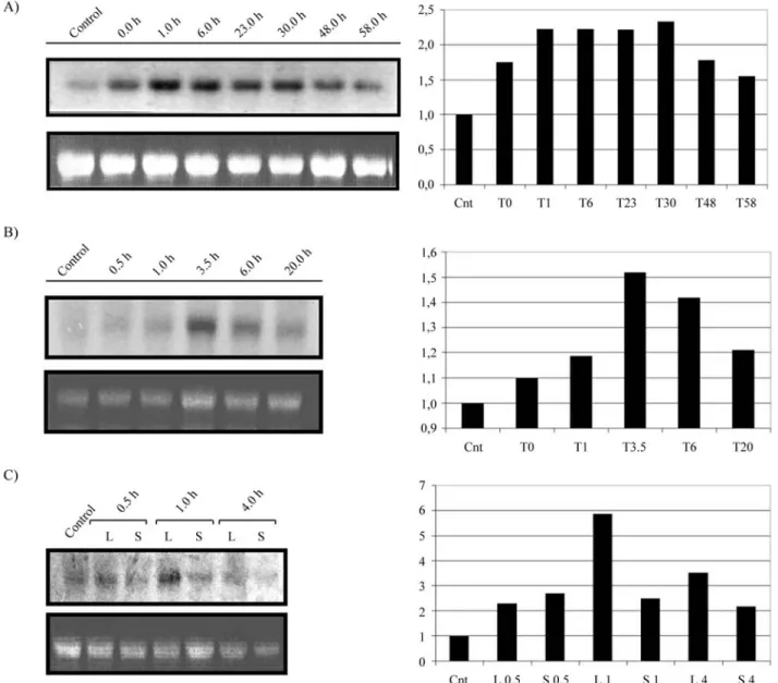
1 h and was maintained at high levels until 6 h after the on-set of the stress. Despite the lower level, after 58 h, the mRNA detected was still higher than that found in non-stressed plants (Figure 1A). Salicylic acid induced AtchitIVtranscripts accumulation inA. thalianaleaves at low level, as revealed by densitometry, to be around 1,5 fold after 3,5 h of treatment (Figure 1B). TheAtchitIV in-duction by salicylic acid, an intermediate compound of the signal transduction pathway of pathogen infection, in addi-tion to its inducaddi-tion by bacteria infecaddi-tion (Gerhardtet al., 1997), reinforces the role of this chitinase in plant defence. In addition to UV light irradiation and SA treatment, wounding also producedAtchitIVtranscript accumulation
in leaves. In stressed leaves, maximal response occurred 1 h after wounding and transcript levels decreased rapidly afterwards (Figure 1C). In contrast to these treatments, heat and cold shock were not able to induceAtchitIVtranscripts (data not shown). ThePvChi4, aPhasealus vulgaris Chia4 chitinase gene that has high identity withAtchitIV(70,2%), is also induced by UV irradiation (Margis-Pinheiroet al., 1993). The non-activation of theAtchitIVtranscript accu-mulation in response to heat stress contrasted to that ob-served on PvChi4 bean chitinase gene, which is highly induced by higher temperatures (Margis-Pinheiro et al., 1994).
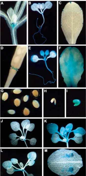
The expression pattern ofAtchitIVpromoter was ana-lysed during plant development using at least five inde-pendent GUS positive transgenic lines of each construct. The general expression pattern of the AtchitIV gene has been reported previously (Passarinhoet al., 2001). In that study, the tissue expression pattern of theAtchitIV/AtEP3 gene was analysed using 1100 bp of the promoter region fused to theguscoding region. Transgenic plants carrying this construct presented GUS activity in the meristematic region, hydathodes, root epidermis, pollen and in stigma during the fertilisation process (Passarinhoet al., 2001). In order to identify possible promoter regions involved in the control ofAtchitIVexpression, in the present study we ana-lysed the tissue expression pattern of transgenic plants con-taining promoter deletions at the positions -1923, -1500, -1083, -600 and -300. The GUS accumulation pattern ob-tained was identical for the transgenic plants lines contain-ing the∆1923, ∆1500 and ∆1083 chimerical constructs, indicating that all regulatory regions needed forAtchitIV tissue expression are present within the 1083 bp of the pro-moter. During seed germination, GUS staining was ob-served in seedling cotyledons and weakly in root, for all analysed constructs. GUS activity was also detected in the meristematic region just before the emergence of the first leaves and remained during plant development (Figure 2A). In seedlings, GUS staining was also observed in stip-ules, hydathodes and root epidermis (Figures 2A and 2B). In leaves of flowering plants, GUS activity was detected in hydathodes at the end of veins and along leaf margins as well (Figure 2C), confirming the constitutive expression of the AtchitIV gene in unstressed leaves of control plants (Figure 1). In flowers, GUS staining was observed in pol-len, in a stage-dependent manner, and in the stigma during the fertilisation process (data not shown). These results confirm a previous study in which the expression of trans-genic Arabidopsis plants containing the AtchitIV/AtEP3 1100 bp promoter was analysed (Passarinhoet al., 2001). In addition, this paper reports that GUS activity was also veri-fied in the floral receptacle, during floral senescence and at the beginning of silique development (Figure 2D). The AtchitIV activation during the senescence process of the floral receptacle corroborates the role of this chitinase in PCD proposed by Passarinhoet al.(2001). During seed de-velopment, GUS activity was detected in the micropyle re-gion (Figure 2G) and in the endosperm (data not shown), next to the radicule pole, whose tissues are addressed to se-nescence and degradation, respectively.
In reproductive organs, transgenic plants harbouring the∆600 and∆300 constructs presented the same GUS ac-tivity pattern observed for the∆1923,∆1500 and ∆1083 constructs. Different from the previous constructs, plants carrying the deletions∆600 and∆300 expressed GUS in the whole leaves and seedlings (Figures 2E and 2F). These re-sults suggest the presence of a restrictive regulatory region between the positions -1083 and -600.
bryos at different stages of development were performed demonstrating that the reporter gene was not expressed at the globular and heart stages (data not shown). TheAtchitIV promoter activity started in the radicule pole at torpedo to late torpedo stage (Figure 2H). The same expression pattern could be observed in late torpedo embryos (data not shown). In mature embryos GUS activity was detected in the whole embryo (Figure 2I). The expression pattern ob-served in zygotic embryos was identical for allAtchitIV promoter deletions studied, indicating that all necessary el-ements to control the embryo tissue expression are present within the 300 bp. However, a more intense activity was verified for embryos containing the ∆300 bp construct, when compared to the∆1923,∆1500,∆1083 and∆600 con-structs, indicating that a second regulatory negative ele-ment could be located between the positions -600 and -300. The regulation ofChia4chitinases during embryogenesis has been demonstrated.Picea glaucabasic chitinase is in-volved in embryogenesis (Dong and Dunstan, 1997). Tran-scripts of this chitinase were highly abundant in embryogenic tissues even in cotylledonary embryos and in plantlets. Nevertheless, the carrot EP3, another Chia4 chitinase, was not detected in zygotic embryos nor in so-matic embryos, although its expression had been
demon-strated during seed development and somatic
embryogenesis process. The EP3 expression suggests a “nursing” function during zygotic and somatic embryo-genesis (Van Hengelet al., 1998). During somatic embryo-genesis, theEP3expression was verified in embryogenic and non-embryogenic cultures (Van Hengelet al., 1998). During somatic embryogenesis, Passarinho and co-workers (2001) found thatAtchitIV/AtEP3expression was restricted to embryogenic cultures in cells close to the developing embryos, but not in the embryos themselves. In contrast to our results, the expression ofguswas not seen during zy-gotic embryo development (Passarinhoet al., 2001). How-ever, Passarinho and co-workers (2001) used the ecotype Wassilewskija to obtainArabidopsistransgenic plants car-ryingAtchitIV/AtEP3promoter-gusfusion, whereas in this study the C24 ecotype was employed for plant transforma-tion. Differences in expression pattern between parental ecotypes ofA. thalianamay explain the contradictory data obtained in this study and by Passarinho and co-workers concerning theAtchitIV/AtEP3expression during embryo development.
In order to map regulatory regions in theAtchitIV pro-moter accounting for the response to abiotic stresses, sev-enteen day-old transgenic plants containing the different promoter deletions were submitted to UV irradiation and salicylic acid treatment. Both treatments were able to in-duce GUS staining in transgenic plants containing the ∆1923,∆1500 and∆1083 chimeric constructions (compare Figure 2J with Figures 2K and 2L). Since the basal GUS ac-tivity in leaves of plants containing the∆600 and∆300 de-letions was already very high, it was not possible to show
further accumulation of GUS activity in response to differ-ent treatmdiffer-ents. These results suggest that the regulatory cis-elements necessary for UV light and salicylic acid sponse are present within the 1083 bp of the 5’ flanking re-gion.
Wound response observed by RNA-blot hybridiza-tion (Figure1C), was also demonstrated in the promoter-gustransgenic lines (Figure 2M). All constructs analysed were able to mediate wound activation of thegusgene, in-dicating that allcis-acting elements necessary for this re-sponse are present within the 300 bp AtchitIV proximal promoter region.
Together these results indicate a combination of posi-tive and negaposi-tive regulatory elements for the tissue-expression pattern of theAtchitIVgene. Negative regula-tory elements seem to be located between -1083 and -600 bp. These elements are responsible for expression me-diated by theAtchitIVpromoter to stipules, hydathodes and roots. The presence of a second negative regulatory region between -600 and -300 positions can also be suggested since a more intense widespread GUS activity was ob-served in∆300 plants. Furthermore, the results presented here indicate that all 5’ regulatory elements, needed for tis-sue expression pattern ofAtchiIVgene are located within 300 bp upstream of the translational start codon. Activation ofAtchitIVpromoter during stress treatments indicate that UV and salicylic acid regulatory regions are located down-stream to 1083 and wounding-activating elements lie be-tween -300 and +1.
The induction ofAtchitIVtranscript accumulation in the early response to several abiotic stresses support the in-volvement of this chitinase in plant defence mechanism. The induction ofAtchitIVtranscript accumulation by sali-cylic acid, an intermediate in the signal transduction path-way during pathogen infection, corroborates the earlier results where this chitinase was triggered by bacteria attack (Gerhardt et al., 1997). In conclusion, we propose that AtchitIV chitinase is involved in the early responses to pathogen attacks such as the establishment of hypersensi-tive reaction. Besides the involvement ofAtchitIVchitinase in plant defence, its expression pattern suggests an addi-tional role in different aspects of plant development such as embryogenesis processes.
Acknowledgments
References
Ancillo G, Witte B, Schmelzer E and Kombrink E (1999) A dis-tinct member of the basic, class I chitinase gene family in potato is specifically expressed in epidermal cells. Plant Mo-lecular Biology 39:1151.
Beerhues L and Kombrink E (1994) Primary structure and expres-sion of mRNAs encoding basic chitinase and 1,3-β -glucanase in potato. Plant Molecular Biology 24:353-367. Bevan M (1984) BinaryAgrobacteriumvectors for plant
transfor-mation. Nucleic Acids Research 12:8711-8721.
Büchter R, Stromberg A, Schmelzer E and Kombrink E (1997) Primary structure and expression of acidic, class II chitinase in potato. Plant Molecular Biology 35:749-761.
Busam G, Kassemeyer H-H and Matern U (1997) Differential ex-pression of chitinases inVitis viniferaL. responding to sys-temic acquired resistance activators or fungal challenge. Plant Physiology 115:1029-1038.
Clarke MC, Wei W and Lindsey K (1992) High-frequency trans-formation of Arabidopsis thaliana by Agrobacterium tumefaciens. Plant Molecular Biology Reporter 10:178-179. de Jong A, Cordewener J, Lo Schiavo F, Terzi M, Vande-kerckhove J, van Kammen A and de Vries SC (1992) A car-rot somatic embryo mutant is rescued by chitinase. The Plant Cell 4:425-433.
de Jong AJ, Heidstra R, Spaink HP, Hartorg MV, Meijer EA, Hendriks T, Lo Schiavo F, Terzi M, Bisseling T and van Kammen A (1993)Rhizobiumlipooligosaccharides rescue a carrot somatic embryo mutant. The Plant Cell 5:615-620. Dong J-Z and Dunstan DI (1997) Endochitinase and β
-1,3-glucanase genes are developmentally regulated during so-matic embryogenesis inPicea glauca. Planta 201:189-194. Gerhardt LBdeA, Sachetto-Martins G, Contarini MG, Sandroni
M, Ferreira RdeP, Lima VMde, Cordeiro MC, de Oliveira D and Margis-Pinheiro M (1997)Arabidopsis thalianaclass IV chitinase is early induced during the interaction with
Xanthomonas campestris. FEBS Letters 419:69-75. Goormachtig S, Lievens S, van de Velde W, Van Montagu M and
Holsters M (1998) Srchi13, a novel early nodulin from
Sesbania rostrata, is related to acidic class III chitinases. The Plant Cell 10:905-915.
Feinberg AP and Vogelstein B (1983) A technique for radio-labelling DNA restriction endonuclease fragments to high specific activity.Anal Biochem132:6-13.
Higushi R, Krummel RK and Saiki RK (1988) A general method of in vitro preparation and specific mutagenesis of DNA fragments: study of protein and DNA interactions. Nucleic Acids Research 16:7351-7367.
Ho SN, Hunt HD, Horton RM, Pullen JK and Pease LR (1989) Site-direct mutagenesis by overlap extension using the poly-merase chain reaction. Genetics 77:51-59.
Jefferson RA (1987). Assaying chimeric genes in plants: the GUS gene fusion system. Plant Molecular Biology Reporter 5:387-405.
Margis-Pinheiro M, Martin C, Didierjean L and Burkard G (1993) Differential expression of bean chitinase genes by virus
in-fection, chemical treatment and UV radiation. Plant Molecu-lar Biology 22:659-668.
Margis-Pinheiro M, Marivet J and Burkard, G (1994) Bean class IV chitinase gene: structure, developmental expression and induction by heat stress. Plant Science 98:163-173. Passarinho PA, Van Hengel AJ, Fransz PF and de Vries SC (2001)
Expression pattern of the Arabidopsis thaliana AtEP3/
AtchitIVendochitinase gene. Planta 212:556-567.
Ragueh F, Lescure N, Roby D and Marco Y (1989) Gene expres-sion inNicotiana tabacumin response to compatible and in-compatible isolates of Pseudomonas solonaciarum. Physiology Molecular Plant Pathology 35:23-33.
Robinson SP, Jacobs AK and Dry IB (1997) A class IV chitinase is highly expressed in grape berries during ripening. Plant Physioogy 114:771-778.
Sachetto-Martins G (1995) Isolation and characterization of gly-cine rich protein genes ofArabidopsis thaliana, atgrps. PhD Thesis, Universidade Federal do Rio de Janeiro, Rio de Ja-neiro, Brazil.
Samac DA, Hironaka CM, Yallaly PE and Shah DM (1990) Isola-tion and characterizaIsola-tion of the genes encoding basic and acidic chitinase inArabidopsis thaliana. Plant Physiology 93:907-914.
Samac DA and Shah DM (1991) Developmental and pathogen-induced activation of theArabidopsisacidic chitinase pro-moter. The Plant Cell 3:1063-1072.
Sambrook J, Fritsch EF, Maniatis T (1989) Molecular cloning: a laboratory manual. 2nd edition. Cold Spring Harbor Labora-tory Press, Cold Spring Harbor, NY.
Staehelin C, Granado J, Muller J, Wiemken A, Mellor RB, Felix G, Regenass M, Broughton WJ and Boller T (1994) Percep-tion ofRhizobiumnodulation factors by tomato cells and in-activation by root chitinases. Proceedings of Natural Academic of Science, USA 91:2196-2200.
Staehelin C, Schultze M, Kondorosi E and Kondorosi A (1995) Lipo-chitooligosaccharide nodulation signals from
Rhyzobium meliloti induce rapid degradation by the host plant alfalfa. Plant Physiology 108:1607-1614.
Valvekens D, Van Montagu M and Van Lijsebettens M (1988)
Agrobacterium tumefaciens-mediated transformation of
Arabidopsisroot explants using kanamicin selection. Pro-ceedings of Natural Academic Science, USA 85:5536-5540. Van Hengel AJ, Guzzo F, van Kammen A and de Vries SC (1998)
Expression pattern of the carrotEP3endochitinase genes in suspension cultures and in developing seeds. Plant Physiol-ogy 117:43-53.
Yeboah NA, Arahira M, Nong VH, Zhang DY, Kadokura K, Watanabe A and Fukazawa C (1998) A class III acidic endochitinase is specifically expressed in the developing seeds of soybean,Glycine max[L.] Merr. Plant Molecular Biology 36:407-415.
Yu L-X, Djebrouni M, Chamberland H, Lafontaine JG and Tabaeizadeh Z (1998) Chitinase: differential induction of gene expression and enzyme activity by drought stress in the wild, Lycopersicon chilense Dun. and cultivated, L. esculentumMill. tomatoes. Plant Phyisiology 153:745-753.