Cleaning-up Synthetic Dyes
from the Environment
Sónia Alexandra Gonçalves Mendes
Supervisor: Prof. Lígia O. Martins
President of the jury: Prof. Cecília Arraiano
| i
Aknowledgments
I would like to express my sincere gratitude to those who have contributed to this thesis and have supported me in one way or another during these past few years.
First of all, my greatest appreciation and gratitude has to go to the person who most generously paved the way to my personal and scientific development, my supervisor Lígia O. Martins. There are no words that could ever express how deeply grateful I am. Lígia is an outstanding professor with an immense scientific knowledge, her perseverance and persistence really make things happen and her high scientific standards and hard work set a reference for me. She has been orienting and supporting me always with promptness and patience, encouraging me in the hardest times, making me permanently see the good side, pushing me forward, believing me and telling me that I could do it, never giving up on me and never giving me up. It has been a pleasure and a great honor to work with you! You have given me so much! You provided me all the conditions, all the financial support, all the research lines, all the publications even though you make me feel so frustrated when everything after your writing become so so much better!! THANK YOU for being who you are and the best supervisor that anyone can have!
I also would like to thank my thesis committee, Manuela Pereira and Karina Xavier for the helpful discussions and insights into the work.
Sanctis and Bruno Correia for PpAzoR crystal structure and a special thanks should be given to Vânia Brissos for everything! In addition thanks to all past and present lab members for the companionship, Paulo Durão, André Fernandes, Rita Catarino, Tânia Rosado, Nádia Gonçalves, Maura Ferreira, Diogo Tavares, Bruna Pinto, Joaquim Madeira, Lúcia Sabala and Ana Fernandes.
I am thankful to Instituto de Tecnologia Química e Biológica António Xavier that provided me all the conditions, facilities and equipment to develop this work; the European projects SOPHIED and BIORENEW; and FCT projects for funding.
| iii
To my father, Manuel Diogo Mendes
| v
Abbreviations
5-ASA–5-aminosalicylic acid 5cHS–5-coordinated high spin
5cQS–5-coordinated quantum mechanically admixed spin 6cHS–6-coordinated high spin
6cLS–6-coordinated low spin
AauDyPI– Dye-decolourising peroxidase from Auricularia auricula-judae
AB194–Acid black 194 AB210–Acid black 210 AB62–Acid blue 62 Abs–Absorbance
ABTS–2,2’-azino bis (3-ethylbenzthiazoline-6-sulfonic acid) AH3517–Escherichia coli strain overexpressing cotA
Ala or A–Alanine
AnaPX– Dye-decolourising peroxidase from Nostoc sp.
AO7–Acid orange 7 APX–Ascorbate peroxidase AQ–Anthraquinonic dye
AQS–Anthraquinone-2-sulfonate AR266–Acid red 266
AR299 or NY1–Acid Red 299 Arg or R–Arginine
Asn or N–Asparagine Asp or D–Aspartate AY49–Acid yellow 49 BQ–1,4-benzoquinone BR–Britton-Robinson
BsDyP– Dye-decolourising peroxidase from Bacillus subtilis
C.I.–Colour index
CcP–Cytochrome c peroxidase
CotA-laccase–Laccase from Bacillus subtilis
Cpd I–Compound I Cpd II–Compound II
Cpop21– Dye-decolourising peroxidase from Polyporaceae sp.
Cys or C–Cysteine DAD–Diode-array DB1 or CSB–Direct blue 1 DB38 or CB– Direct black 38 DB71–Direct blue 71 DNA– Deoxyribonucleic acid DR80–Direct red 80 DRR–Direct red R DY106–Direct yellow 106
DyP–Dye-decolourising peroxidase
DyP2– Dye-decolourising peroxidase from Amycolatopsis sp.
DypA– Dye-decolourising peroxidase from Rhodococcus jostii
DypB– Dye-decolourising peroxidase from Rhodococcus jostii
EDTA–Ethylenediaminetetraacetic acid
EfeB/YcdB– Dye-decolourising peroxidase from Escherichia coli
EPO–Eosinophil peroxidase
EPR–Electron paramagnetic resonance
ESI-MS–Electrospray ionization-mass spectrometry ɛ– Molar extinction coefficient
FAD–Flavin adenine dinucleotide FMN–Flavin mononucleotide
GC/MS–Gas chromatography-mass spectrometry GdnHCl–Guanidine hydrochloride
| vii
His or H–Histidine
HPLC–High-performance liquid chromatography HRP–Horseradish peroxidase
HS–High spin
IBD–Inflammatory bowel disease
IPTG–Isopropyl-β-D-thiogalactopyranoside KatG–Bacterial catalase-peroxidase KCl–Potassium chloride
LB– Luria-Bertani
LC/MS–Liquid chromatography-mass spectrometry Leu or L–Leucine
LiP–Lignin peroxidase
LOM528–Escherichia coli strain overexpressing ppAzoR
LOM529–Escherichia coli strain co-expressing ppAzoR and cotA
LOM530–Pseudomonas putida MET94 strain overexpressing ppAzoR
Lot6p–Flavin-dependent quinone reductase from Saccharomyces cerevisiae
LPO–Lactoperoxidase LS–Low spin
MALDI-TOF MS – Matrix assisted laser desorption ionization-time of flight-mass spectrometry
MB17–Mordant black 17 MB3–Mordant black 3 MB9–Mordant black 9 MCO–Multi-copper oxidase min–minute(s)
MnP–Mangenese peroxidase MPO–Myeloperoxidase MR–Methyl red
MS/MSn–Tandem mass spectrometry
Msp1– Dye-decolourising peroxidase from Marasmius scorodonius
MxDyP– Dye-decolourising peroxidase from Myxococcus xanthus
MYspDyP–Dye-decolourising peroxidase from Mycobacterium sp.
N–Native enzyme
NADH –Nicotinamide adenine dinucleotide
NADPH –Nicotinamide adenine dinucleotide phosphate NaOCl–Sodium hypochloride
NMR–Nuclear magnetic resonance NQO1–Mammalian quinone reductase OD–Optical density
PaDyP–Dye-decolourising peroxidase from Pseudomonas aeruginosa
PCR–Polymerase chain reaction PDB–Protein data bank
PEG 400–poly(ethylene glycol) 400 molecule Phe or F–Phenylalanine
PpAzoR–Azoreductase from Pseudomonas putida MET94
PpDyP –Dye-decolourising peroxidase from Pseudomonas putida MET94
Pro or P–Proline
QS–Quantum mechanically admixed spin RB222–Reactive blue 222
RB5–Reactive black 5
RR–Resonance Raman spectroscopy RR195–Reactive red 195
RR4–Reactive red 4 RY145–Reactive yellow 145 RY81–Reactive yellow 81 s–second(s)
SavDyP– Dye-decolourising peroxidase from Streptomyces avermitilis
SBP–Soybean peroxidase
Sco3963– Dye-decolourising peroxidase from Streptomyces coelicolor A3(2)
SDS–Sodium dodecyl sulfate
| ix
SOG–Sudan orange G
SviDyP – Dye-decolourising peroxidase from Saccharomonospora viridis
T1–Type 1 copper site T2–Type 2 copper site T3–Type 3 copper site
TAP– Dye-decolourising peroxidase from Termitomyces albuminosus
TEMPO–2,2,6,6-tetramethyl-piperidine-1-oxyl
TfuDyP– Dye-decolourising peroxidase from Thermobifida fusca
Tm–Melting temperature
Topt–Optimal temperature
TPO–Tyroid peroxidase Tyr or Y–Tyrosine
TyrA–Dye-decolourising peroxidase from Shewanella oneidensis
UV–Ultraviolet Val or V–Valine
| xi
Abstract
Synthetic dyes are xenobiotic compounds widely used in several industries from textile, paper, pharmaceutical and food, to leather or cosmetics. During the dyeing processes in the textile industry, millions of tons of dyes are discharged in industrial effluents causing serious environmental and health hazards. The current physico-chemical methods used in the treatment of dye-containing wastewaters are not cost-effective and may result in the formation of hazardous by-products. Therefore, there is an urgent need in the development of efficient and environmentally friendly technologies for the treatment of dye-containing wastewaters. Enzymatic processes can offer several advantages such as specificity to attack the dye molecules while keeping intact dyeing additives or fibers, water recycling, easiness of engineering to improve their robustness, environmentally friendly nature and non-production of sludge. Azoreductases, laccases and peroxidases are among the few enzymes reported in the literature with the ability to degrade synthetic dyes. Hence, the purpose of this thesis was to study the enzymatic properties, mechanisms and toxicity of dye-degradation products of different bacterial enzymes to set-up multi-enzymatic systems for cleaning synthetic dyes from the environment.
A new bacterial strain, Pseudomonas putida MET94, was selected among 48
bacterial strains for its ability to decolourise at high extent structurally different synthetic azo dyes. P. putida is an ubiquitous bacterium engaged in
important metabolic activities in the environment, including element cycling and degradation of biogenic and xenobiotic pollutants. Therefore, the physiology of synthetic dyes decolourisation processes in growing and resting cells of P. putida MET94 was addressed. Whole-cell systems were
showed that the P. putida system is highly competent in the biological
degradation of model dyestuff wastewaters. The involvement of an azoreductase in the decolourisation process was suggested by in silico
screening using the genome of P. putida KT2440. To gain molecular insight
into the bacterial decolourisation system, the gene coding for an azoreductase was cloned and expressed in Escherichia coli and the
recombinant enzyme was produced, purified and characterized at the spectroscopic, structural and kinetic level. The P. putida azoreductase
(PpAzoR) was shown to be a 40 kDa homodimer FMN dependent NAD(P)H:dye quinone oxidoreductase with broad substrate specificity for azo dye reduction under anoxic conditions. The PpAzoR enzyme was also shown to be able to use several quinones as well as O2 as oxidizing substrates. A P. putida strain overexpressing the gene coding for the
PpAzoR enzyme was constructed and exposed to the presence of quinones. The results have shown that, most likely the biological role of this enzyme is related to providing general protection against the oxidative stress caused by quinones. The X-ray crystal structure of the enzyme was determined in collaboration with the Crystallography Unit of ITQB showing that the monomer adopts a flavodoxin-like fold, with a central twisted β-sheet formed by five parallel β-strands connected by α-helices.
High-performance liquid chromatography was used to identify aromatic amines as the PpAzoR azo dye degradation products. With the aim of setting-up a non-toxic enzymatic decolourisation system, the toxicity of intact dyes and of reaction mixtures treated with PpAzoR was assessed by measuring the growth inhibition of Saccharomyces cerevisiae and
reproduction inhibition of the nematode Caenorhabditis elegans. However,
| xiii
treated with PpAzoR. The addition of CotA-laccase resulted in a significant drop in the final toxicity. As a consequence, a recombinant E. coli strain
co-expressing both ppAzoR and cotA genes was constructed and whole-cell
assays used in the treatment of model dye wastewaters. Decolourisation levels above 80% and detoxification levels up to 50% were obtained. The high attributes of this engineered strain, make it a promising candidate for both decolourisation and detoxification of azo dye-contaminated effluents. In a second stage of my PhD working programme, we have performed the cloning, purification and characterization of two new dye-decolourising peroxidases (DyPs) from the soil bacteria Bacillus subtilis (BsDyP) and P. putida MET94 (PpDyP). DyPs are haem peroxidases belonging to a new
family of microbial peroxidases. These enzymes are known to successfully degrade a wide variety of different substrates including high redox synthetic dyes. DyPs have primary sequence, structural and apparently, mechanistic features, unrelated to those of other known as “classical” peroxidases. DyPs have been classified into four phylogenetically distinct subfamilies on the basis of sequence comparison, with bacterial enzymes constituting A-C subfamilies and fungal enzymes belonging to D subfamily. The genes encoding BsDyP and PpDyP, belonging to subfamilies A and B, respectively, were cloned and heterologously expressed in E. coli. The
purified BsDyP was shown to be a single 48 kDa monomer (Topt 20 – 30ºC) whereas PpDyP is a 120 kDa homotetramer with a peculiar flat and broad temperature profile (Topt 10 – 30ºC). The results have shown that the two enzymes have distinct spectroscopic, kinetic and stability properties. PpDyP exhibits higher activities and a wider scope of substrates, including apart from synthetic dyes, phenolic and non-phenolic lignin units as well as metal ions, showing potential for industrial applications.
establishment of the structural determinants of DyPs catalytic mechanism. Therefore, the structural determinants that underlie the mechanistic properties of PpDyP and BsDyP enzymes were studied by using site directed mutagenesis. The distal residues (Asp, Arg, Asn) were substituted and a combination of transient and steady-state kinetics, spectroscopic and electrochemical approaches allowed to assess the impact of the point mutations on structural, redox and catalytic properties of the enzymes. In BsDyP, none of the studied residues was shown to be individually indispensable for promoting the (de)protonation of hydrogen peroxide and the cleavage of peroxide group. In contrast, our data point to the importance of the distal arginine in the catalytic mechanism of PpDyP, as also observed in other B-type DyPs, but not in DyPs from A and D subfamilies. Clearly more members of DyP-type peroxidase superfamily need to be thoroughly characterized and more solid kinetic, thermodynamic and structural information provided for the full understanding of the molecular determinants of catalytic mechanisms of DyPs. These insights will allow us to fully exploit quite unique properties of DyP-type peroxidases in biotechnological applications.
| xv
Resumo
Os corantes sintéticos são compostos xenobióticos amplamente usados em várias indústrias, desde a indústria têxtil, farmacêutica e alimentar até à indústria dos cosméticos e dos cortumes. Na indústria têxtil, durante os processos de tingimento, milhares de toneladas de corantes são libertados para os efluentes industriais. A presença de cor é o primeiro indicador de contaminação das águas residuais e, desta forma, a libertação de substâncias fortemente coradas sem qualquer tratamente prévio, podem causar sérios problemas ambientais de saúde pública. Os métodos fisico-químicos que existem actualmente para tratar águas residuais contaminadas com corantes, para além de caros, não são totalmente eficazes, devido à complexidade e elevada estabilidade das estruturas químicas que os corantes apresentam. Assim sendo, existe uma grande necessidade de desenvolver métodos mais eficientes e ambientalmente sustentáveis para o tratamento de águas residuais contaminadas com corantes. Os processos enzimáticos, poderão ser uma solução interessante neste contexto. Este tipo de processos apresentam inúmeras vantagens, nomeadamente a sua especificidade para as moléculas de corante, mantendo intactos outros componentes do corantes, tais como aditivos ou fibras, permitir a reciclagem de água, a facilidade de aplicação de técnicas de engenharia com vista ao seu melhoramento, e a sua natureza ambientavelmente sustentável. As azoreductases, as lacases e as peroxidases são das poucas enzimas descritas na literatura como sendo capazes de degradar corantes sintéticos. O objectivo desta dissertação foi estudar as propriedades e mecanismos enzimáticos, e a toxicidade dos produtos de degradação dos corantes de forma a estabelecer sistemas multi-enzimáticos eficazes para remover corantes sintéticos do meio ambiente. Uma estirpe bacteriana nova, Pseudomonas putida MET94, foi seleccionada pela sua
diversas actividades metabólicas no meio ambiente, incluíndo, sequestrar elementos nocivos poluentes biogénicos e xenobióticos do meio ambiente, degradá-los e libertá-los novamente para o meio ambiente. Estudámos a fisiologia dos processos de descoloração de corantes sintéticos em células em crescimento e em células inteiras (em fase estacionária) da estirpe P. putida MET94. Sistemas de células inteiras foram testados e optimizados
não só para a redução de moléculas de corantes simples individualmente, mas também para a degradação de misturas-modelo de corantes com elevada força iónica que mimitizam efluentes reais produzidos durante os processos de tingimento de algodão e de lã. Mostrámos que o sistema P. putida, é
altamente competente na degradação biológica de modelos de águas residuais contaminadas com corantes. O envolvimento de uma azoreductase no processo de descoloração foi sugerido por rastreamento in silico usando o
genoma da estirpe P. putida KT2440. Com o objectivo de analisar em maior
pormenor o sistema de descoloração bacteriano, clonámos e expressámos o gene que codifica para a azoreductase em Escherichia coli e produzimos,
purificámos e caracterizámos ao nível espectroscópico, cinético e estrutural a enzima recombinante. A azoreductase de P. putida (PpAzoR) mostrou ser
| xvii
performance (HPLC), os produtos de degradação resultantes da acção enzimática da PpAzoR revelaram ser aminas aromáticas. Numa tentativa de estabelecer e desenvolver um sistema de descoloração enzimática, avaliámos a toxicidade dos corantes e dos produtos de degradação, quantificando a inibição do crescimento da levedura Saccharomyces cerevisiae e inibição de
reprodução do nemátode Caenorhabditis elegans. Os resultados mostraram
que, para a maioria dos corantes testados, os produtos de degradação enzimática, eram cerca de 2 a 3 vezes mais tóxicos que o corante inicial. Desta forma, às misturas reaccionais com a enzima PpAzoR, decidimos adicionar uma outra enzima muito bem estudada no nosso laboratório com capacidade comprovada na oxidação de compostos aromáticos, a CotA-lacase. Esta sequência resultou num decréscimo significativo dos valores de toxicidade finais. Como consequência, construímos uma estirpe recombinante de E. coli a co-expressar os genes ppAzoR e cotA e usámos
células inteiras deste sistema para tratar modelos de águas resisuais contaminadas com corantes. Usando este sistema, obtivémos níveis de descoloração acima de 80% e níveis de destoxificação acima dos 50%. Os elevados atributos desta estirpe “engenheirada” tornam-na numa promissora candidata para descolorar e destoxificar efluentes contaminados com corantes.
Numa segunda etapa do meu Doutoramento, endereçámos a clonagem, purificação e caracterização de duas novas peroxidases capazes de descolorar corantes (“dye-decolourising peroxidases”, DyPs), uma da bactéria do solo Bacillus subtilis (BsDyP) e a outra da bactéria P. putida
clássicas. As DyPs foram classificadas em 4 subfamílias filogeneticamente distintas, sendo que as enzimas bacterianas constituem as subfamílias A a C e as enzimas fúngicas pertencem à subfamília D. Os genes que codificam para as enzimas BsDyP e PpDyP, pertencentes à subfamília A e B, respectivamente, foram clonados e heterologamente expressos em E. coli. A
| xix
D. Estes resultados mostram claramente, ser necessário uma caracterização mais aprofundada de mais membros da superfamília das DyP-peroxidases, para que exista um conhecimento alargado acerca dos determinantes moleculares do mecanismo catalítico das DyPs. Esta informação, é essencial à exploração deste tipo de peroxidases DyP com vista à sua aplicação biotecnológica.
| xxi
List of publications
Sónia Mendes, Luciana Pereira, Carlos Batista and Lígia O. Martins (2011).
Molecular determinants of azo reduction activity in the strain Pseudomonas putida MET94. Appl. Microbiol. Biotechnol. 92, 393-405.
Bruno Correia, Zhenjia Chen, Sónia Mendes, Lígia O. Martins and Isabel Bento
(2011). Crystallization and preliminary X-ray diffraction analysis of the azoreductase PpAzoR from Pseudomonas putida MET94. Acta Crystallogr. Sect. F Struct. Biol. Cryst. Commun. 67, 121-123.
Sónia Mendes, Ana Farinha, Christian G. Ramos, Jorge H. Leitão, Cristina A.
Viegas and Lígia O. Martins (2011). Synergistic action of azoreductase and laccase leads to maximal decolourization and detoxification of model dye-containing wastewaters. Bioresour. Technol. 102, 9852–9859.
Ana Maria D. Gonçalves, Sónia Mendes, Daniele de Sanctis, Lígia O. Martins and
Isabel Bento (2013). The crystal structure of Pseudomonas putida PpAzoR: the active site revisited. FEBS J. 280:6643-6657.
Ana Santos, Sónia Mendes, Vânia Brissos and Lígia O. Martins (2014). New dye
decolourising peroxidases from Bacillus subtilis and Pseudomonas putida: towards biotechnological applications. Appl. Microbiol. Biotechnol. 98:2053-2065.
Sónia Mendes, Vânia Brissos, Antonieta Gabriel, Teresa Catarino, David L. Turner,
Smilja Todorovic and Lígia O. Martins (2015). An integrated view of redox and catalytic properties of B-type PpDyP from Pseudomonas putida MET94 and its distal variants. Arch. Biochem. Biophys. doi: 10.1016/j.abb.2015.03.009.
Sónia Mendes, Teresa Catarino, Célia Silveira, Smilja Todorovic and Lígia O.
| xxiii
Thesis outline
The diversity and complexity of synthetic dyes mixtures present in dye-containing effluents poses serious problems on the design of technically feasible and cost-effective treatment methods. The characterization of enzymes that make a discernible contribution to the degradation of synthetic dyes paves the way for the improvement of multi-enzymatic systems, through protein engineering strategies, to maximize their biodegradation, biotransformation and also valorization potential. Therefore in this thesis we have studied enzymes that are known to be involved in degradation of synthetic dyes: the azoreductase PpAzoR from P. putida MET 94, the
CotA-laccase from B. subtilis and the dye-decolourising peroxidases PpDyP from P. putida MET94 and BsDyP from B. subtilis.
This Doctoral Thesis is composed by six chapters.
In Chapter 1 the state of the art on dyes and their degradation methods and mechanisms is presented.
In Chapter 2 we have focused in i) the screening for dye decolourisation using a collection of 48 bacterial strains and the selection of Pseudomonas putida MET94 strain on the basis of its superior ability to degrade dyes with
high efficiency; ii) the cloning, expression, production and kinetic and structural characterization of a potential enzyme responsible for decolourisation activity in the strain P. putida MET94, the azoreductase
PpAzoR.
real dye-containing wastewaters. Finally, an E. coli strain co-producing both
PpAzoR and CotA-laccase enzymes was constructed, and a whole-cell system was developed for decolourisation and detoxification of the model dye-containing wastewater baths.
In Chapter 4 the study of bacterial dye-decolourising peroxidases (DyP-type) was addressed. We have performed the cloning, expression, production, purification and characterization of two new bacterial DyPs: one from P. putida MET94 (PpDyP) and the other from B. subtilis (BsDyP). The results
show that the two enzymes have distinct spectroscopic, kinetic and stability properties. The B subfamily PpDyP, is a more versatile and in general, a more active biocatalyst, than BsDyP from subfamily A. The spectroscopic data suggested distinct haem microenvironments in the two enzymes that might account for the distinct catalytic behavior.
Table of contents
Aknowledgments ... i
Abbreviations ... v
Abstract ... xi
Resumo ... xv
List of publications ... xxi Thesis outline ... xxiii
Chapter 1 ... 1 Introduction
Synthetic dyes ... 3 Traditional physico-chemical methods of dye removal from dye-containing wastewaters ... 6 Biological degradation methods of dye removal from dye- containing
wastewaters: microorganisms and enzymatic systems ... 10 Azoreductases ... 17 Laccases ... 23 Peroxidases ... 30
Chapter 2 ... 41 Molecular determinants of azo reduction activity in the strain
Pseudomonas putida MET94
Abstract ... 43 Introduction ... 44 Material and methods ... 46
Bacterial strains, media, and plasmids ... 46
Chemicals ... 46
Isolation of Pseudomonas putida MET94 clones with higher ability to decolourise dyes ... 47
Culture conditions ... 47
Preparation of resting whole cells ... 48
Preparation of cell-free extracts ... 48
Cloning and overexpression of PpAzoR in P. putida and in E. coli ... 50
Effect of quinone on the growth of P. putida wild-type and P. putida
overexpressing PpAzoR+ strains ... 51 Survival of P. putida wild-type and P. putida overexpressing PpAzoR+ strains upon exposure to 1,4-benzoquinone... 51
Identification of the prosthetic group... 52
UV-visible spectrum of PpAzoR and molar extinction coefficient ... 53
Enzyme assays ... 53
Analysis of reductive and oxidative half-reactions by stopped-flow spectroscopy .... 55
Identification of azo dye products ... 56
Crystallization ... 56
Data collection and structural determination ... 57
Other methods ... 58 Results and discussion ... 59
Screening for dye decolourisation... 59
Effect of oxygen and dye concentration on growth and decolourisation
parameters ... 62
Decolourisation using resting whole cells ... 65
Identification of the P. putida azoreductase gene ... 67
Overproduction and purification of recombinant PpAzoR ... 68
Spectroscopic properties of the recombinant protein ... 70
Enzymatic properties of PpAzoR ... 72
The catalytic mechanism of PpAzoR ... 76
Crystal analysis ... 81
Overall structure of the native enzyme ... 82
The active site: comparison and conformational differences ... 84
Putative physiological role of PpAzoR in P. putida MET94 ... 86
Chapter 3 ... 89 Synergistic action of azoreductase and laccase leads to maximal decolourisation and detoxification of model dye-containing wastewaters
Abstract ... 91 Introduction ... 92 Material and methods ... 94
Chemicals ... 94
Construction of an E. coli strain co-expressing cotA and ppAzoR ... 96
Overproduction of enzymes in heterologous host... 96
Enzyme assays using purified enzymes ... 97
Enzyme assay using recombinant whole cells ... 98
Toxicity analysis ... 98 Results and discussion ...100
Degradation of dyes using the reductive PpAzoR and the oxidative CotA-laccase . 100
Toxicity of azo dyes over S. cerevisiae growth and C. elegans reproduction ... 100
Sequential enzymatic treatment of model wastewaters ... 106
Construction of an E. coli strain co-expressing both PpAzoR and cotA genes ... 108
Enzymatic treatment of model wastewaters using whole cells co-expressing both ppAzoR and cotA... 110
Chapter 4 ... 113 New dye-decolourising peroxidases from Bacillus subtilis and
Pseudomonas putida MET94: towards biotechnological
applications
Abstract ... 115 Introduction ... 116 Material and methods ... 118
Cloning and over expression of ppDyP and bsDyP in Escherichia coli ... 118
Purification of recombinant PpDyP and BsDyP ... 119
Spectroscopic analysis ... 120
Kinetic analysis ... 120
Enzyme stability ... 122
Other methods ... 123
Results and discussion ...124
PpDyP and BsDyP are members of the DyP family of enzymes ... 124
Biochemical and spectroscopic characterization of PpDyP and BsDyP ... 129
Kinetic properties of purified enzymes ... 131
Stability of PpDyP and BsDyP ... 140
Chapter 5 ... 143 Distal haem pocket residues of A and B-type dye-decolorizing peroxidase: an integrated view of spectroscopic, redox and catalytic properties
Abstract ... 145 Introduction ... 147 Material and methods ... 150
Construction, overproduction and purification of BsDyP and PpDyP variants ... 150
Spectroscopic analysis ... 151
Cyclic voltammetry ... 152
Stopped-flow experiments ... 153
Analysis of transient kinetic data for BsDyP WT and variants ... 154
Analysis of transient kinetic data for PpDyP WT ... 154
Steady-state kinetic assays ... 156
5.1 Distal haem pocket residues of A-type Bacillus subtilis
dye-decolourising peroxidase: neither aspartate nor arginine is individually essential for peroxidase activity ... 159
Results and discussion ...159
Biochemical and spectroscopic characterization of BsDyP mutants ... 159
Redox properties of BsDyP variants ... 164
Effect of pH in Compound I formation and spontaneous decay ... 165
Steady-state kinetic characterization of BsDyP variants ... 171
5.2 An integrated view of redox and catalytic properties of B-type
Ppdyp from Pseudomonas putida MET94 and its distal variants ... 177
Results and discussion ...177
Stopped-flow kinetic data and standard reduction potential of Compound I/native PpDyP couple ... 177
Biochemical and UV-Vis characterization of PpDyP mutants ... 182
Resonance Raman spectroscopy ... 183
Redox properties of Fe3+/Fe2+ couple of PpDyP variants ... 188 Steady-state kinetic characterization of PpDyP variants ... 190
Chapter 6 ... 199 Concluding remarks
Chapter 1 | 1
Chapter 1
Chapter 1 | 3
Introduction
Environmental pollution is one of the major and most important problems of the modern world. In order to fulfill the needs and demands of the overgrowing human population, developments in agriculture, medicine, energy sources, and all chemical industries are necessary (Ali 2010). Over the last century, the increased industrialization and continued population growth led to an augmented production of environmental pollutants that are released into air, water, and soil, with significant impact in the degradation of various ecosystems (Ali 2010, Khan et al. 2013). Xenobiotics are characterized as non-natural compounds foreign to specific ecological systems at unusually high concentrations (Kandelbauer & Guebitz 2005, Varsha et al. 2011). The potential health hazard of xenobiotics is a function of its persistence and toxicity in the environment. Munitions waste, pesticides, organochlorides, polychlorinated biphenyls, polycyclic aromatic hydrocarbons, wood preservatives, synthetic polymers and synthetic dyes are recalcitrant compounds that mostly contribute to environmental pollution (Ali 2010).
Synthetic dyes
chromophores, as well as electron withdrawing or donating substituents that cause or intensify the colour of the chromophores, called auxochromes. Common chromophores include azo (–N=N–), carbonyl (–C=O), methane (–CH=), nitroso (NO or N–OH), nitro (–NO2 or NO–OH), and sulphur (C=S). The most important auxochromes include amine (–NH3
),
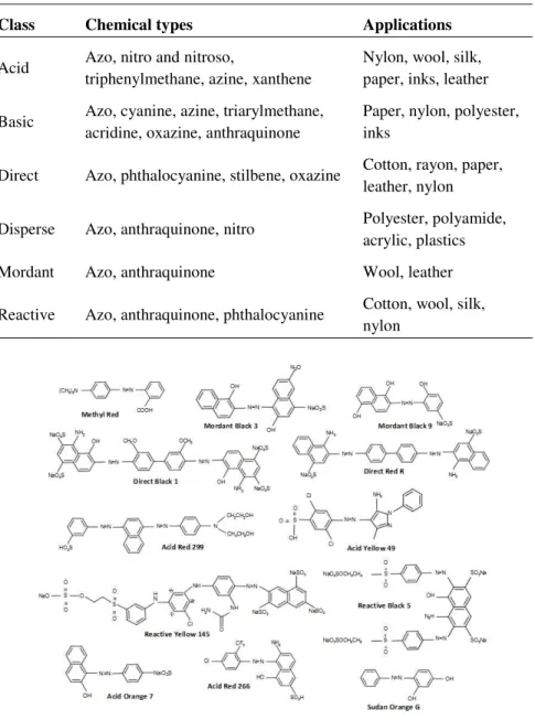
carboxyl (–COOH), sulfonate (–SO3H) and hydroxyl (–OH) (dos Santos et al. 2007, Pereira & Alves 2012). These auxochromes can belong to the classes of reactive, acid, direct, basic, disperse, or mordant dyes, among others (Rodriguez-Couto 2009). Additionally, dyes are composed by an aromatic structure, typically benzene, naphthalene or an anthracene ring, called chromogen.Chapter 1 | 5
Table 1.1.
Classes, chemical types and applications of dyes adapted from (Husain2006).
Class Chemical types Applications
Acid Azo, nitro and nitroso,
triphenylmethane, azine, xanthene
Nylon, wool, silk, paper, inks, leather
Basic Azo, cyanine, azine, triarylmethane, acridine, oxazine, anthraquinone
Paper, nylon, polyester, inks
Direct Azo, phthalocyanine, stilbene, oxazine Cotton, rayon, paper, leather, nylon
Disperse Azo, anthraquinone, nitro Polyester, polyamide,
acrylic, plastics
Mordant Azo, anthraquinone Wool, leather
Reactive Azo, anthraquinone, phthalocyanine Cotton, wool, silk, nylon
Figure 1.1
In fact, apart from the aesthetic problems, the greatest environmental concern with the dye-containing effluents is their absorption and reflection of the sunlight entering the water, affecting severely the photosynthetic activity which leads to a decrease in dissolved oxygen concentration and water quality, which can result in acute toxic effects on aquatic flora and fauna (Pearce et al. 2003, Rodriguez Couto 2009, Ali 2010, Saratale et al. 2011, Khan et al. 2013). In addition, many synthetic dyes and their metabolites are toxic, carcinogenic or mutagenic, leading to potential health hazard to humankind due to possible accumulation in the food chain (Rai et al. 2005, Khan et al. 2013). Health disorders such as nausea, hemorrhage, ulceration of skin and mucous membranes, kidney, reproductive system, liver, brain, and central nervous system damages, constitute some of the harsh side effects that have been reported (Sarayu & Sandhya 2012). Thus, it is clear that dye-containing wastewaters must be treated prior to their discharge into natural environments.
Traditional physico-chemical methods of dye removal from dye-containing wastewaters
Chapter 1 | 7
Figure 1.2
Physico-chemical and biological treatment methods for the removal of dyes from dye-containing wastewaters.
Adapted from (Saratale et al. 2011).
Chapter 1 | 9
reactive dyes. These methods consist in the destabilization of electrostatic interactions that exist between dye molecules and water through the addition of a coagulant. Noteworthy, that are some reports in literature showing decolourisation levels above 90% employing Al2SO4, FeCl3, MgSO4, MnCl2, as coagulants (Hsu et al. 1998, Venkat et al. 1999, Tan et al. 2000). Flocculation reagents are synthetic polymers with a linear structure that used along with chemicals such as aluminium sulphate, ferrous and ferric sulphate, ferric chloride, copper sulphate, allows for colour removal which is accomplished by aggregation/precipitation and sorption of colouring substances onto the polynuclear coagulant species and hydrated flocks (Alinsafi et al. 2005). However, in addition to the low colour removal efficiency, the large amount of sludge produced limits the application of this technique (Saratale et al. 2011). Adsorption methods for colour removal are based on the high affinity of dyes for diverse adsorbent materials. The selection of an adsorbent is based not only in the affinity for target compounds but also on the possibility for its regeneration. The use of activated carbon is by far, the most commonly used method with confirmed success for removal of mordant and acid dyes and lower effectiveness for disperse, direct and reactive dyes (Robinson et al. 2001). However, activated carbon has to be regenerated offsite and is relatively expensive. To make the process economically feasible, some efforts on low-cost adsorbents materials, like peat, bentonite clay, fly ash, polymeric resins, ion exchangers, and many biological materials, such as corn/maize cobs, maize stalks, and wheat straw, were reported (El-Geundi & Aly 1992, Ho & Mckay 1998, Janos et al. 2003, Wang et al. 2005, Hu & Qiao 2006). However, the practical application of these adsorbents has been limited by problems associated with their regeneration or disposal, high sludge production, long contact time and low adsorption capacity (Gupta & Suhas 2009).
formation of hazardous by-products or intensive energy or chemical requirements (Stolz 2001, Husain 2006). This reinforced the interest in the use of biological systems for the treatment of dye-containing wastewaters (Stolz 2001). Therefore, microbial or enzymatic decolourisation and degradation become very attractive alternatives to physico-chemical processes as they produce less sluge, are eco-friendly and cost-competitive (Rai et al. 2005, Saratale et al. 2011, Leelakriangsak 2013).
Biological degradation methods of dye removal from dye-containing wastewaters: microorganisms and enzymatic systems
The use of microorganisms and their enzymatic systems (Fig. 1.2) for the removal of synthetic dyes from industrial effluents offers considerable advantages. The process set-up and the running costs are relatively inexpensive, thus biodegradation is a promising approach for the remediation of dye-containing wastewaters (Forgacs et al. 2004, Pandey et al. 2007, Ali 2010). Biological methods involve the utilization of fungi, algae, yeast or bacteria that are able to decolourise and even completely mineralize synthetic dyes under selected environmental conditions (Pandey et al. 2007, Sarayu & Sandhya 2012). Azoreductases, laccases and peroxidases are reported as the mainly responsible enzymes for dye decolourisation in microbial cells (Kandelbauer & Guebitz 2005).
White-rot fungi represents the group of microorganisms that seems to be the most efficient in the degradation of complex aromatic dyes (Pointing 2001). Fungal cultures such as Trametes versicolor (Yemendzhiev et al. 2009), Trametes hirsute, Marasmius sp. (Jadhav et al. 2010), Phanerochaete chrysosporium (Ghasemi et al. 2010), Bjerkandera adusta (Anastasi et al.
2011), Aspergilus niger (Ali et al. 2009) are able to decolourise dyes at
Chapter 1 | 11
unspecific nature of their extracellular ligninolytic oxidoreductive degrading enzymatic systems, such as peroxidases (lignin peroxidase (LiP), manganese peroxidase (MnP) and versatile peroxidase (VP)) and laccases (Kandelbauer & Guebitz 2005, Rodriguez Couto 2009). However, the long growth cycle and time for complete decolourisation constitute the major impairments for their utilization as decolourisation agents (Khan et al. 2013).
Microalgae are also suitable for the bioremediation of coloured wastewaters (Solís et al. 2012). Microalgae such as Spirogyra sp. (Mohan et al. 2008), Chlorella vulgaris, Chlorella ellipsoidea, Chlorella kessleri, Scenedesmus bijuga, Scenedesmus bijugatus and Scenedesmus obliquus (Omar 2008) were
reported to possess azoreductases and other oxidative enzymes responsible for the decolourisation of dyes. Yeast strains showing high growth rates and ability to resist unfavourable environments are also good candidates for decolourisation purposes (Ali 2010, Solís et al. 2012, Khan et al. 2013). Dye degradation processes have been associated with the yeast growth metabolism and are thus dependent on the presence of glucose or other easily metabolized carbon and energy source (Lucas et al. 2006, Yang et al. 2008). The presence of azo dyes induces the production of oxidoreductases such as MnP, azoreductases and laccases and, some yeast strains such as
Trichosporon beigelii (Saratale et al. 2009), Galactomyces geotrichum
(Jadhav et al. 2008), Candida krusei and Pseudozyma rugulosa (Yu & Wen
2005), have been reported as capable of degrading dyes at considerable levels.
microscopic view of what is happening in the system, and the results are often not reproducible leading to difficulties in the interpretation of the results (Pearce et al. 2003). Efforts to isolate pure bacterial cultures capable of degrading azo dyes started in the 1970s with reports of Aeromonas hydrophila (Idaka et al. 1978), B. cereus (Horitsu et al. 1977) and B. subtilis
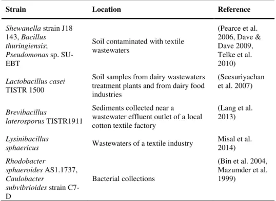
(Wuhrmann et al. 1980) strains. The use of a pure culture system ensures not only better interpretation of experimental observations, but also allows to assess detailed mechanisms of biodegradation using biochemistry and molecular biology tools, and this information can be useful to regulate and improve the enzymatic systems involved (Pearce et al. 2003). Therefore, in recent years, there has been an increased interest in the isolation of good decolourising species, especially bacteria, either in pure cultures or in consortia of known composition, for the treatment of dye containing wastewaters from several different locations (Table 1.2). In general, the mechanism of microbial degradation of azo dyes involves the reductive cleavage of azo bonds (–N=N–) with the help of NAD(P)H-dependent azoreductases under anaerobic conditions. Most azo dyes have sulphonated substituent groups and a relative high molecular weight and are unlikely to pass through cell membranes. Therefore, the reducing activity is not likely dependent on their intracellular uptake. One hypothesis involves the
Chapter 1 | 13
Table 1.2.
Bacterial strains capable of dye decolourisation.Strain Location Reference
Bacillus sp. SF,
Pseudomonas sp. SUK1
Wastewater drain of a
textile finishing industry (Maier et al. 2004, Kalyani et al. 2009)
Xenophilus azovorans KF46F, Pigmentiphaga kullae K24
Soil after a prolonged enrichment with Orange II or carboxy-Orange I as sources of carbon and energy
(Blümel et al. 2002, Bürger & Stolz 2010, Zimmermann et al. 1982, Blümel & Stolz 2003)
Pseudomonas
aeruginosa Soil near a tannery site
(Nachiyar & Rajakumar 2005)
Enterobacter
agglomerans Dye-contaminated sludge
(Moutaouakkil et al. 2003)
Pseudomonas luteola
Activated-sludge system utilized to treat wastewater from a dyeing factory
(Chang et al. 2001, Hsueh & Chen 2007)
Shewanella decolorationis S12
Activated-sludge of
textile-printing wastewaters (Hong et al. 2007)
Bacillus latrosporus
RRK1 Municipal wastewater treatment plant (Sandhya et al. 2008)
Aquiflexum sp. DL6, Bacillus badius
Alkaline Crater Lake of
Lonar (Misal et al. 2013, Misal et al. 2011)
Aeromonas hydrophila Fountain springs (Hsueh et al. 2009)
Bacillus velezensis Effluent of a textile industry
(Bafana et al. 2008a)
Bacillus sp. strain B29, Bacillus sp. strain
OY1-2 Soil
Strain Location Reference
Shewanella strain J18 143, Bacillus
thuringiensis; Pseudomonas sp. SU-EBT
Soil contaminated with textile wastewaters
(Pearce et al. 2006, Dave & Dave 2009, Telke et al. 2010)
Lactobacillus casei TISTR 1500
Soil samples from dairy wastewaters treatment plants and from dairy food industries
(Seesuriyachan et al. 2007)
Brevibacillus
laterosporus TISTR1911
Sediments collected near a
wastewater effluent outlet of a local cotton textile factory
(Lang et al. 2013)
Lysinibacillus
sphaericus Wastewaters of a textile industry
Misal et al. 2014)
Rhodobacter
sphaeroides AS1.1737, Caulobacter
subvibrioides strain C7-D
Bacterial collections
(Bin et al. 2004, Mazumder et al. 1999)
The degradation using these systems involves the transfer of four-electrons (reducing equivalents) to the azo linkage, in two stages, resulting in the reduction of the chromophore and formation of colourless solutions containing aromatic amines (Fig. 1.3) (van der Zee & Villaverde 2005, dos Santos et al. 2007, Saratale et al. 2011). Aromatic amines are the products of azoreductases reactions. These compounds are biorefractory and highly toxic because they can react easily in the blood to convert haemoglobin into methaemoglobin, thereby preventing oxygen uptake (Isik & Sponza 2007).
Chapter 1 | 15 N N= NH2 NH2 X X X X Azoreductase Redox mediatorox Redox mediatorred
NAD(P)H NAD(P)+
Carbon complexes
Oxidation products Enzyme liberating e
-Coloured solution containing the azo dye
Colourless solution containing aromatic amines
Cell
Figure 1.3
Proposed mechanism for reduction of azo dyes by whole bacterial cells.
Adapted from (dos Santos et al. 2007)
Aromatic amines are difficult to remove via traditional wastewater treatments and inevitably tend to be persistent because they are usually not further metabolized under anaerobic conditions (Chen et al. 2009, Kandelbauer & Guebitz 2005, Khan et al. 2013, Stolz 2001). Further mineralization of these compounds has been reported to occur only under aerobic conditions (Mendez-Paz et al. 2005). Therefore the combination of the anaerobic cleavage of the azo dyes with an aerobic treatment system has successfully been implemented (Gonçalves et al. 2005, van der Zee & Villaverde 2005, Lourenço et al. 2006).
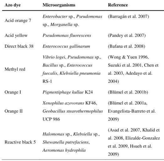
been often reported to inhibit the azo bond reduction activity. Aerobic respiration dominate the utilization of NAD(P)H, which are also electron donors of azoreductases (Pearce et al. 2003, Saratale et al. 2011, Solís et al. 2012). However in the past few years, several bacterial strains that can decolourise azo dyes in the presence of O2 were isolated (Table 1.3); in particular, Xenophilus azovorans KF46 and Pigmentiphaga kullae K24 were
reported to have the ability to grow aerobically on azo compounds as the sole carbon source (Table 1.3) (Blümel et al. 2001a; Blümel et al. 2001b).
Table 1.3.
Various aerobic bacteria capable of dye decolourisation adapted from(Sarayu & Sandhya 2012, Khan et al. 2013).
Azo dye Microorganisms Reference
Acid orange 7 Enterobacter sp., Pseudomonas sp., Morganella sp.
(Barragán et al. 2007)
Acid yellow Pseudomonas fluorescens (Pandey et al. 2007)
Direct black 38 Enterococcus gallinarum (Bafana et al. 2008)
Methyl red
Vibrio logei, Pseudomonas sp., Bacillus sp., Enterococcus faecalis, Klebsiella pneumonia RS-1
(Wong & Yuen 1996, Suzuki et al. 2001, Chen et al. 2003, Adedayo et al. 2004)
Orange I Pigmentiphage kullae K24 (Blümel et al. 2001b)
Orange II
Xenophilus azovorans KF46, Geobacillus stearothermophilus UCP 986
(Blümel et al. 2001a, Evangelista-Barreto et al. 2009)
Reactive black 5
Halomonas sp., Klebsiella sp., Shewanella putrefaciens, Aeromonas hydrophila
Chapter 1 | 17
Interestingly, azoreductases are involved in site-specific delivery of azo pro-drugs, which are therapeutically inactive in their intact form and rely on azo reduction by azoreductases of intestinal microflora for activation (Liu et al. 2009; Ryan et al. 2010; Wang et al. 2007). Sulfasalazine, balsalazide and olsalazine constitute some of the pro-drugs used to treat inflammatory bowel disease (IBD) (Wang et al. 2007). These pro-drugs are most commonly used in the treatment of IBD and have been developed with the aim of delivering the active anti-inflammatory agent 5-aminosalicylic acid (5-ASA) at the site of action being reductively cleaved in situ (Hanauer 1996).
In conclusion, enzymatic systems are particularly sought for the treatment of dye-containing effluents mainly because of their specificity and relatively ease of engineering towards improved robustness; enzymes only attack the dye molecules, while valuable dyeing additives or fibers are kept intact and can potentially be re-used (Kandelbauer & Guebitz 2005). Likewise, new recycling technologies will allow the reduction of enormous water consumption in the textile finishing industry. Importantly, although dye molecules display high structural variety, they are only degraded by few enzymes that share common mechanistic features as they all catalyse redox reactions exhibiting relatively wide substrates specificities.
Azoreductases
Azoreductases is the generic name given to enzymes involved in the reduction of azo bonds. The vast majority of azoreductases are FMN or FAD dependent oxidoreductases that require either NADH or NADPH as electron donors (Table 1.4) (Stolz 2001, Blümel & Stolz 2003, Chengalroyen & Dabbs 2013, Khan et al. 2013).
aureus, Geobacillus staerothermophilus, Clostridium perfringens and Brevibacillus laterosporus TISTR1911 (Bin et al. 2004, Chen et al. 2005,
Chen et al. 2004, Lang et al. 2013, Maier et al. 2004, Matsumoto et al. 2010, Morrison et al. 2012, Nachiyar & Rajakumar 2005; Nakanishi et al. 2001, Suzuki et al. 2001). Flavin-free azoreductases were also identified in
Pigmentiphaga kullae K24 and Xenophilus azovorans KF46F (Blümel et al.
2002, Bürger & Stolz 2010, Chen et al. 2010a), and more recently in
Lysinibacillus sphaericus (Misal et al. 2014). Most of these azoreductase
were heterologously produced in Escherichia coli and kinetically and
biochemically characterized (Table 1.4). Azoreductases can be broadly divided into two groups depending on oxygen sensitivity. Those from
Enterococcus faecalis (Chen et al. 2004, Chen et al. 2008), Pigmentiphaga kullae (Blümel & Stolz 2003), Rhodobacter sphaeroides (Bin et al. 2004,
Liu et al. 2007a), Xenophilus azovorans (Blümel et al. 2002), Bacillus sp.
OY1-2 (Suzuki et al. 2001), Escherichia coli (Nakanishi et al. 2001), Pseudomonas aeruginosa (Nachiyar & Rajakumar 2005, Wang et al. 2007)
have been shown to decolourise azo dyes in the presence of oxygen while enzymes from Pseudomonas luteola (Chang et al. 2001) and Bacillus sp.
Chapter 1 | 19
Table 1.4.
Bacteria containing genes coding for azoreductases and characteristics of recombinant azoreductases in Escherichia coli. Adaptedfrom (Leelakriangsak 2013).
Bacteria Gene Molecular mass (kDa) Cofactor(s) Reference
Bacillus sp. B29 azrA 48 (dimer) FMN, NADH (Ooi et al. 2007)
azrB 48 (dimer) FMN, NADH (Ooi et al. 2009)
azrC 48 (dimer) FMN, NADH (Ooi et al. 2009)
Bacillus sp. OY1-2 20 NADPH (Suzuki et al. 2001)
Bacillus subtilis yvaB (azoR2) 45 (dimer) NADH (Nishiya & Yamamoto 2007)
yhdA 76 (tetramer) FMN, NADPH (Deller et al. 2006)
Brevibacillus laterosporus RRK1 58 NADH (Sandhya et al. 2008)
Clostridium perfringens azoC 90 (tetramer) FAD, NADH (Morrison et al. 2012)
Enterococcus faecalis azoA 43 (dimer) FMN, NADH (Chen et al. 2004)
Enterococcus faecium acpD 23 NAD(P)H (Macwana et al. 2010)
Escherichia coli acpD (azoR) 46 (dimer) FMN, NADH (Nakanishi et al. 2001)
Geobacillus stearothermophilus azrG 23 (dimer) FMN, NADH (Matsumoto et al. 2010)
Pigmentiphaga kullae K24 azoB 22 NADPH (Chen et al. 2010a)
Pseudomonas aeruginosa paazor1 110 (tetramer) FMN, NAD(P)H (Wang et al. 2007)
paazor2 23 NADH (Ryan et al. 2010)
paazor3 26 NADH (Ryan et al. 2010)
Figure 1.4
Mechanism for reduction of
azo dyes by azoreductases. Adapted from (dos Santos et al. 2007)
Four bacterial azoreductases have the three-dimensional structure solved: the azoreductases from E. coli (Protein Data Bank (PDB) code 2Z98) (Ito et al.
2006), P. aeruginosa (PDB code 2V9C) (Wang et al. 2007), E. faecalis
(PDB code 2HPV) (Liu et al. 2007b) and Salmonella typhimurium (PDB
Chapter 1 | 21
Figure 1.5
Ribbon diagram of the P. aeruginosa azoreductase monomer.
Each monomer contains one FMN, which is bound to the C-terminal end of the β-sheet. β-strands are shown in yellow,
α-helices in red and loop regions are depicted in green. (Wang et al. 2007)
The FMN prosthetic groups bind on the C-terminal end of the central β-sheet at the dimer interface (in the case of dimeric proteins). Each FMN cofactor binds non-covalently to both monomers; 15 hydrogen bonds are formed with one monomer, whereas hydrophobic contacts are made between both monomers. The isoalloxazine moiety of FMN interacts with residues involved in loops L7 and L11 of one monomer and and loop L3’ of the other monomer (Fig. 1.6) (Ito et al. 2006).
Flavin-dependent azoreductases share strong similarities with regard to sequence, structure, and reaction mechanism with the larger family of flavin-dependent quinone reductases that includes Lot6p from Saccharomyces cerevisiae and the mammalian NQO1 presenting quinone reductase activity
Figure 1.6
Overall structure of the azoreductase from Escherichia coli.
Ribbon diagram of the homodimer of AzoR. The two subunits
Chapter 1 | 23
Quinones, characteristic of two carbonyl groups in an unsaturated six-member carbon ring, constitute an important class of ubiquitous and naturally occurring compounds present in the environment as well as in prokaryotic and eukaryotic cells (Deller et al. 2008, Liu et al. 2008a, Sollner & Macheroux 2009). Quinones are metabolites that can participate in deleterious redox cycling, which can lead to the accumulation of reactive oxygen species such as superoxide, hydrogen peroxide and hydroxyl radical, and can impair lipids, proteins and nucleotides (Liu et al. 2009). Azoreductases with quinone reductase activity may have a protective effect against quinone-based oxidative cell damage and may be involved in the detoxification of quinones (Liu et al. 2009, Liu et al. 2008a; Sollner & Macheroux 2009). Therefore, these enzymes are assumed to take part in the organism’s enzymatic detoxification systems; e.g., the azoreductases from E. coli and B. subtilis were implicated in the cellular response to thiol-specific
stress (Leelakriangsak et al. 2008, Liu et al. 2009, Towe et al. 2007) and Lot6p, the azoreductase homologue in S. cerevisiae has been implicated in
the response to oxidative stress (Sollner et al. 2009, Sollner et al. 2007). The flavin-containing enzymes were also shown to use other substrates such as nitroaromatic substrates (Liu et al. 2007). Further study of the genes coding for these xenobiotic-metabolizing enzymes may help to reveal factors that govern the horizontal transfer of genetic information among bacterial species in the environment. Overall, as additional members of this family of enzymes are discovered, the list of transformed substrates will continue to grow.
Laccases
Laccases are multi-copper oxidoreductases (MCOs) that are present in all domains of life: Archaea, Bacteria and Eukarya (Martins et al. 2015).
of rapid hardening of the latex from Japanese lacquer trees (Rhus vernicifera) in the presence of air (Yoshida 1883). Their catalytic centres
consist of three structurally and functionally distinct copper (Cu) sites; type 1 (T1) is a mononuclear centre involved in the substrate oxidation, whereas type 2 (T2) and the binuclear type (T3) form a trinuclear centre involved in the dioxygen reduction to water (Solomon et al. 1996) (Fig. 1.7). The oxidation of four substrate molecules at the T1 copper centre (the primary electron acceptor site), leads to the reductive cleavage of dioxygen to two molecules of water at the trinuclear site (Solomon et al. 1996). Most MCOs are composed of three Greek key β-barrel cupredoxin domains (domains 1, 2 and 3) that come together to form the three spectroscopically distinct types of Cu sites (Solomon et al. 1996, Martins et al. 2015) (Fig. 1.7). The T1 mononuclear copper centre shows an intense absorption band at ca. 600 nm,
which is responsible for the blue colour of the protein, and is due to the ligand-to-metal charge transfer between the cysteine sulphur and the copper atom. This site also shows a characteristic electron paramagnetic resonance (EPR) signal that is due to the high covalency at the copper site. The T2 copper site also exhibits a characteristic EPR signal, but no observable bands in the absorption spectra. The pair of T3 copper ions is EPR silent, a fact attributed to their antiferromagnetic coupling by the presence of a bridging ligand, normally assumed to be hydroxyl. The T3 site also shows an absorption band at ca. 330 nm that has been attributed to the charge transfer
Chapter 1 | 25
Figure 1.7
Overall three-dimensional structure of Bacillus subtilis CotA-laccase.
A) Three-dimensional structure of CotA with each cupredoxin domain coloured in different colour (domain 1 in blue, domain 2 in green and
domain 3 in orange). The copper atoms are shown as brown spheres, with the fifth regulatory Cu ion identified near the T1 Cu site.
B) Structural detail of the catalytic copper centres, the mononuclear
type 1 copper centre (T1) where the copper atom is coordinated by a cysteine and two histidines, and the trinuclear centre which comprises a type 2 copper atoms (T2) and two type 3 (T3) copper atoms. The cysteine residue (C492) that coordinates the T1 copper atom is bound to two of the histidine residues (H491 and H493) that
coordinate the two T3 coppers in the trinuclear centre. Adapted from (Bento et al. 2010, Martins et al. 2015).
The use of laccases for dye decolourisation has been extensively studied (Molina-Guijarro et al. 2009, Pereira et al. 2009a, Pereira et al. 2009b, Moya et al. 2010, Loncar et al. 2013, Martins et al. 2015). These enzymes decolourise azo dyes without direct cleavage of the azo bond through a highly nonspecific free radical mechanism, thereby avoiding the formation of toxic aromatic amines (Chivukula & Renganathan 1995, Zille et al. 2005a, Pereira et al. 2009a, Pereira et al. 2009b). Fungal laccases are the enzymes used in the vast majority of the studies but bacterial enzymes shows advantages for biotechnological processes due to the lack of post-translational modifications, their higher yields of production, easiness of manipulation and improvement by protein engineering approaches. The first study using bacterial laccases for dye decolourisation was performed with the recombinant CotA-laccase from Bacillus subtilis, which is a
thermoactive and intrinsically thermostable enzyme (with half-life of 2 h at 80°C) showing the predictable robustness for biotechnological applications (Martins et al. 2002, Pereira et al. 2009a, Pereira et al. 2009b). The lack of a strict requirement for redox mediators exhibited by bacterial CotA-laccase constitutes an advantage over other laccases from a technological perspective. Indeed, in spite of the proven efficiency of mediators, such as 2,2-azino-bis (3-ethylbenzothiazoline-6-sulfonic acid (ABTS), 1-hydroxybenzotriazole, violuric acid or 2,2,6,6-tetramethyl-piperidine-1-oxyl radical (TEMPO), in the degradation of recalcitrant aromatic compounds their application is strongly impeded by its high cost, and generation of toxic species leading to biocatalyst inactivation (Astolfi et al. 2005). The decolourisation of synthetic dyes was reported for two additional bacterial laccases from Streptomyces ipomoea CECT 3341 (SilA) (Molina-Guijarro et
al. 2009) and Streptomyces cyaneus CECT 3335 (Moya et al. 2010) in the
Chapter 1 | 27
pathway of the azo dye Sudan orange G (SOG) by CotA-laccase was studied in detail (Pereira et al. 2009b) (Fig. 1.8A). The laccase oxidizes the phenolic group of the dye with the participation of one electron, generating a phenoxy radical, which is sequentially followed by the oxidation to a carbonium ion. The nucleophilic attack by water on the phenolic ring carbon bearing the azo linkage causes NC-bond cleavage. The one-electron oxidation of SOG molecule by the enzyme resulted therefore, in the formation of unstable radical molecules and in the concomitant destruction of the dye chromophoric structure. The products obtained during the azo dye degradation reactions, can undergo further reactions and can be polymerized or coupled among them or with the unreacted dye producing a large amount of coupled and polymeric products as identified by ESI-MS and MALDI-TOF MS (Fig. 1.8B) leading to a darkening of the solution (Pereira et al. 2009b, Mendes et al. 2015c).
formation of an azo bond in (4), responsible for the colour observed in the reaction mixtures (Pereira et al. 2009a, Mendes et al. 2015c).
Figure 1.8
Proposed mechanism for the biotransformation of SOG by CotA-laccase (A) and
proposed structures (1)-(7) for the oxidation products (B).
Chapter 1 | 29
Figure 1.9
Proposed mechanism of AB62 biotransformation by laccases. Two oxidative routes are
Peroxidases
Chapter 1 | 31
Figure 1.10
Catalytic cycle of haem peroxidases.
On the basis of sequence similarity, the super-family of fungal, plant and bacterial peroxidases has been classified into: Class I (intracellular peroxidases) that includes yeast cytochrome c peroxidase (CcP, a mitochondrial soluble protein that exerts a protective function against toxic peroxides (Dunford 1999, Everse et al. 2011)), ascorbate peroxidase (APX, which removes hydrogen peroxide in chloroplasts and cytosol of higher plants (Dunford 1999, Everse et al. 2011)) and the bacterial catalase-peroxidase (KatG, which feature both catalase-peroxidase and catalase activities (Dunford 1999, Smulevich et al. 2006)); Class II (secretory fungal peroxidases) that includes lignin peroxidase (LiP), manganese peroxidase (MnP), versatile peroxidase (VP), all involved in the oxidative degradation of lignin and the peroxidase from Arthromyces ramous (also known as Coprinus cinereus, ARP or CiP) (Banci 1997); and Class III (secretory
senescence and protection against pathogens and are also involved in different tissue-specific functions, such as removal of H2O2 from chloroplasts and cytosol, oxidation of toxic compounds, biosynthesis of cell walls and defense towards wounding (Dunford 1999, Hofrichter et al. 2010). The peroxidase-cyclooxygenase superfamily evolved independently. It is constituted of soluble mammalian peroxidases, such as myeloperoxidase (MPO), eosinophil peroxidase (EPO), lactoperoxidase (LPO) and thyroid peroxidase (TPO), that are effective antimicrobial oxidants, oxidizing small anions such as halides (chloride and bromide), thiocyanate, and nitrate to the corresponding oxidation products and take part in the innate immune system (Zederbauer et al. 2007, Zamocky & Obinger 2010).
The interest in peroxidases for industrial applications has been increasing rapidly, particularly in ligninolytic peroxidases, harbouring the highest redox potential among peroxidases, for the selective delignification of lignocellulosic materials (Martinez et al. 2009, Ruiz-Duenas & Martinez 2009). These enzymes are also suitable for environmental applications including the treatment of toxic effluents, such as those containing synthetic dyes, generated in various industrial processes. In particular, LiP, VP, MnP, horseradish peroxidase (HRP), soybean peroxidase (SBP) are able to oxidize dyes leading to bleaching of coloured compounds (Heinfling et al. 1998, Moldes et al. 2003, Perez-Boada et al. 2005, Souza et al. 2007, Ruiz-Duenas & Martinez 2009, Ali et al. 2013, Kalsoom et al. 2013).
Chapter 1 | 33
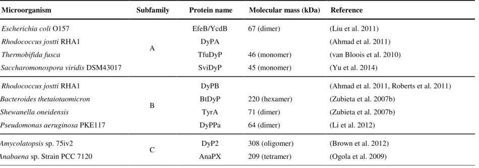
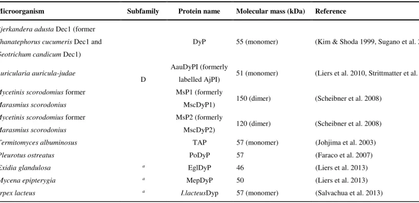
shown in Table 1.5. These enzymes show different molecular masses and oligomeric states ranging from monomers to hexamers although sharing the same non-covalently bound haem (proto-haem IX) as cofactor (Colpa et al. 2014).
DyPs have been classified into four phylogenetically distinct subfamilies, with bacterial enzymes constituting A-C subfamilies and fungal enzymes belonging to D subfamily (Ogola et al., 2009) (Table 1.5 and Fig. 1.11), but their physiological function is at present unclear.