Interaction with dsDNA and Camptothecin Derivatives
Gary S. Laco*
Laboratory of Computational and Molecular Biochemistry, Lake Erie College of Osteopathic Medicine, Bradenton, Florida, United States of America
Abstract
Human topoisomerase I (Top1) relaxes supercoiled DNA during cell division. Camptothecin stabilizes Top1/dsDNA covalent complexes which ultimately results in cell death, and this makes Top1 an anti-cancer target. There are two current models for how camptothecin and derivatives bind to Top1/dsDNA covalent complexes (Staker, et al., 2002, Proc Natl Acad Sci USA 99: 15387–15392; and Laco, et al., 2004, Bioorg Med Chem 12: 5225–5235). The interaction energies between bound camptothecin, and derivatives, and Top1/dsDNA in the two models were calculated. The published structure-activity-relationships for camptothecin and derivatives correlated with the interaction energies for camptothecin and derivatives in the Laco et al. model, however, this was not the case for several camptothecin derivatives in the Stacker et al. model. By defining the binding orientation of camptothecin and derivatives in the Top1/dsDNA active-site these results allow for the rational design of potentially more efficacious camptothecin derivatives.
Citation:Laco GS (2011) Evaluation of Two Models for Human Topoisomerase I Interaction with dsDNA and Camptothecin Derivatives. PLoS ONE 6(8): e24314. doi:10.1371/journal.pone.0024314
Editor:Rakesh K. Srivastava, The University of Kansas Medical Center, United States of America ReceivedMay 27, 2011;AcceptedAugust 4, 2011;PublishedAugust 30, 2011
Copyright:ß2011 Gary S. Laco. This is an open-access article distributed under the terms of the Creative Commons Attribution License, which permits unrestricted use, distribution, and reproduction in any medium, provided the original author and source are credited.
Funding:This work was supported solely by an intramural grant from the Lake Erie College of Osteopathic Medicine. The funders had no role in study design, data collection and analysis, decision to publish, or preparation of the manuscript.
Competing Interests:The authors have declared that no competing interests exist. * E-mail: [email protected]
Introduction
Camptothecin (CPT) is a natural product that was isolated from the Chinese treeCamptotheca acuminataby Wall and Wani and was shown to inhibit the growth of cancer cells in cell culture [1]. Derivatives of CPT, including topotecan and irinotecan, have been approved by the Food and Drug Administration for the treatment of cancer. Several groups have shown that CPT specifically targets human topoisomerase type IB (Top1) [2,3]. Top1 binds dsDNA and attacks the backbone scissile bond phosphate with the active-site Tyr723 to make a covalent tyrosyl-phosphate bond to the -1 scissile strand deoxyribonucleotide (-1 nucleotide). This results in a +1 deoxyribonucleoside (+1 nucleoside) with a free 59-OH. CPT stabilizes the Top1/dsDNA covalent complex [4]. When cells replicate DNA containing CPT stabilized Top1/dsDNA covalent complexes the single-strand breaks are converted into dsDNA breaks and it is thought that this ultimately results in cell death [5–7]. Numerous derivatives of CPT have been designed and synthesized to increase specificity and bioavailability, reviewed in [8,9]. However, one problem associated with the use of CPT derivatives in the treatment of breast cancer is the development of drug resistance [9]. Breast cancer resistance has been linked to the over expression of the breast cancer resistance protein, the substrates for which are planar with conjugated pi-orbitals and with an OH or amino group at the A-ring 10 position of CPT derivatives [10]. A model of CPT binding in the Top1/dsDNA active-site would allow for the rational design of CPT derivatives which are not substrates for the breast cancer resistance protein.
A key result in the Top1 field was obtained when Redinbo et al. solved the X-ray crystal structure of Top1 in covalent complex
with a suicide-DNA [11]. Since Top1 has a low affinity for relaxed dsDNA, a suicide dsDNA oligonucleotide (suicide-DNA) was used to generate the Top1/suicide-DNA covalent complexes [11]. The suicide-DNA differed from native dsDNA in that the backbone scissile bond 59oxygen (O59) was replaced with a sulfur. When the Top1 active-site Tyr723 attacked the suicide-DNA it generated a native tyrosyl-phosphate bond to the 39end of the -1 nucleotide while leaving a non-native 59-SH on the free end of the +1 nucleoside [11,12]. And while the 59-SH can attack the tyrosyl-phosphate bond resulting in religation of the scissile strand DNA backbone, the equilibrium is shifted,7-fold towards the Top1/
dsDNA covalent complex [13]. As a result, Top1 remained trapped in covalent complex with the suicide-DNA in the crystals [11]. The Redinbo et al. structure allowed for the structure-based modeling of CPT and derivatives in the Top1/suicide-DNA active site and resulted in models by Redinbo et al. [11], Kerrigan et al. [14], and Laco et al. [15], the last of which is described here as the Rotated+1 Nucleoside model.
into the helix allowing the +1 nucleoside 59OH to attack the tyrosyl-phosphate bond, religate the DNA backbone, and allow Top1 to dissociate from the fully relaxed dsDNA [15]. Rotation of the +1 nucleoside out of the helix leaves a cavity in the Top1/ dsDNA active-site which can be occupied by CPT and derivatives. When CPT and derivatives bind in the Rotated +1 Nucleoside model Top1/dsDNA active-site they block the+1 nucleoside from re-entering the helix thus stabilizing the Top1/dsDNA covalent complex [15].
The Rotated+1 Nucleoside model is significantly different from the Staker et al. X-ray crystallography based models of Top1 in covalent complex with suicide-DNA and either topotecan [18] or CPT [19]. In both Staker et al. structure based models the +1 nucleoside was based-paired in the helix, and the inhibitors intercalated in between the -1 and+1 base pairs, forcing the+1 base pairs,3.4 A˚ further away from the -1 base pairs [18,19]. As
a result, the suicide-DNA +1 nucleoside 59SH was 11.6 A˚ away from the tyrosyl-phosphate bond preventing religation of the DNA [18,19]. The Staker et al. structure-based model of Top1 in covalent complex with dsDNA and bound inhibitor is described here as the ‘‘Intercalated model’’. The Rotated +1 Nucleoside model and the Intercalated model were evaluated in silicousing Discovery Studio computational chemistry software (Accelrys, San Diego, CA). This approach allowed for evaluation of the binding orientations of CPT and derivatives in the Rotated+1 Nucleoside and Intercalated models, to see which binding orientation correlated best with the reported in vitro structure-activity-relationships for CPT and derivatives.
Results and Discussion
CPT and derivatives selected for this study, along with the respectivein vitroinhibition results for each, are shown in Figure 1 and Table 1 [15,16,20]. There were three selection criteria for the inhibitors used in this study: 1) inhibitors had been tested in anin vitro Top1 dsDNA oligonucleotide assay in which the dsDNA oligonucleotide had a single high affinity Top1 cleavage site; 2) the assay measured the accumulation of Top1/dsDNA covalent complexes over time; and 3) inhibitors were tested at three or more concentrations to quantitate inhibition. As a result, topotecan was not included in the study due to the lack of published reports documenting the in vitro inhibition activity of topotecan using the same assay and conditions as for the inhibitors in Table 1 [21,22]. The original Laco et al. model of Top1 in covalent complex with DNA and inhibitor [15] was based on the Redinbo et al. X-ray crystal structure of Top1 in covalent complex with suicide-DNA [11]. A reconstituted Top1 was used to generate the Redinbo et al. Top1/suicide-DNA covalent-complex structure and the following Top1 residues were not resolved in the
functional domain which includes the DNA binding and enzymatic domains of Top1 (aa 201–265) [17]: residues 201– 214; core residues 627–634, linker residues 635–697; and C-terminal residues 698–719 which are part of the Top1 active-site [11]. A more recent structure by Staker et al. of a Top1/suicide-DNA/topotecan ternary complex resolved all main-chain Top1 atoms in the functional domain [18]. However, in that structure the intercalated topotecan, a CPT derivative with an A-ring 9-CH2N(CH3)2and 10-OH, displaced the+1 bases,3.4 A˚ further
away from the cleavage-site -1 base pairs. In order to generate a structurally complete model of Top1 in covalent-complex with native dsDNA and no inhibitor the displaced DNA strands were replaced in the Stacker et al. Top1/suicide-DNA/topotecan structure [18] with the corresponding DNA strands from the Redinbo et al. Top1/suicide DNA structure [11] using Discovery Studio and the Consistent Force Field (CFF) which has parameters for protein, DNA, and small ligands (Methods). Next, the +1 nucleoside was rotated out of the helix until it was trapped in a network of H-bonds/electrostatic interactions with Arg488, Asp533, Arg590 as previously reported [15]. CPT was then docked into the active-site of the Rotated+1 Nucleoside model and modified into the indicated CPT derivatives (Figure 1 and Table 1); with 10-OH CPT shown in the Rotated+1 Nucleoside model active-site in Figure 2. The orientations of CPT and derivatives in this structurally complete Rotated +1 Nucleoside model were similar to the Laco et al. model [15] that was based on the incomplete Top1/suicide-DNA structure by Redinbo et al. [11]. In addition, the suicide-DNA backbone sulfur, that is found as a free 59SH on the+1 nucleoside, was changed to the native oxygen [11,18,19]. Explicit waters were added to the Rotated+1 Nucleoside model presented here followed by minimization, however, individual waters did not make any bridging H-bonds/ electrostatic interactions between 10-OH CPT and the Top1/ dsDNA active-site (data not shown). This result is consistent with previously published results on the Rotated+1 Nucleoside model in which individual explicit waters did not make bridging H-bond interactions between ligands and the Top1/dsDNA active-site [15]. As a result, all Rotated+1 Nucleoside model Top1/dsDNA/ inhibitor complexes were minimized with an implicit solvent model (Methods).
The Staker et al. structure based Intercalated model of Top1 in covalent complex with suicide-DNA and topotecan was modified Figure 1. Structure of CPT and hCPT E-ring.CPT left, hCPT E-ring
right; hCPT and derivatives differ from CPT in that they contain an additional E-ring carbon between the 20-OH and the adjacent carbonyl oxygen to give a seven-member E-ring.
doi:10.1371/journal.pone.0024314.g001
Table 1.Relativein vitroinhibition of Top1 by CPT, hCPT, and derivatives.
Ligand Position Inhibition
10 11 20
CPT H H OH 0.9*
hCPT H H OH 1.0*
620-deoxy CPT H H H 0.03**
620-Cl CPT H H Cl 0.29**
10-OH CPT OH H OH 3.5*
10,11-diF hCPT F F OH 12.0*
Modified A-ring ring positions (10, 11) and E-ring position (20) are indicated for CPT/hCPT derivatives; modifications shown in italics (see Fig. 1 for CPT and hCPT structures).
*As reported by Laco et al. [15,16].
[18], topotecan was converted into CPT and derivatives including 10-OH CPT, and the suicide-DNA backbone sulfur was changed to the native oxygen (Fig. 1 and 3, Table 1, Methods). In the Staker et al. structure of topotecan in ternary complex with Top1 and suicide-DNA two crystallographic waters were found to make bridging H-bonds between the topotecan carboxylate and Top1 active-site residues [18], however, carboxylate forms of CPT derivatives have been shown to be inactive [15]. One additional water was found to be within 2.9 A˚ of the topotecan A-ring 10-OH, but there were no Top1/dsDNA H-bond donor/acceptor residues within 4.4 A˚ of that water [18]. Here the Intercalated
model was initially solvated with explicit waters followed by minimization, and a water was found close to the OH of 10-OH CPT, however, the only other H-bond donors/acceptors within 4.4 A˚ were other waters (data not shown). Since no individual waters were found to make bridging H-bond/ electrostatic interactions between the inhibitor and the Top1/ dsDNA active-site, all Intercalated model Top1/dsDNA/inhibitor complexes were minimized with an implicit solvent model (Methods). 10-OH CPT is shown in the Intercalated model Top1/dsDNA active-site in Figure 3. The Top1/suicide-DNA/ topotecan X-ray crystallography based model was used here because Top1 in the Top1/suicide-DNA/CPT model was not structurally complete [19].
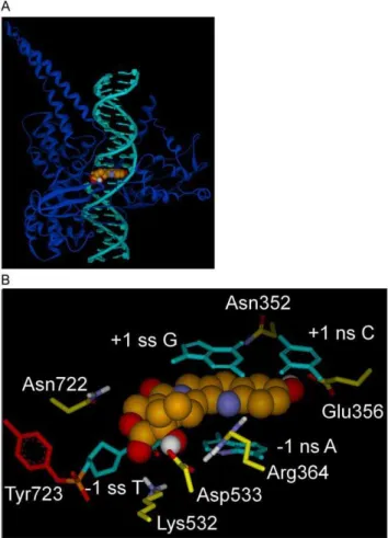
The inclusion of explicit water molecules during an interaction energy calculation between an inhibitor and Top1/dsDNA would artificially make the electrostatic component of the interaction energy score more negative (i.e., stronger) when individual waters Figure 2. Rotated+1 Nucleoside model for Top1 interaction
with dsDNA and 10-OH CPT.A) Rotated+1 Nucleoside model, Top1
shown as blue ribbon, dsDNA in teal with the rotated+1
deoxygua-nosine left of 10-OH CPT (CPK rendering; carbon, green; oxygen, red; nitrogen, blue; hydrogen, white). B) Rotated+1 Nucleoside model, close
up of Fig. 2A active-site. 10-OH CPT with E-ring in foreground, bound in the Top1/dsDNA active-site in which the+1 scissile strand G is rotated
out of the helix to the left until trapped in a network of H-bonds/ electrostatic interactions with Asp533 (center) and Arg488/Arg590 (not shown for clarity, see Fig. 4A). Selected atoms involved in H-bonds/ electrostatic interactions are colored: nitrogen, blue; oxygen, red; hydrogen, white. Top1 side-chain carbons are yellow, except for Tyr723 (red) in tyrosyl-phosphate bond (phosphorus, orange) to the -1 scissile strand T. 10-OH CPT interactions: 10-OH CPT D-ring stacks over the -1 scissile strand T; 20-OH H-bonds to -1 scissile strand T carbonyl oxygen; A-ring 10-OH oxygen makes electrostatic interaction with Asn352 nitrogen (3.6 A˚ ); E-ring carbonyl oxygen H-bonds to Lys532 nitrogen; C-ring carbonyl oxygen H-bonds to Asn722 nitrogen. Scissile strand rotated+1 G 59OH H-bonds to Asp533. Arg364 nitrogens H-bond to+1 non-scissile strand C carbonyl oxygen and -1 non-scissile strand A nitrogen. Scissile strand, ss; non-scissile strand, ns. For flat image see Fig. 4A.
doi:10.1371/journal.pone.0024314.g002
do not mediate significant bridging H-bonds/electrostatic inter-actions between the inhibitor and the Top1/dsDNA active-site.
Similarities and differences between Rotated+1
Nucleoside and Intercalated Models
While the orientation of 10-OH CPT and DNA in the two models is overall strikingly different, there are similarities and they are described first. Top1 active-site residues Arg364, Lys532, and Arg533 are close to the E-ring in both models, while Asn722 faces the D-ring carbonyl oxygen in both models. In addition, the -1 scissile strand thymine base stacks under the D-ring in both models (Fig. 2 and 3).
In terms of the differences between the two models: 1) In the Rotated +1 Nucleoside model the A-ring 10-OH makes an electrostatic interaction with Asn352 (Fig. 2B and 4A), while in the Intercalated model the A-ring 10-OH H-bonds to Glu356 (Fig. 3B and 4B); 2) In the Rotated+1 Nucleoside model 10-OH CPT only stacks with the -1 scissile strand T (Fig. 2), while in the Intercalated model 10-OH CPT stacks with the -1 and+1 base pairs (Fig. 3). And while Asn430, Pro431, Leu721, and Lys751 are within 4– 7.7 A˚ of 10-OH CPT in the Rotated+1 Nucleoside model, those residues are 1–4 A˚ further away from 10-OH CPT in the Intercalated model (data not shown). By determining the orientation of CPT and derivatives in the Top1 active-site, CPT
derivates could be designed to make additional H-bonds and van der Waals interactions with one or more of the above residues. The default Discovery Studio 2.1 definition for H-bonds was used with H-bonds distances reported here between 3.0–3.1 A˚ (Accelrys, San Diego, CA).
In order to evaluate the orientation of CPT and derivatives in the two models, and compare them to the reportedin vitroactivities for those same compounds (Figure 1 and Table 1), the interaction energy scores between CPT and derivatives and the active-sites of the Rotated +1 Nucleoside model (Fig. 2 and 4A) and the Intercalated model (Fig. 3 and 4B) were calculated (Fig. 5). For both models, the CPT interaction energy score (Rotated +1 Nucleoside model: vdW 48.6, E 4.67; Intercalated model: vdW -69.22, E -5.9) was subtracted from that for 10-OH CPT, 20-deoxy CPT, and 20-Cl CPT, while the hCPT interaction energy score
Figure 4. Flattened images of two models for Top1 interaction with dsDNA and 10-OH CPT.A) Flat image of Rotated+1 Nucleoside
model from Fig. 2, H-bonds and electrostatic interactions within 3.6 A˚ are indicated with dashed lines. B) Flat image of Intercalated model from Fig. 3, H-bonds and electrostatic interactions within 3.6 A˚ are indicated with dashed lines.
doi:10.1371/journal.pone.0024314.g004
Figure 5. Relative interaction energy in kcal/mol between CPT/ hCPT-derivatives and the Top1/dsDNA active-site.The interac-tion energy scores for the derivatives of CPT and hCPT were subtracted from the score of the respective parent inhibitor with the resulting difference values plotted on the graph. The CPT and hCPT interaction energy scores were set to zero. A negative kcal/mol score indicates that a derivative bound tighter than the parent inhibitor, while a positive kcal/mol score indicates that it bound weaker than the parent inhibitor. A) Interaction energy values for CPT, hCPT and derivatives when bound in the Rotated +1 Nucleoside model Top1/dsDNA active-site. B)
Interaction energy values for CPT, hCPT and derivatives bound in the Intercalated model Top1/dsDNA active-site.
(Rotated+1 Nucleoside model: vdW -49.33, E -1.92; Intercalated model: vdW -68.63, E -5.54) was subtracted from that for 10,11-diF hCPT. The resulting difference values for 10-OH CPT, 20-deoxy CPT, 20-Cl CPT, and 10,11-diF hCPT in the two models were then graphed to visualize whether an inhibitor bound either stronger (negative kcal/mol), or weaker (positive kcal/mol), than either CPT, or hCPT, which were set to zero (Fig. 5). For each of the following ligands the relative increase/decrease in the van der Waals (vdW) and electrostatic energy (E) is indicated. It has been reported that 10-OH CPT and 10,11-diF hCPT were 3.5-fold and 12-fold more potent in vitro than CPT and hCPT, respectively (Table 1) [15,16]. It was also reported that the racemic 20-deoxy CPT had 3%, and racemic 20-Cl CPT had 29%, of the inhibition of CPT (Table 1) [20].
Ligand scores for Rotated+1 Nucleoside model
In the Rotated+1 Nucleoside model, the 10-OH CPT value was -3.23 kcal/mol (vdW -2.16, E -1.07) stronger than CPT, and the 10,11diF hCPT value was 2.03 kcal/mol (vdW 0.16, E -1.87) (Fig. 5A) stronger than hCPT due to either the A-ring 10-OH, or 10,11 diF, interacting with Asn352 (Fig. 2B and 4A). These additional interactions explained why 10-OH CPT and 10,11-diF hCPT were more potentin vitrothan CPT and hCPT, respectively (Table 1) [15,16]. The 20-deoxy CPT value was 1.81 kcal/mol (vdW 1.5, E 0.31) weaker than CPT (Fig. 5A) due to the loss of the 20-OH interaction with the Top1/dsDNA active-site (Fig. 5A). This is consistent with the racemic 20-deoxy CPT having lowin vitro activity (Table 1) [20], with the 1.81 kcal/mol value (vdW 1.5, E 0.31) representing a loss of 6% of the total electrostatic interaction energy for CPT. Interestingly, the 20-Cl CPT value was only 0.49 kcal/mol (vdW 0.2, E 0.29) weaker than CPT (Fig. 5A). This result for 20-Cl CPT is consistent with the lack of a H-bond between the E-ring 20-Cl and the -1 scissile strand thymidine carbonyl oxygen (see E-ring 20-OH hydrogen in Fig. 2B and 4A). However, the 20-Cl CPT is significantly more potentin vitro than 20-deoxy CPT (Table 1), indicating that the 20-Cl is contributing to interactions between 20-Cl CPT and Top1/ dsDNA through electrostatic interactions with Arg364 and Lys532. In the Rotated +1 Nucleoside model the Arg364 side-chain nitrogens make H-bonds to Asp533 and the -1 non-scissile strand cytosine and the +1 non-scissile strand adenine, and electrostatic interactions with the E-ring 20-OH (Fig. 4A). These interactions by Arg364 would contribute to the stabilization of the Top1/dsDNA/inhibitor complex (Fig. 2 and 4A). When Arg364 was mutated to His, the mutant Top1 became resistant to CPT and derivatives [23]. The shorter His side chain can neither make H-bonds to Asp533 and the -1/+1 non-scissile strand bases, nor electrostatic interactions with the E-ring 20-OH (data not shown). These in silico results support the binding orientation for CPT, hCPT, and derivatives in the Rotated+1 Nucleoside model (Fig. 2). However, it is important to note that while the 10,11 diF hCPTin vitroinhibition (Table 1, 12-fold) follows the respective Rotated+1 Nucleoside model difference value of -2.03 kcal/mol (Fig. 5A), the two values do not correlate in magnitude with the corresponding values for 10-OH CPT (Table 1, 3.5-fold; and Fig. 5A, -3.23). Fluorinated compounds have increased hydrophobicity [24], which in vitrolikely results in 10,11-diF hCPT partitioning more efficiently out of the solvent phase and into the Top1/dsDNA binding cavity than does 10-OH CPT which is more hydrophilic due to the A-ring 10-OH. However, the interaction energy calculations in this study did not take that into account. Consistent with the reported 12-fold inhibition enhancement for 10,11-diF hCPT (Table 1), an inhibitor of glycogen phosphorylase, in which two hydroxyls were replaced with fluorines, was 10-fold more
potent than the non-fluorinated inhibitor [25]. And while the addition of a hydroxyl to an inhibitor can be used to probe the active-site of an enzyme for a H-bond donor/acceptor, the substitution of a hydroxyl on an inhibitor with fluorine can be used to probe the active-site of an enzyme for the presence of just a H-bond donor [24,25]. Thein vitrodata in Table 1 indicates that 10-OH CPT, 10,11-diF hCPT, and 20-Cl CPT interact with H-bond donors in the Top1 active-site. The Rotated+1 Nucleoside model supports this conclusion since Asn352 can donate H-bond/ electrostatic interactions to 10-OH CPT and 10,11-diF hCPT (Fig. 2B and 4A), and Arg364 and Lys532 which are H-bond donors can make electrostatic interactions with 20-Cl CPT at
,4 A˚ (data not shown).
Ligand scores for Intercalated model
In the Intercalated model, the 10-OH CPT value of -3.31 kcal/ mol (vdW -2.06, E -1.25) (Fig. 5B) was stronger than CPT due to the 10-OH CPT A-ring 10-OH making a H-bond to Glu356 (Fig. 3B and 4B). In the Staker et al. structure based Top1/suicide-DNA/ topotecan model, the Glu356 side-chain oxygen was 3.8 A˚ from the topotecan A-ring 10-OH with a reported B-factor of 48 [18]. The topotecan A-ring 9-CH2-N-(CH3)2 may have disrupted the
interaction between Glu356 and the adjacent A-ring 10-OH. In contrast, the 10,11-diF hCPT value of -0.2 kcal/mol (vdW -0.34, E 0.13) was only slightly stronger than hCPT (Fig. 5B), due to an electrostatic clash between the A-ring 10,11-diF and the Glu356 side-chain oxygens (note positive E value in the above score). These in silicoresults for 10,11-diF hCPT appear inconsistent with the reportedin vitro 12-fold increase in inhibition (Table 1) [15]. As pointed out above, fluorine has been used to probe the active-site of enzymes for H-bond donors, however, the Glu356 side-chain oxygens can not donate a H-bond to fluorine. In the Intercalated model, the 20-deoxy CPT value was 6.03 kcal/mol (vdW 1.91, E 4.12) weaker than CPT with the electrostatic component repre-senting a loss of 70% of the total electrostatic interaction energy for CPT (see above, Fig. 5B). The decrease in electrostatics for 20-deoxy CPT can be explained by the lack of a H-bond between the 20-deoxy CPT E-ring and Asp533; the racemic 20-deoxy CPT had lowin vitroactivity (Table 1) [20]. The 20-Cl CPT value of 5.64 kcal/mol (vdW 0.74, E 4.9) was also significantly weaker than CPT due again to the lack of a H-bond between the 20-Cl CPT E-ring and the Asp533 side-chain oxygen (see 20-OH group of 10-OH CPT in Fig. 3B and 4B). In the Intercalated model, the CPT 20-OH H-bond to Asp533 is the dominant electrostatic interaction between the inhibitor and the Top1/dsDNA active-site. However, in the Intercalated model the 20-Cl makes an electrostatic clash with Asp533 (see 20-OH of 10-OH CPT in Fig. 3B and 4B). Thein silico results for 10,11-diF hCPT and 20-Cl CPT in the Intercalated model (Fig. 5B) do not follow the trend for the known in vitro inhibition by these CPT/hCPT derivatives (Table 1). It is important to note that thesein silicoresults allow for further testing of both models. For the Intercalated model to be biologically relevant (Fig. 3 and 4B) mutation of Top1 Glu356 to Ala should result in a Top1 mutant that could not distinguish between CPT and 10-OH CPT due to the lack of a H-bond between an Ala356 and the A-ring 10-OH. In contrast, for the Rotated+1 Nucleoside model, the same Top1 Glu356Ala mutant should still be inhibited,3.5-fold more by
10-OH CPT, than by CPT, because in the Rotated+1 Nucleoside model the 10-OH CPT A-ring does not interact with Glu356 (Fig. 2B).
hCPT, respectively (Table 1 and Fig. 5B). The results for the Intercalated model suggest that the use of suicide-DNA, and high inhibitor-to-enzyme ratios (,34:1) to generate
Top1/suicide-DNA/inhibitor ternary complexes resulted in non-biologically relevant binding modes for CPT and derivatives [18,19]. This is supported by the fact that the Top1/suicide-DNA/topotecan active-site was found to contain both the lactone (63%) and carboxylate (37%) forms of topotecan (the same ratio was also found for topotecan in mother liquor alone) [18]. The Top1/ suicide-DNA/CPT active-site was also found to contain both the lactone and carboxylate forms of CPT, for which the percentages were so close they could not be differentiated [19]. In contrast, Top1 and dsDNA oligonucleotides were reported in vitro to preferentially stabilize the active lactone form of CPT (97.1%) versus the inactive carboxylate form (2.9%) [15]. The carboxylate form of hCPT, which can not reverse to the lactone form, has been shown to have no detectable inhibitory activityin vitro[15]. It can be argued that the Staker et al. Top1/suicide-DNA active-site could not distinguish between active and inactive forms of CPT and derivatives because the+1 nucleoside was not rotated out of the helix to open a biologically important inhibitor binding cavity (Fig. 2) [15]. The Rotated +1 Nucleoside model H-bond/ electrostatic interactions between Arg364 and -1 non-scissile strand A and +1 non-scissile strand C, Arg488 and +1 scissile strand G, Lys532 and -1 scissile strand T, and Arg590 and +1 scissile strand G (Fig. 2B and 4A) are consistent with CPT stabilized Top1/dsDNA covalent-complexes occurring preferen-tially at sequences containing a -1 scissile strand T/+1 scissile strand G and followed by other combinations of -1 pyrimidines and+1 purines/pyrimidines [26].
Here it was also found that the lactone forms of CPT and derivatives made direct H-bonds to the Top1/dsDNA active-site in both the Rotated+1 Nucleoside and Intercalated models which is consistent with: 1) a previous report for a solvated Rotated+1 Nucleoside model [15]; 2) for the X-ray crystallography based Intercalated model [18]; and 3) molecular dynamic simulations of the topotecan lactone in the solvated Top1/dsDNA active-site by Mancini et al. [27].
The probing of the Top1 active-site with CPT derivatives containing chlorine and fluorine substitutions for hydroxyls revealed that 20-Cl CPT was able to make an electrostatic interaction with the Top1/dsDNA active-site, and 10,11-diF hCPT interacted with active-site H-bond donors near the A-ring. Only the binding of these inhibitors in the Rotated+1 Nucleoside model allows for 20-Cl CPT to maintain electrostatic interactions with Top1/dsDNA via Arg364 and Lys532, and for Asn352 to interact with fluorines on 10,11-diF hCPT (see 10-OH CPT in Fig. 2B and 4A). Together these findings further define the orientation of CPT and derivatives in the Top1 active-site, and allow for the rational design of CPT derivatives that make additional interactions with the Top1 active-site residues, including Asn430, Pro431, Leu721, and Lys751. This approach, in combination with the rational design of CPT derivatives which avoid cancer cell drug resistance mechanisms [9,10], has the potential to result in more efficacious CPT derivatives for the treatment of drug resistant cancers and cancers that are currently not treated with CPT derivatives.
Methods
Structure based Top1/dsDNA Rotated+1 Nucleoside
model
All Top1/dsDNA and inhibitor modifications, minimizations, and calculations, were carried out using Discovery Studio 2.0 with conditions and parameters that were optimized to mimic general
Gradient algorithms until either a RMS Gradient of 0.1, or Energy Change of 0.0, was reached.
Structure based Top1/dsDNA Intercalated model
All modifications and minimizations were carried out using Discovery Studio 2.0 (Accelrys, San Diego, CA). 1) In the Staker et al. 1K4T.pdb [18] the topotecan carboxylate, mercury, PEG, and all waters were deleted. 2) All incomplete Top1 side-chains were built out and the scissile strand+1 nucleoside 59sulfur (S59) was changed to the native oxygen. 3) The topotecan A-ring 9-N(CH3)2
was replaced with a hydrogen to give 10-OH CPT, and then the A-ring 10-OH was replaced with a hydrogen to give CPT. CPT was then modified into the following derivatives: 20-deoxy CPT, 20-Cl CPT, hCPT and 9,10 diF-hCPT. Inhibitors were first typed with CFF and then the C-ring C15 and C16 atom types in hCPT and derivatives, and C10 and C11 atoms in CPT and derivatives, were manually corrected from cpb (bridge carbon biphenyl functional group) to cp (Sp2 aromatic carbon in either five- or six-member ring) and minimized with an Implicit Solvent Model using Implicit Distance-Dependent Dielectrics and no restraints using Steepest Descent and then Conjugate Gradient algorithms to a Final Convergence of 0.1. 4) Next the entire Top1/dsDNA/ inhibitor covalent complex was typed with CFF (inhibitor C-ring atom types were corrected from cpb to cp), and then minimized with an Implicit Solvent Model (implicit distance-dependent dielectrics, dielectric constant of 3), Electrostatics spherical cutoff, Nonbond List Radius of 14 (nonbond higher cutoff distance 12,
nonbond lower cutoff distance 10), and no restraints using the Steepest Descent and then Conjugate Gradient algorithms until either a RMS Gradient of 0.1, or Energy Change of 0.0, was reached.
Interaction energy calculations
All calculations were carried out using Discovery Studio 2.0 (Accelrys, San Diego, CA).
The interaction energy scores between inhibitors and Top1/ dsDNA were calculated using an Implicit Solvent Model (implicit distance-dependent dielectrics, dielectric constant of 3) and a Nonbond List Radius of 14 (nonbond higher cutoff distance 12, nonbond lower cutoff distance 10). A dielectric constant of 3 was used to approximate the direct H-bonds between the ligands and the Top1/dsDNA active-site residues which excluded the presence of water (see Results and Discussion) [28,29].
Acknowledgments
The author thanks both Dr. Sidney Hecht and Dr. Venkat Mathura for thoughtful discussions and review of the manuscript.
Author Contributions
Conceived and designed the experiments: GSL. Performed the experi-ments: GSL. Analyzed the data: GSL. Contributed reagents/materials/ analysis tools: GSL. Wrote the paper: GSL.
References
1. Wall ME, Wani MC, Cook CE, Palmer KH, McPhail AT, et al. (1966) Plant antitumor agents. I. The isolation and structure of camptothecin. A novel alkaloidal leukemia and tumor inhibitor fromcamptotheca acuminata. Journal of the American Chemical Society 88: 3888–3890.
2. Hsiang YH, Hertzberg R, Hecht S, Liu LF (1985) Camptothecin induces protein-linked DNA breaks via mammalian DNA topoisomerase I. J Biol Chem 260: 14873–14878.
3. Nitiss J, Wang JC (1988) DNA topoisomerase-targeting antitumor drugs can be studied in yeast. Proc Natl Acad Sci U S A 85: 7501–7505.
4. Hertzberg RP, Caranfa MJ, Hecht SM (1989) On the mechanism of topoisomerase I inhibition by camptothecin: Evidence for binding to an enzyme-DNA complex. Biochemistry 28: 4629–4638.
5. Hsiang YH, Lihou MG, Liu LF (1989) Arrest of replication forks by drug-stabilized topoisomerase I-DNA cleavable complexes as a mechanism of cell killing by camptothecin. Cancer Res 49: 5077–5082.
6. Bendixen C, Thomsen B, Alsner J, Westergaard O (1990) Camptothecin-stabilized topoisomerase I-DNA adducts cause premature termination of transcription. Biochemistry 29: 5613–5619.
7. Li TK, Liu LF (2001) Tumor cell death induced by topoisomerase-targeting drugs. Annu Rev Pharmacol Toxicol 41: 53–77.
8. Du W (2003) Towards new anticancer drugs: A decade of advances in synthesis of camptothecins and related alkaloids. Tetrahedron 59: 8649–8687. 9. Teicher BA (2008) Next generation topoisomerase I inhibitors: Rationale and
biomarker strategies. Biochem Pharmacol 75: 1262–1271.
10. Nakagawa M, Schneider E, Dixon KH, Horton J, Kelley K, et al. (1992) Reduced intracellular drug accumulation in the absence of p-glycoprotein (mdr1) overexpression in mitoxantrone-resistant human mcf-7 breast cancer cells. Cancer Res 52: 6175–6181.
11. Redinbo MR, Stewart L, Kuhn P, Champoux JJ, Hol WG (1998) Crystal structures of human topoisomerase I in covalent and noncovalent complexes with DNA. Science 279: 1504–1513.
12. Burgin AB, Jr., Huizenga BN, Nash HA (1995) A novel suicide substrate for DNA topoisomerases and site-specific recombinases. Nucleic Acids Res 23: 2973–2979.
13. Henningfeld KA, Arslan T, Hecht SM (1996) Alteration of DNA primary structure by DNA topoisomerase I. Isolation of the covalent topoisomerase I-DNA binary complex in enzymatically competent form. J Am Chem Soc 118: 11701–11714.
14. Kerrigan JE, Pilch DS (2001) A structural model for the ternary cleavable complex formed between human topoisomerase I, DNA, and camptothecin. Biochemistry 40: 9792–9798.
15. Laco GS, Du W, Kohlhagen G, Sayer JM, Jerina DM, et al. (2004) Analysis of human topoisomerase I inhibition and interaction with the cleavage site+1
deoxyguanosine, via in vitro experiments and molecular modeling studies. Bioorg Med Chem 12: 5225–5235.
16. Laco GS, Collins JR, Luke BT, Kroth K, Sayer JM, et al. (2002) Human topoisomerase I inhibition: Docking camptothecin and derivatives into a structure-based active site model. Biochemistry 41: 1428–1435.
17. Laco GS, Pommier Y (2008) Role of a tryptophan anchor in human topoisomerase I structure, function and inhibition. Biochem J 411: 523–530. 18. Staker BL, Hjerrild K, Feese MD, Behnke CA, Burgin AB, Jr., et al. (2002) The
mechanism of topoisomerase I poisoning by a camptothecin analog. Proc Natl Acad Sci 99: 15387–15392.
19. Staker BL, Feese MD, Cushman M, Pommier Y, Zembower D, et al. (2005) Structures of three classes of anticancer agents bound to the human topoisomerase I-DNA covalent complex. J Med Chem 48: 2336–2345. 20. Wang X, Zhou X, Hecht SM (1999) Role of the 20-hydroxyl group in
camptothecin binding by the topoisomerase I-DNA binary complex. Biochem-istry 38: 4374–4381.
21. Kingsbury WD, Boehm JC, Jakas DR, Holden KG, Hecht SM, et al. (1991) Synthesis of water-soluble (aminoalkyl)camptothecin analogues: Inhibition of topoisomerase I and antitumor activity. J Med Chem 34: 98–107.
22. Tanizawa A, Kohn KW, Kohlhagen G, Leteurtre F, Pommier Y (1995) Differential stabilization of eukaryotic DNA topoisomerase I cleavable complexes by camptothecin derivatives. Biochemistry 34: 7200–7206. 23. Urasaki Y, Laco GS, Pourquier P, Takebayashi Y, Kohlhagen G, et al. (2001)
Characterization of a novel topoisomerase I mutation from a camptothecin-resistant human prostate cancer cell line. Cancer Res 61: 1964–1969. 24. Biffinger JC, Kim HW, DiMagno SG (2004) The polar hydrophobicity of
fluorinated compounds. Chembiochem 5: 622–627.
25. Street IP, Armstrong CR, Withers SG (1986) Hydrogen bonding and specificity. Fluorodeoxy sugars as probes of hydrogen bonding in the glycogen phosphorylase-glucose complex. Biochemistry 25: 6021–6027.
26. Porter SE, Champoux JJ (1989) Mapping in vivo topoisomerase I sites on simian virus 40 DNA: Asymmetric distribution of sites on replicating molecules. Mol Cell Biol 9: 541–550.
27. Mancini G, D’Annessa I, Coletta A, Sanna N, Chillemi G, et al. (2010) Structural and dynamical effects induced by the anticancer drug topotecan on the human topoisomerase I - DNA complex. PLoS One 5: e10934. 28. Fersht AR, Shi JP, Knill-Jones J, Lowe DM, Wilkinson AJ, et al. (1985)
Hydrogen bonding and biological specificity analyzed by protein engineering. Nature 314: 235–238.