Article
J. Braz. Chem. Soc., Vol. 22, No. 2, 308-318, 2011. Printed in Brazil - ©2011 Sociedade Brasileira de Química 0103 - 5053 $6.00+0.00
A
*e-mail: [email protected]
Synthesis and Biological Evaluation of Novel Conjugates of Camptothecin and
5-Flurouracil as Cytotoxic Agents
Liu Yang,a Chun-Yan Zhaob and Ying-Qian Liu*,b
aEnvironmental and Municipal Engineering School, Lanzhou Jiaotong University, Lanzhou 730000,
People’s Republic of China
bSchool of Pharmacy, Lanzhou University, Lanzhou 730000, People’s Republic of China
Uma série de novos conjugados de camptotecina e 5-luorouracil foi sintetizada pela primeira vez e suas atividades citotóxicas contra duas linhagens de células humanas tumorais (SGC-7901 e A-549) assim como a determinação farmacocinética in vitro da estabilidade de lactona foram
investigadas. Dentre estes compostos, a maioria apresentou atividades citotóxicas comparáveis ou superiores a 2, mas menos potentes quando comparados a 1. Em particular, os conjugados 10b e 10d foram altamente ativos contra A-549 com valores de IC50 de 0,45 e 0,38 µmol L-1, respectivamente. Além disto, a determinação farmacocinética in vitro dos níveis de lactona do
composto representativo 10b mostraram que o tempo de vida biológico de suas formas de lactona em plasma humano e de rato aumentaram signiicativamente quando comparados com o composto-mãe 1. O método de relações quantitativas de estrutura-atividade (QSAR) foi então aplicado para o desenvolvimento de modelos lineares para prever as atividades citotóxicas destes derivados que ainda não foram sintetizados ou testados experimentalmente. Além disto, ‘docking’ molecular foi utilizado para esclarecer o modo de ligação destes derivados a DNA topoisomerase humana I. Ligações de hidrogênio importantes foram observadas entre estes derivados e seu receptor. Os resultados de modelagem molecular e estudos QSAR podem ser usados para guiar o design de novos conjugados com maior atividade antitumoral.
A series of novel conjugates of camptothecin and 5-luorouracil were irst synthesized and their cytotoxic activities against two human tumor cell lines (SGC-7901 and A-549) as well as in vitro
pharmacokinetic determination of lactone stability were studied. Among these compounds, most tested conjugates showed comparable or superior cytotoxic activities to 2, but less potent compared with 1. Particularly, conjugates 10b and 10d were highly active against A-549 with IC50 values of 0.45 and 0.38 µmol L-1, respectively. Also, the in vitro pharmacokinetic determination of lactone levels of representative compound 10b showed that the biological life span of their lactone forms in human and mouse plasma signiicantly increased compared with their mother compound 1. Quantitative structure-activity relationship (QSAR) method was then applied for developing linear models to predict the cytotoxic activities of these derivatives that have not yet been synthesized or experimentally tested. In addition, molecular docking was used to clarify the binding mode of these derivatives to human DNA topoisomerase I. The important hydrogen-bonding interactions were observed between these derivatives and their receptor. The results from molecular modeling and QSAR study can guide the design of novel conjugates with higher antitumor activity.
Keywords: camptothecin, 5-luorouracil, cytotoxic agents, conjugates
Introduction
A challenging research focus in current cancer therapy is the discovery of molecules that could be selective for tumor cells and that could be characterized by reduced undesirable effects. Among various strategies to improve
we explored the syntheses and evaluation of heterodimer conjugates by combination of an antimetabolite together with one or more different anticancer agents through various linkages, which has led to a large increase in potency over their monomeric counterparts.2,3,7
Camptothecin (CPT) (1) is a topoisomerase I targeting cytotoxic alkaloid with signiicant antineoplastic activity.8-10 The therapeutic use of unmodiied CPT was however suspended because of high toxicity stemming in part from instability of the active lactone form due to preferential binding of the carboxylate to serum albumin and problems with delivery due to poor water solubility. Included in approaches to improving the biological proile of CPTs are the development of prodrugs for preferential cellular uptake into tumor cells, enhanced solubility and lipophilicity, and stabilization of the camptothecin E-ring lactone. These efforts have led to the food and drug administration (FDA) approval of topotecan (2) and irinotecan (3) for colon and ovarian cancers treatment, respectively, and to the synthesis of several novel CPT derivatives that are currently in various stages of clinical trials.11-13
Antimetabolite 5-luorouracil (5-FU, 4) is one of the major anticancer agents used clinically for the treatment of stomach, colorectal, head, and neck cancers.14 But 5-FU is poorly selective toward tumor, so its therapy causes high incidences of toxicity in the bone marrow, gastrointestinal tract, central nerve system and skin. To optimize the eficacy of 5-FU, it is often administrated by continuous infusion as well as in combination with other cytotoxic or with biochemical modulators.15,16 Moreover, the considerable data from clinical studies showed that the combined use of CPT-related analogues and 5-FU resulted in higher response rates than for either agent alone and simultaneously circumvent some faults and decreased repair of camptothecin-induced DNA damage and increased induction of apoptosis, or increased DNA adduct formation in cancer cells.16,17
Herein, the aim of our work was to investigate the cytotoxicity, stability of the lactone ring and SAR studies of
such novel conjugates and the effect of different dipeptide spacer linkers at the C-20-position.
Experimental
Melting points were taken on a Koler melting point apparatus and are uncorrected, infrared (IR) spectra were obtained on NIC-5DX spectrophotometer, mass spectral analysis was performed on a ZAB-HS and Bruker Daltonics APEXII49e instrument. Optical rotations were determined on PerkineElmer Model 341 spectropolarimeter. Nuclear magnetic resonance (NMR) spectra were recorded on a Bruker AM-400 spectrometer at 400 MHz using tetramethylsilane (TMS) as reference (Bruker Company, USA). The starting camptothecin was isolated from a Chinese medicinal plant C. acuminata and was puriied before being used. The [(5-fluorouracil-1-yl)acetyl]-L-amino acids 7a-7g used for the experiments were prepared by following a modiied previous procedure.18,19
Synthesis of camptothecin-20-glycinate TFA salt (9)
t-Boc-glycine (377 mg, 2.1 mmol) was dissolved in 200 mL of anhydrous dichloromethane at room temperature, and to this solution were added CPT (250 mg, 0.72 mmol), dicyclohexylcarbodiimide (DCC) (444 mg, 2.1 mmol) and 4-dimethylaminopyridine (DMAP) (87 mg, 0.72 mmol) at 0 oC. The reaction mixture was stirred for 1 h and subsequently allowed to room temperature and left overnight. The solution was washed with 0.1 mol L-1 hydrochloric acid (HCl), dried and evaporated under reduced pressure to yield a white solid, which was recrystallized from methanol to give camptothecin-20-ester of t-Boc-glycine 8, The t-Boc protection group was removed by dissolving in a mixture of methylene chloride (CH2Cl2) (15 mL) and TFA (triluoroacetic acid) (15 mL) and stirred at room temperature for 1 h. Solvent was removed and the solid was recrystallized from CH2Cl2 and ether to give
N N
O O
OHO
Topotecan (3): R1 = OH, R2 = (CH
3)2NHCH2, R3 = H
R1
R2 R3
N
N ,R2 = H, R3 = CH2CH3
Irinotecan (2): R1 =
HN
N H O
O F
N N
O O
OH O
Camptothecin (1)
5-Fluorouracil (4)
O O–
camptothecin-20-glycinate TFA salt 9. Spectral data for 9 are identical to the reported by Greenwald.20
General procedure for synthesis of target compounds (10a-10g)
[(5-Fluorouracil-1-yl)acetyl]-L-amino acids 7a-7g (0.047 mol) are dissolved in 50 mL of anhydrous dimethylformamide (DMF) and cooled to 0 °C. 1-Hydroxy-benzotriazole (HOBt) (0.07 mol) and (0.056 mol) of N-ethyl-N-(3-dimethylaminopropyl)carbodiimide hydrochloride (EDCI) are added, and the mixture is stirred for 30 min at 0 °C. Subsequently, (0.039 mol) of camptothecin-20-glycinate TFA salt 9 and inally 24.3 mL N-ethyl diisopropylamine are added. The mixture is stirred for 16 h at room temperature. The solvent was removed under reduced pressure and the residue was separated by lash-column chromatography (gradient elution with mixtures of chloroform-methanol) on silica gel and monitored by TLC. Synthesized target compounds 10a-10g were characterized by mp, IR, 1H NMR, 13C NMRand HRMS analyses.
Spectral data of compound 10a
Yield: 73%; mp: 186-188 oC; [α]
D20 –79o (c 0.5, DMF); IR (KBr) νmax/cm-1 463(NH), 3073, 1619, 1595, 1559 (ArH), 1762, 1702, 1664 (C=O), 1236 (C-F), 1184, 1155 (ester linkage-O-C); 1H NMR: (DMSO-d
6, 400 MHz) d 0.89 (3H, t, H-18), 2.13 (2H, q, H-19), 4.04 and 4.23 (4H, 2d, 2×CH2-Gly-Gly), 4.29 (2H, s, 5-Fu-N-CH2), 5.47 (2H, s, H-17), 5.26 (2H, s, H-5), 7.14 (1H, s, H-14), 7.70 (1H, t, H-11), 7.84 (1H, s, 5-Fu-ring-H-6), 7.94 (1H, t, H-10), 8.12 (d, 1H, H-12), 8.46 (d, 1H, H-9), 8.66 (s, 1H, 1H, H-7), 11.80 (s, 1H, 5-Fu-ring-NH); 13C NMR (DMSO-d6,400 MHz) d 169.05, 168.81, 166.95, 157.58, 157.33, 156.45, 152.23, 149.66, 147.82, 145.92, 144.99, 138.07 and 140.36 (5-Fu-ring-5-C, JCF 229 Hz), 131.52, 131.02, 130.69, 130.04, 129.64, 128.85,128.46, 127.90 and 127.64 (5-Fu-ring-C-6, JCF 26 Hz), 118.96, 95.11, 76.27, 66.31, 50.11, 49.45, 41.77, 30.25, 7.45. HRMS (m/z) calculated for C30H25N6O9F: 633.1740 [M+ H]+. Found: 633.1740.
Spectral data of compound 10b
Yield: 69%; mp 189-191 oC; [α]
D20 –80o (c 0.5, DMF); IR (KBr) νmax/cm-1 3359 (NH), 3067, 1600, 1558 (ArH), 1749, 1698, 1662 (C=O),1237 (C-F),1187, 1158 (ester linkage-O-C); 1H NMR (DMSO-d
6) d 0.92 (3H, t, H-18), 1.24 (3H, d, L-Alanine-CH3), 2.15 (2H, q, H-19), 4.05 (2H, d, CH2-Gly), 4.25 (1H, m, L-alanine-CH),
4.35 (2H, s, 5-Fu-N-CH2), 5.31 (2H, s, H-5), 5.50 (2H, s, H-17), 7.17 (1H, s, H-14), 7.73 (1H, t, H-11), 7.85 (1H, s, 5-Fu-ring-H-6), 7.89 (1H, t, H-10), 8.15 (1H, d, H-12), 8.48 (1H, d, H-9), 8.71 (1H, s, H-7), 11.80 (1H, s, 5-Fu-ring-NH); 13C NMR (DMSO-d
6) d 168.62, 167.17, 166.93, 157.56, 157.30, 156.42, 152.23, 149.54, 147.81, 145.95,144.93, 138.15 and 140.42 (5-Fu-ring-5-C, JCF 228 Hz), 131.47, 130.84, 130.50, 130.32, 129.62, 128.83, 128.42, 127.88 and 127.60 (5-Fu-ring-C-6, JCF 28 Hz), 118.91, 95.08, 76.33, 66.28, 50.10, 49.37, 40.26, 30.37, 7.42. HRMS (m/z) calculated for C31H27N6O9F: 647.1896 [M+ H]+. Found: 647.1896.
Spectral data of compound 10c
Yield: 56%; mp 190-192 oC; [α]
D20 –71o (c 0.5, DMF); IR (KBr) νmax/cm-1 3293 (NH), 3065, 1598, 1559 (ArH), 1750, 1699, 1662 (C=O), 1237 (C-F), 1188, 1161 (ester linkage-O-C); 1H NMR (DMSO-d
6 400 MHz)
d 0.92 (3H, t, H-18), 2.15 (2H, q, H-19), 3.03 (2H, m,
L-phenylalanine-CH2), 4.08 (2H, d, CH2-Gly), 4.35 (2H, s, 5-Fu-N-CH2), 4.56 (1H, m, L-phenylalanine-CH), 5.30 (2H, s, H-5), 5.51 (2H, s, H-17), 7.19 (1H, s, H-14), 7.23 (m, 5H, L-phenylalanine-ArH), 7.73 (1H, t, H-11), 7.85 (1H, s, 5-Fu-ring-H-6), 7.88 (1H, t, H-10), 8.15 (1H, d, H-12), 8.54 (1H, d, H-9), 8.70 (1H, s, H-7), 11.77 (1H, s, 5-Fu-ring-NH); 13C NMR (DMSO-d
6) d 171.25, 168.77, 166.91, 166.35, 157.49, 157.23, 156.45, 152.29, 149.50, 147.83, 145.95, 144.98, 137.92, 137.45, 131.50, 130.91, 130.58, 130.35, 129.69, 129.08, 128.87, 128.46, 127.98, 127.90, 127.64, 126.20, 118.94, 95.12, 76.27, 66.28, 53.85, 50.13, 49.13, 40.30, 37.65, 30.39, 7.43. HRMS (m/z) calculated for C37H31N6O9F: 745.209 [M+ Na]+. Found: 745.2014.
Spectral data of compound 10d
Yield: 52%; mp 186-188 oC; [α]
D20 –90o (c 0.5, DMF); IR (KBr) νmax/cm-1 3311(NH), 3067, 1599, 1557 (ArH), 1751, 1699, 1661 (C=O), 1236 (C-F), 1186, 1156 (ester linkage-O-C); 1H NMR (DMSO-d
6 400 MHz) d 0.83 (6H, dd, L-valine-CH(CH3)2), 0.92 (3H, t, H-18), 1.97 (1H, m, L-valine-CH(CH3)2), 2.15 (2H, q, H-19), 4.14 (2H, d, CH2-Gly), 4.36 (2H, s, 5-Fu-N-CH2), 4.42 (1H, m, L-valine-CH), 5.31 (2H, s, H-5), 5.50 (2H, s, H-17), 7.17 (1H, s, H-14), 7.73 (1H, t, H-11), 7.87 (1H, s, 5-Fu-ring-H-6), 7.89 (1H, t, H-10), 8.16 (1H, d, H-12), 8.60 (1H, d, H-9), 8.71 (1H, s, H-7), 11.78 (1H, s, 5-Fu-ring-NH); 13C NMR (DMSO-d
131.99, 131.66, 131.07,130.26, 129.15, 129.11, 128.57 and 128.33 (5-Fu-ring-C-6, JCF 24 Hz), 119.61, 109.99, 95.58, 76.96, 66.99, 56.73, 50.80, 31.39, 31.20, 19.18, 18.62, 8.18. HRMS (m/z) calculated for C33H31N6O9F: 675.2209 [M+ H]+. Found: 675.2219.
Spectral data of compound 10e
Yield: 45%; mp 176-178 oC; [α]
D20–77o (c 0.5, DMF); IR (KBr) νmax/cm-1 3291(NH), 3069, 1598, 1557 (ArH), 1751, 1700, 1661 (C=O), 1237 (C-F), 1188, 1160 (ester linkage-O-C); 1H NMR (DMSO-d
6 400 MHz) d 0.81 (6H, m, 2×L-leucine-CH3), 0.91 (3H, t, H-18), 1.48 (2H, m, L-leucine-CH2), 1.59 (1H, m, L-leucine-CH(CH3)2), 2.13 (2H, q, H-19), 4.06 (2H, d, CH2-Gly), 4.19 (1H, m,
L-leucine-α-CH), 4.37 (2H, s, 5-Fu-N-CH2), 5.31 (2H, s, H-17), 5.50 (2H, s, H-5), 7.17 (1H, s, H-14), 7.73 (1H, t, H-11), 7.97 (1H, s, 5-Fu-ring-H-6), 8.17 (1H, t, H-10), 8.40 (1H, d, H-9), 8.59 (1H, d, H-12), 8.71 (1H, s, H-7),11.79 (1H, s, 5-Fu-ring-NH); 13C NMR (DMSO-d
6) d 169.53, 167.67, 167.04, 158.29, 158.04, 157.17, 152.96, 150.32, 148.55, 146.68, 145.80, 140.98 and 138.71 (5-Fu-ring-5-C, JCF 227 Hz), 132.23, 131.92, 131.59, 131.07, 130.36, 129.61, 129.17, 128.62 and 128.36 (5-Fu-ring-C-6, JCF 26 Hz), 119.62, 95.79, 76.98, 66.99, 56.72, 51.58, 50.16, 41.91, 31.16, 24.72, 23.58, 22.22, 19.20, 8.20. HRMS (m/z) calculated for C34H33N6O9F: 689.2366 [M+H]+. Found: 689.2366.
Spectral data of compound 10f
Yield: 68%; mp 192-194 oC; [α]
D20–67o (c 0.5, DMF); IR (KBr) νmax/cm-1 3307 (NH), 3066, 1599, 1541 (ArH), 1752, 1702, 1660 (C=O), 1236 (C-F), 1180, 1154 (ester linkage-O-C); 1H NMR (DMSO-d
6 400 MHz) d 0.91 (3H, t, H-18), 1.92 (3H, s, L-methionine-CH3), 2.13 (3H, m, L-methionine-CH2SCH3 and H-19), 2.41 (2H, m,
L-methionine-CH2CH2SCH3), 4.09 (2H, d, CH2-Gly), 4.30 (1H, m, L-methionine-α-CH), 4.38 (2H, s, 5-Fu-N-CH2), 5.29 (2H, s, H-17), 5.48 (2H, s, H-5), 7.15 (1H, s, H-14), 7.72 (1H, t, H-11), 7.94(1H, s, 5-Fu-ring-H-6), 8.15 (1H, t, H-10), 8.44(1H, d, H-9), 8.54 (1H, d, H-12), 8.68 (1H, s, H-7),11.78 (1H, s, 5-Fu-ring-NH); 13C NMR (DMSO-d
6)
d 171.32, 168.80, 166.91, 166.57, 157.56, 157.30, 156.44, 152.23, 149.63, 147.82, 145.94, 144.99, 140.28 and 138.01 ((5-Fu-ring-5-C, JCF 217 Hz), 131.49, 131.15, 130.81, 130.35, 129.64, 128.873 and 128.61 (5-Fu-ring-C-6, JCF 26 Hz),127.89, 127.63, 118.92, 95.07, 76.28, 66.27, 51.73, 50.111, 49.52, 40.34, 32.09, 30.43, 29.35, 14.47, 7.46. HRMS (m/z) calculated for C33H31N6O9SF: 729.1749 [M+Na]+. Found: 729.1738.
Spectral data of compound 10g
Yield: 62%; mp 192-194 oC; [α]
D20 –75o (c 0.5, DMF); IR (KBr) νmax/cm-1 3421, 3308 (NH,OH), 3072, 1589, 1562 (ArH), 1755, 1699, 1659 (C=O), 1238 (C-F), 1182, 1162 (esterlinkage-O-C); 1H NMR (DMSO-d
6 400 MHz) d 0.91 (3H, t, H-18),1.05 (3H, m, L-threonine-CH(OH)CH3), 2.17 (2H, q, H-19), 4.07 (2H, d, CH2-Gly), 4.12 (1H, m,
L-threonine-CH(OH)CH3), 4.19 (m, 1H, L -threonine-α-CH), 4.28 (2H, s, 5-Fu-N-CH2), 5.50 (2H, s, H-17), 5.31 (2H, s, H-5), 7.19 (1H, s, H-14), 7.75 (1H, t, H-11), 7.87 (1H, s, 5-Fu-ring-H-6), 7.95 (1H, t, H-10), 8.18 (1H, d, H-12), 8.38 (1H, d, H-9), 8.71 (1H, s, H-7), 11.79 (1H, s, 5-Fu-ring-NH); 13C NMR (DMSO-d
6) d 170.30, 168.81, 166.96, 166.79, 157.56, 157.30, 156.46, 152.28, 149.61, 147.82, 145.93, 144.99, 137.95 and 140.22 (5-Fu-ring-5-C, JCF 227 Hz), 131.54, 131.22, 130.88, 130.38, 129.68, 128.87, 128.47, 127.91 and 127.66 (5-Fu-ring-C-6, JCF 25 Hz), 118.96, 95.20, 76.23, 66.52, 58.24, 50.14, 49.47, 40.43, 30.50, 19.81, 7.45. HRMS (m/z) calculated for C32H29N6O10F: 699.1821 [M+Na]+. Found: 699.1825.
Cytotoxicity assays
Cytotoxicity assays are performed on human gastric tumor SGC-7901 and human lung carcinoma A-549. Cells (6000-10.000) in 100 µL culture medium per well were seeded into 96 well microtest plates (Falcon, CA). Cells were treated in triplicate with gradient concentration of test compounds and incubated at 37 oC for 48 h (or 72 h). The microculture [3-(4,5-dimethylthiazol-2-yl)-2,5-diphenyl tetrazolium bromide, MTT (Sigma, St. Louis, MO) assay was performed to measure the cytotoxic effects. The drug concentration required for 50% growth inhibition (IC50) of tumor cells was determined from the dose-response curve.
In vitro determination of lactone levels in human and mouse plasma for 1 and 10b
Camptothecin and 10b were detected (detector: DAB) at 254 nm. The percent of lactone was determined by the ratio of lactone levels measured at different time points to the lactone levels measured at starting point (t = 0 h).
Computational details
General
The crystal structure for complex CPT-DNA-Topo I (3.0 Å resolution, Rcryst = 0.244) was obtained from the Brookheaven protein databank with PDB code 1T8I. All docking calculations were performed with discovery studio 2.1 software package and QSAR models were performed with Codessa software. All performances were completed on Pentium IV computer.
Docking analysis
The protein atoms were then typed using the CharMm force ield. Ligand conformations were randomly generated and energy minimized using the CharMm force ield. We set the maximum number of conformations generated to 300 (MaxConfs 300) with a 20 kcal mol-1 energy window to generate lowest energy conformers per ligand. The maximum number of conformations was chosen to ensure adequate coverage of conformational space. For protein, the binding site can be found using as volume of selected ligand from protein complex with a value of 9 Å distance for the site opening based on binding site module. Then, LibDock procedure was applied to position conformation of these compounds correctly in the active site. The procedure was performed using LibDock module. The binding results could be displayed by scoring ligand poses and several scoring functions can be used for measuring the goodness of a docking study to ind a top ranked pose for ligands. In this study, relatively energy, absolute energy and Libdock score can be obtained and the latter was used as the inal criteria.
QSAR study
The structures of the compounds were drawn with the ISIS DRAW 2.3 program. The inal geometries were obtained with the semi-empirical PM3 method in the HYPERCHEM program. All calculations were carried out at restricted Hartree-Fock level with no coniguration interaction. The molecular structures were optimized using the Polak-Ribiere algorithm until the root-mean-square gradient was 0.001, the resulted geometry was transferred into software Codessa, which can calculate 470 descriptors for each compound. Then, heuristic method
was employed to select the optimal subset from original calculated descriptors. The step involves correlation of the given property with (i) the top descriptor in the above list with each of the remaining descriptors and (ii) the next one with each of the remaining descriptors, etc. The best pairs, as evidenced by the highest F-values in the two-parameter correlations, are chosen. Then, as shown above, three descriptors were inally obtained. Thus, the linear functions can be developed based on the three descriptors using multiple linear regression module of Codessa software.
Results and Discussion
Reaction of 5-fluorouracil 4 with chloroacetic acid in the presence of potassium hydroxide gave the corresponding 5-luorouracil-1-acetic acid 5 in almost excellent yields,18 which upon treatment with p-nitrophenol using N,N-dicyclohexylcarbodiimide (DCC) afforded activated ester 6. Sequential treatment of 6 with a series of L-amino acids in alkaline DMF-H2O solvent produced the corresponding [(5-luorouracil-1-yl)acetyl]-L-amino acids 7a-7g (Scheme 1).19 Camptothecin 1 was isolated from a Chinese medicinal plant Camptotheca acuminata and served as the starting material for the preparations of all the derivatives. Conversion of the isolated available camptothecin to camptothecin-20-ester of N-Boc-glycine derivative 8 in the presence of N,N-dicyclohexylcarbodiimide (DCC) and 4-dimethylaminopyridine (DMAP) as catalyst was accomplished by a modiied version of Greenwald’s method,20 followed by removal of the N-Boc group of 8 with TFA in CH2Cl2 (1:1), forming the TFA salt 9. The desired compounds 10a-10g were obtained by treating the TFA salt 9 with the corresponding [(5-luorouracil-1-yl) acetyl]-L-amino acids 7a-7g using EDCI as the coupling agent (Scheme 2). The desired compounds 10a-10g were puriied by standard lash chromatography on silica gel and characterized by mp, IR, 1H NMR, 13C NMRand HRMS analyses.
Cytotoxic activities
The cytotoxicities of compounds 10a-10g were measured on two different human cancer cell lines (human gastric tumor SGC-7901 and human lung carcinoma A-549) using MTT assay in vitro. Irinotecan (2)and CPT (1) were used as reference compounds. The IC50 values are shown in Table 1.
or superior cytotoxic activities to irinotecan 2. Their cytotoxic activity IC50 values were in the range of several micromole. In particular, compounds 10b, 10c and 10d exhibited more potent cytotoxicities than irinotecan 2 against A-549 cell line with IC50 value of < 1 µmol L-1. The different cytotoxic activity range of compounds 10a-10g indicated that substituent of dipeptides linkages obviously affected the activity proiles of this compound class and this difference could be ascribed to a combination of factors, like nature of the substituent (which may depend on the size of substituent, electronic characteristics of substituent, and other factors) or by a different interaction at the site. As seen with compounds 10f and 10g, inclusion of moieties HN N O O F O OH HN N O O F O
O NO2
HN N H O O F HN N O O F O H N R O OH 4 5
7a-7g
ClCH2COOH
10% KOH, reflux
p-NO2C6H5OH
DCC/DMF, r.t., stir
RCH(NH2)COOH, 5% NaOH
DMF-H2O, stir
6
a. R = H b. R = CH3 c. R = CH2Ph d. R = CH(CH3)2 e. R = CH2CH(CH3)2 f. R = CH2SCH3 g. R = CH(OH)CH3
Scheme 1. Synthesis of [(5-luorouracil-1-yl)acetyl]-L-amino acids 7a-7g.
a. R = H b. R = CH3 c. R = CH2Ph d. R = CH(CH3)2 e. R = CH2CH(CH3)2 f. R = CH2SCH3 g. R = CH(OH)CH3
N N O O O O O N H O H N O R HN N O O F N N O O OH O N N O O O O O NHBoc N N O O O O O
NH2TFA
1
8
9
10a-10g
DCC/DMAP, CH2Cl2, r.t.
50%TFA/CH2Cl2
r.t.
7a-7g
EDCI/HOBt/DMF, stir Boc-NHCH2CO2H
Scheme 2. Synthesis of the target compounds 10a-10g.
Table 1. In vitro cytotoxicity assay against SGC-7901and A-549 cell lines
Compound Cytotoxic activities (IC50, µmol L-1)
SGC-7901 A-549
10a 3.10 2.45
10b 2.35 0.45
10c 4.62 0.57
10d 2.92 0.38
10e 2.38 1.43
10f 5.99 5.21
10g 8.01 5.37
1 0.625 0.091
containing hydroxyl and methylthio groups in the dipeptide side chains decreased the activity signiicantly, this decrease may be caused by hydrogen bonding between the nitrogen or oxygen atoms and the enzyme/DNA might cause the loss of activity, or the relatively polar moieties might impede an important hydrophobic interaction between the molecules and topoisomerase I/DNA. This investigation also further highlighted the fact that the constitution of the dipeptide spacers has a major impact on cytotoxic activity of such analogues. Hence, a systemic, predictable correlation could be made between the nature of dipeptides and anticancer activities. Further pharmacological and toxicological evaluation of these promising compounds is in progress. The detailed explanation of SAR would be discussed in the following QSAR studies.
Molecular docking
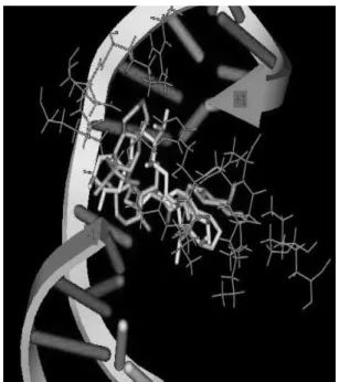
Human DNA topoisomerase I (Topo I) is molecular target of CPT derivatives. These compounds can bind to a transient Topo I-DNA covalent complex, leading to an accumulation of DNA strand breaks upon replication, ultimately causing cell death.21 Thus, we were interested in understanding the interactions of camptothecin derivatives with topoisomerase I by investigating the binding behavior of the two compounds 10b and 10g,which can be treated as the representation for these derivatives based on their cytotoxicities on two different human cancer cell lines listed in Table 1. In the present paper, a ligand-receptor docking studies were carried out to explore the probable binding modes within the active site of protein topoisomerase I.
The crystal structure of topoisomerase I (code: 1T8I) was extracted from Protein Data Bank. The hydrogen atoms, missing atoms and residues were added to complete the protein chain. Compounds 10b and 10g were minimized and the lowest energy conformation of each ligand was then located. The whole docking studies were performed using Libdock method based on discovery studio 2.1 software in our molecular modeling process. As shown in Figure 2, it can be seen that the two compounds can intercalate at the site of DNA cleavage with base-stacking interactions with both the −1 (upstream) and +1 (downstream) base pair. The orientations of 10b and 10g in the active site cavity are perpendicular to the main axis of the DNA while the direction of camptothecin moiety is reverse. Previous studies indicated that hydrogen-bonding interaction between ligand and receptor always played key roles during the formation of drug-DNA-enzyme ternary complex.22,23 As shown in Figure 3, ternary complex of compound 10b-DNA-enzyme formed four hydrogen bonds, while the 10g-DNA-enzyme
Figure 2. Base pairs forming stack interaction between Topoisomerase I and compounds 10b and 10g. Amino acid residues are colored purple. Compounds 10b and 10g are represented by green and yellow color respectively. The backbone of DNA is expressed as white arrows and the base pairs as ladders.
ternary complex formed two H-bonds, respectively. For the complex with compound 10b (Figure 3A), camptothecin and 5-luorouracil moieties formed H-bonds with amino ARG364 and ASN722, in addition, dipeptides linkage moiety produced other two H-bonds with nucleotides. For the complex with compound 10g (Figure 3B), the two H-bonds formed between the dipeptides linkage moiety with amino acid residues, THR718 and ARG364. As we know, the more hydrogen bond interactions always indicated stronger binding afinity. Thus, compound 10b has higher potency to form the hydrogen bonding with protein compared with compound 10g.
From above it can be seen that amino acid residue ARG364 is the necessary amino acid when hydrogen bond formed for both compounds 10b and 10g. Furthermore, it can be deduced that amino acid residue ARG364 is likely to play a key role for the process of compounds binding to topoisomerase I.
QSAR study
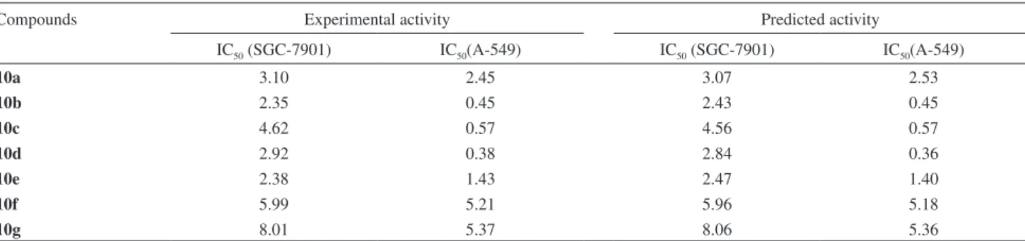
To gain insight into the structural and biological activities of these derivatives, QSAR (quantitative structure-activity relationships) analysis has been widely accepted to account for the various types of activities of the molecule and they are found useful in predicting the activities of other derivatives hitherto unknown. In the present paper, heuristic method (HM)24 available in the framework of the Codessa program was undertaken to perform a complete search for the best multilinear correlations with a multitude of descriptors.
A large pool of 470 descriptors, including constitutional, topological and quantum chemistry properties for compounds 10a-10g was calculated using Codessa software. After correlation analysis of the descriptors, some of them were removed to eliminate redundant information. Three descriptors were then obtained to develop the predictive model and their physical-chemical meanings were listed in Table 2. Thus, in the present paper, the cytotoxic activities against human gastric tumor cell SGC-7901and human lung carcinoma A-549 were regarded as the dependent variable and the selected descriptors were treated as independent variable for constructing the linear regression function 1 and 2 with statistical results squared correlation coeficient (R2), and squared correlation coeficient for leave-one-out
cross-validation R2
cv. In addition, the predicted cytotoxicity
activities based on QSAR models were shown in Table 3 andFigure 4.
For cytotoxic activity against human gastric tumor cell SGC-7901 (equation 1):
IC50(SGC-7901) = 2.77×10⁴ + 3.86×10⁵MaxREHN – 2.01×101FNSA
2 + 5.37×10⁻²HASA2 n = 7, R2 = 0.9989, R2
cv = 0.9893, F = 598.82 (1)
For cytotoxic activity against human gastric tumor cell A-549 (equation 2):
IC50(a-549) = –7.03×10³ + 1.69×10⁻¹HASA1 + 1.42×10³MinEE
C-N – 2.05×101ABIC2 n = 7, R2 = 0.9975, R2
cv = 0.9903, F = 403.54 (2)
Table 2. Physical-chemical meanings for selected descriptors
Dependent variable Independent variable Chemical-physical meaning
IC50 (SGC-7901) MaxPCN Max partial charge for a N atom [Zeirov’s PC]
FNSA2 FNSA-2 Fractional PNSA (PNSA-2/TMSA) [Zeirov’s PC]
HASA2 HASA-2 [Quantum-Chemical PC]
IC50(A-549) HASA1 HASA-1 [Zeirov’s PC]
MinEEC−N Min exchange energy for a C−N bond ABIC2 Average Bonding Information content (order 2)
Table 3. Experimental and predicted activities
Compounds Experimental activity Predicted activity
IC50 (SGC-7901) IC50(A-549) IC50 (SGC-7901) IC50(A-549)
10a 3.10 2.45 3.07 2.53
10b 2.35 0.45 2.43 0.45
10c 4.62 0.57 4.56 0.57
10d 2.92 0.38 2.84 0.36
10e 2.38 1.43 2.47 1.40
10f 5.99 5.21 5.96 5.18
Among the selected descriptors which were used to build QSAR model, FNSA2, HASA2 and HASA1 were all related with charged partial surface area (CPSA) descriptors, which were originally designed to capture information about the features of molecules responsible for polar intermolecular interactions in terms of functional group portions.By combining the atomic surface areas and charges for speciic atom types, CPSA descriptors
can convey information about the speciic atom types (such as N, O) or encode the hydrogen-bonding ability of a molecule. In addition, as we know, hydrogen bonds are formed when a hydrogen atom is shared between two molecules. A hydrogen atom covalently attached to a very electronegative atom (N, O, or P) shares its partial positive charge with a second electronegative atom (N, O, or P). Thus, for the parameter “Max partial charge for N atom”, it is also related with the ability of hydrogen-bonding.
Then, from inspection of predictive model, it indicated that the hydrogen bonding interactions may be the most relevant factor controlling the binding behavior and the cytotoxicity activity. Considering the good statistical results, the development QSAR model is suitable to do a good prediction for cytotoxicity activity and it can give us some insight into the factors that govern the cytotoxicity activity and binding behavior with topoisomerase I.
In vitro determination of lactone levels in human and mouse plasma for 1 and 10b
The results of in vitro determination, based on literature method,25 of lactone levels in human and mouse plasma buffer for 10b and CPT (1) are shown in Table 4.
As shown in Table 4, the percent lactone of CPT (1) in human blood is 28.4% after 4 h, 10.9% after 8 h, and 3.8% after 24 h. In other words, the active form of 1 is signiicantly decreased in a relative short time period after oral administration. This is the opposite to what is observed in mice, in which the active drug form (i.e., the closed lactone form) lasts for a relative long time period. For example, the lactone form of 1 in mouse plasma is still 40.5% even after 4 h. The lactone form of 10b in human plasma is much more stable than its mother compound 1. For example, 70.3% of 10b is still detected as the lactone form even after 4 h. CPT 1, in terms of lactone level, shows a difference between mouse and human. Similarly, this kind of difference is also observed for prodrug 10b. These results further show that the biological life span of lactone Figure 4. Plot of calculated versus experimental cytotoxicity activity
against human gastric tumor SGC-7901(A) and human lung carcinoma A-549 (B).
Table 4. Comparison of percent lactone of the representative prodrug 10b and 1 in human and mouse plasmas
time (h)
0 2 4 6 8 24
Human plasma
lactone for 10b (%) 100.0 81.9 70.3 54.3 42.5 10.5
lactone for CPT (%) 100.0 61.4 28.4 19.4 10.9 3.8
Mouse plasma
lactone for 10b (%) 100.0 83.8 80.7 73.1 52.6 17.9
forms of these compounds in human and mouse plasma signiicantly increased when compared with their mother compound CPT 1. Meanwhile, the cleavage of 10b in both mouse and human plasmas was monitored by HPLC that clearly showed that compound 10b was cleaved to afford mainly its parental compound of CPT in both mouse and human plasmas. The cleavage of 10b is the result of the combination of chemical and enzymatic effects in both mouse and human plasmas. It was also the case in pH 7.4 phosphate buffer.
Another important aspect, which has to be addressed, is that these compounds would be cleaved to release active camptothecin and 5-luorouracil in both mouse and human plasma and such an assumption was supported by employing HPLC chromatography experimental method, which simultaneously implied that these conjugates might act as mutual prodrugs and exert their cytotoxicities by hydrolyzing to the parent compounds camptothecin and 5-luorouracil, and they could have synergetic effects in biological systems.
Conclusions
In summary, we irstly synthesized a series of novel conjugates of camptothecin and 5-luorouracil joined by dipeptide linkages based on the effective combination principle. Most of compounds exhibited potent cytotoxic activity against tumor cell replication. Particularly, conjugates 10b and 10d were highly active against A-549 with IC50 values of 0.45 and 0.38 µmol L-1 respectively. Compound 10b selected for the in vitro determination of lactone levels showed that their biological life span in human and mouse plasma was much longer than that of their parent compounds. Moreover, some interesting QSAR equations have been developed for cytotoxic activity and the correlation between the actual versus predicted activities are in good agreement particularly by taking into account the molecular graphic models. Molecular docking study was further used to clarify the binding mode of these derivatives. Three hydrogen bonds are formed between the dipeptide linkages of conjugates and the active site of Topo I. The results obtained from QSAR studies indicated that the hydrogen bonding interactions may be the most relevant factor controlling the binding behavior and the cytotoxic activity. Considering the good statistical results, the development of QSAR model is suitable to do a good prediction for cytotoxicity activity and it allows for the rational design of more potent conjugates.
These results are encouraging and suggested that the design and syntheses of these compounds should be beneicial for therapeutic values of camptothecin analogues and the approach should be applicable for other antitumor
agents, and it is worthwhile to explore the antitumor potential of these and similar types of compounds. In-depth mechanistic studies and the development of new camptothecin/5-fluorouracil conjugates are actively underway in our laboratory.
Acknowledgments
We thank for NeroTrident Technology Limited Company for providing Discovery Studio software. This work was inancially supported by the National Natural Science Foundation of China (30800720); the Key Laboratory of Environmental Chemistry and Ecotoxicology Fund of Chinese Academy of Sciencer (KF2008-17); the Post-Doctor Research Foundation (20090450142); the Interdisciplinary Innovation Research Foundation For Young Scholars, Lanzhou University (LZUJC2007018).
Reference
1. Tietze, L. F.; Bell, H. P.; Chandrasekhar, S.; Angew. Chem., Int. Ed.2003, 42, 3996.
2. Liu, Y. Q.; Yang, L.; Tian, X.; Curr. Bioact. Compd.2007, 3, 37.
3. Liu, Y. Q.; Liu, Y. Q.; Tian, X.; Yang, L.; Nat. Prod. Res. 2008, 22, 285.
4. Shi, Q.; Wang, H. K.; Bastow, K. F.; Tachibana, Y.; Chen, K. F.; Lee, Y.; Lee, K. H.; Bioorg. Med. Chem. 2001, 9, 2999. 5. Ohtsu, H.; Nakanishi, Y.; Bastow, K. F.; Lee, F. Y.; Lee, K. H.;
Bioorg. Med. Chem. 2003, 11, 1851.
6. Tatsuzaki, J.; Taniguchi, M.; Bastow, K. F.; Nakagawa-Goto, K.; Morris-Natschke, S. L.; Itokawa, H.; Baba, K.; Lee, K. H.; Bioorg. Med. Chem.2007, 15, 6193.
7. Chen, S. W.; Xiang, R.; Liu, J.; Tian, X.; Bioorg. Med. Chem. 2009, 173, 111.
8. Slichenmyer, W. J.; Rowinsky, E. K.; Donehower, R. C.; Kaufmann, S. H.; J. Nat. Cancer Inst.1993, 85, 271. 9. Takimoto, C. H.; Wright, J.; Arbuck, S. G.; Biochim. Biophys.
Acta 1998, 1400, 107.
10. Liew, S. T.; Yang, L. X.; Curr. Pharm. Des.2008, 14,1078. 11. Lorence, A.; Nessler, C. L.; Phytochemistry2004, 65, 2735. 12. Li, Q. Y.; Zu, Y. G.; Shi, R. Z.; Yao, L. P.; Curr. Med. Chem.
2006, 13, 2021.
13. Verma, R. P.; Hansch, C.; Chem. Rev.2009, 109, 213. 14. Campos, J.; Domínguez, J. F.; Gallo, M. A.; Espinosa, A.; Curr.
Pharm. Des. 2000, 6, 1797.
15. Longley, D. B.; Harkin, D. P.; Johnston, P. G.; Nat. Rev. Cancer 2003, 3, 331.
17. Guichard, S.; Hennebelk, I.; Bugat, R.; Cana, P.; Biochem. Pharmacol. 1998, 55, 667.
18. Liu, Y. Q.; Yang, H.; Tian, X.; Chin. J. Chem.2006, 24, 785. 19. Chen, S. W. ; Tian, X.; Tu, Y. Q.; Bioorg. Med. Chem. Lett.
2004, 14, 5063.
20. Greenwald, R. B.; Pendri, A.; Conover, C. D.; Lee, C.; Choe, Y. H.; Gilbert, C.; Martinez, A.; Xia, J.; Wu, D.; Hsue, M.; Bioorg. Med. Chem.1998, 6, 551.
21. Miao, Z. Y.; Sheng, C. Q.; Zhang, W. N.; Ji, H. T.; Zhang, J.; Shao, L. C.; You, L.; Zhang, M.; Yao, J. Z.; Che, X. Y.; Bioorg. Med. Chem. 2008, 16, 1493.
22. Staker, B. L.; Hjerrrild, K.; Feese, M. D.; Behnke, C. A.; Burgin, A. B.; Stewart, L.; Proc. Natl. Acad. Sci. U. S. A.2002, 99, 15387.
23. Staker, B. L.; Feese, M. D.; Cushman, M.; Pommier, Y.; Zembower, D.; Stewart, L.; Burgin, A. B.; J. Med. Chem. 2005, 48, 2336.
24. Katritzky, A. R.; Lobanov, V. S.; Karelson, M.; CODESSA; Reference Manual; University of Florida, Gainesville, 1994. 25. He, X. G.; Lu, W.; Jiang, X. Q.; Cai, J. C.; Zhang, X. W.; Ding,
J.; Bioorg. Med. Chem.2004, 12, 4003.
Submitted: May 8, 2010