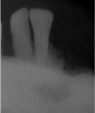
Clinical, histological, and features of a mandibular metastasis from a primary cardiac angiosarcoma
Texto
Imagem


Documentos relacionados
Ousasse apontar algumas hipóteses para a solução desse problema público a partir do exposto dos autores usados como base para fundamentação teórica, da análise dos dados
i) A condutividade da matriz vítrea diminui com o aumento do tempo de tratamento térmico (Fig.. 241 pequena quantidade de cristais existentes na amostra já provoca um efeito
didático e resolva as listas de exercícios (disponíveis no Classroom) referentes às obras de Carlos Drummond de Andrade, João Guimarães Rosa, Machado de Assis,
The probability of attending school four our group of interest in this region increased by 6.5 percentage points after the expansion of the Bolsa Família program in 2007 and
Despercebido: não visto, não notado, não observado, ignorado.. Não me passou despercebido
The fourth generation of sinkholes is connected with the older Đulin ponor-Medvedica cave system and collects the water which appears deeper in the cave as permanent
Isto é, o multilateralismo, em nível regional, só pode ser construído a partir de uma agenda dos países latino-americanos que leve em consideração os problemas (mas não as
aging to dementia must be considered a con- tinuum, both in terms of the slow manifesta- tion of the impairment of cognitive functions, as well as functional limitation. The
