Page 1 of 8
What is the role of inflammatory mediators on energy
metabolism?
Simone Fátima Gomes1, Fernanda Cacilda Silva2, Ana Carolina Pinheiro Volp3
1
Nutrition and Health MSc Student (Research Line: Nutrition Biochemistry and Pathophysiology), Federal University of Ouro Preto, Ouro Preto – Minas Gerais, Brazil
2
PhD in Biological Science and Postdoctoral researcher in Cardiovascular Physiology Laboratory at Federal University of Ouro Preto, Ouro Preto – Minas Gerais, Brazil
3
PhD in Food Science and Technology, and Adjunct Professor at Federal University of Ouro Preto, Ouro Preto – Minas Gerais, Brazil
Correspondence: Ana Carolina Pinheiro Volp E-mail: anavolp@gmail.com
Received: January 13, 2016 Published online: February 22, 2016
The subclinical and low intensity inflammation, oxidative stress, high calorie and high fat diet patterns are striking features of the obesity. The adipose tissue, through its endocrine function, is associated with the cytokines secretion, such as: IL-4, IL-13, IL-15 and IFN-γ, which trigger metabolic changes and possibly modulate the energy metabolism by modifying the biochemical, anthropometric and body composition parameters. This review summarizes scientific evidences about the relationship between such cytokines and mediators which alter the energy metabolism, predisposing or preventing the obesity.
Keywords: Inflammation; obesity; interleukin-4; interleukin-13; interleukin-15; interferon-gamma.
To cite this article: Simone Fátima Gomes, et al. What is the role of inflammatory mediators on energy metabolism? Inflamm Cell Signal 2016; 3: e1189. doi: 10.14800/ics.1189.
Copyright:© 2016 The Authors. Licensed under a Creative Commons Attribution 4.0 International License which allows users including authors of articles to copy and redistribute the material in any medium or format, in addition to remix, transform, and build upon the material for any purpose, even commercially, as long as the author and original source are properly cited or credited.
Introduction
The obesity, a disease with a complex and multifactorial etiology [1, 2], is associated with an imbalance in the energy balance, the altered oxidative status [3, 4] and the subclinical and low intensity systemic inflammation [3-7].
Such metabolic changes stimulate the white adipose tissue (WAT) to secrete cytokines, for example, interleukin 4 (IL-4 ), interleukin 13 (IL-13), interleukin 15 (IL-15) and IFN-γ (interferon-gamma), or other inflammatory markers which could modulate the energy metabolism and,
consequently, modify anthropometric, biochemical and body composition parameters [5]. However, so far, it is not known the exactly role, as well as the magnitude of the effect that each one of these mediators can have on the predisposition or prevention of obesity.
It has been shown that during obesity, elevated concentrations of IFN-γ favor the body weight gain and
impair the glucose homeostasis [8-10]. However, the
Page 2 of 8 decreased food intake, suppressing the orexigenic and stimulating the anorexigenic route[8]. So, it is plausible to infer that there is correlation between such mediator and mechanisms which alter the energy metabolism.
In other hand, the IL-4, IL-13 and IL-15 with a predominantly anti-inflammatory effect on WAT, can modulate the energy metabolism by increasing insulin sensitivity and reducing the body weight and fat which contributes to the obesity prevention [6, 11-14].
Given the importance of the subject and the scarcity in literature about the modulation of inflammatory mediators
that interfere with energy metabolism, this review aims to summarize studies that analyzed such mediators on the energy metabolism, focusing on IL-4, IL-13, IL-15 and IFN-γ biomarkers.
Obesity, energy metabolism and inflammation
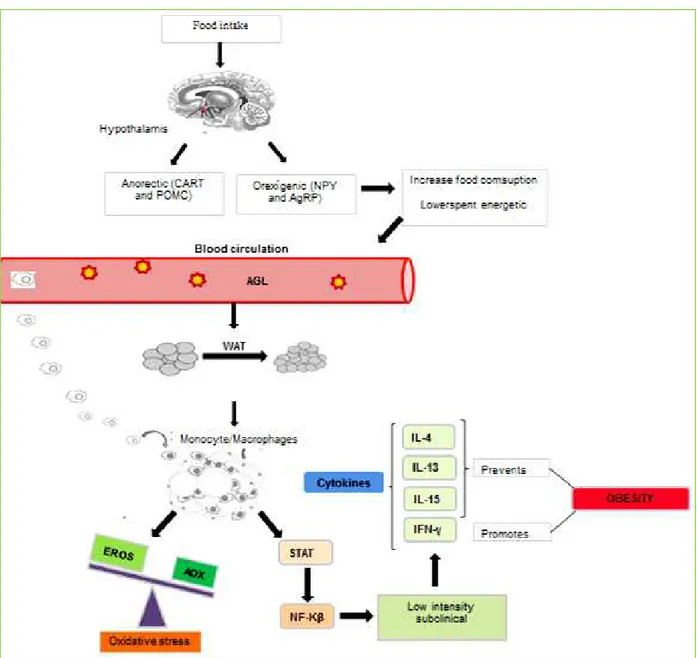
Food intake is regulated in the hypothalamus, primarily by two large groups of neuropeptides: the anorexigenic, Melanocyte-stimulating hormone - Pro-opiomelanocortin (POMC) and amphetamine-regulated transcript (CART); the other group is the orexigenic: Neuropeptide Y (NPY) and Agouti-related peptide (AgRP) [15, 16]. In the presence of the
Figure 1. Endocrine effect of the adipose tissue and signaling pathways of the oxidative stress and inflammatory response in obesity. The food
Page 3 of 8 obesity, through the resistance to leptin, the anorexigenic neuropeptides do not perform their normal functions while the orexigenic become active, favoring the food intake increase [17]
. This consequently promotes metabolic changes, such as: adiposity, oxidative stress and increased secretion of inflammatory mediators [16, 17].
Thus, the typical standard diet in obesity consists of high calorie and fat consumption and low intake of antioxidant nutrients [6, 18, 19] which promote an increase in free fatty acids plasma concentrations. Therefore, in the WAT, mitochondria do not perform their normal functions, getting
overloaded and resulting in incomplete β-oxidation of fatty acids. This predisposes the homeostasis loss and increases the reactive oxygen species (ROS) production, triggering oxidative stress status. These changes lead to cells deterioration, resulting in insulin resistance or in a greater lipid accumulation in adipocytes, due to the increased nicotinamide adenine dinucleotide phosphate- reduced (NADPH) supply, predisposing to obesity by changes in energy metabolism [4, 20].
Hypoxia, from adiposity excess significantly alter the adipocytes vital parameters, such as: the increase in ROS (432.53%), lipids and proteins oxidation (376.6% and 566.6%, respectively), the antioxidant enzymes and inflammatory mediators release reduction [21].
Thus, the adiposity excess and oxidative stress observed in obesity trigger an inflammatory cascade by phosphorylation of signal transduction and activation of transcription (STAT) pathways - Jun N-terminal kinase proteins (JNK); Nuclear Factor kappa Beta (NF-KB) [22]. They are stimulated by the macrophages influx in adipocytes, inducing the inflammatory cytokines synthesis that characterizes a low intensity and subclinical inflammatory process. Such process is derived of the hypoxia in adipose tissue, adipocytes hypertrophy and hyperplasia, cytokine synthesis and adipose tissue endocrine signaling [4-6].
According to Xu et al.[23] in obesity, some cytokines can activate STAT3 gene that binds to the enhancer binding protein (C/EBP) gene target, promoting the adipogenesis. In this way, cytokines may regulate lipid metabolism through STAT pathway activation and play a promoter role to obesity [23, 24]
. In a Barbarroja et al. [25] study, obese individuals had significantly higher STAT3 levels in comparison with lean controls, which links strongly the STAT3 to obesity (Figure 1).
On the other hand, there are evidences that the inflammatory response occurs to mobilization of energy reserves and anti-inflammatory cytokines in adipose tissue,
such as: IL-4, IL-13 and IL-15, which elicit positive effects on metabolic homeostasis in nutrient overload situation. Moreover, they contribute to inhibition of pro-inflammatory cytokines expression and, consequently, reduce the inflammatory process in adipose tissue. So, these interleukins action is essential to modulate energy metabolism should be hence prevent obesity [26] .Another protective mechanism in obesity is the brown adipose tissue (BAT) action by thermogenesis, with uncoupling proteins (UCPs) active expression when fatty acid concentrations are high. Thus, there is the homeostasis restoration and promotion of protective mechanism against the increased lipid metabolism and fat storage. So, this favors an increase in energy expenditure and WAT reduction, besides modulates the body composition and, therefore, prevents the obesity and disorders related to such disease [27-29]. In this way, in a study proposed by Ye [30], the IL-15 administration increased the UCPs expression, promoting thermogenesis – one of the mechanisms which induces energy expenditure, and consequently inhibit obesity [20, 31].
IFN-γ
IFN-γ is a cytokine produced by T and natural killer cells (NK). It has pro-inflammatory effects in the immune system and also in adipose tissue and is able to increase the ability of macrophages and endothelial cells to present antigens and stimulate the production of other inflammatory mediators,
such as: Tumor Necrosis Factor-Alpha (TNF-α) and
interleukin-1 (IL-1), which amplify the inflammation [10, 32].
In a study proposed by Khazen et al. [33] both in rodents and in humans, the IFN-γ presence had a particular effect in adipocytes leading to a rapid and extensive decline in phosphoenolpyruvate carboxykinase expression (PEPCK) in glyceroneogenesis, increasing the release of plasma free fatty acids, which could be involved in insulin resistance induced by obesity. Waite et al.[34] proposed another mechanism, in which IFN-γ inhibited the peroxisome proliferator-activated receptor (PPAR) expression in adipocytes and, consequently, decreased the lipoprotein Lipase (LPL) activity, resulting in the development of insulin resistance and lipolysis increase.
Recent studies have shown that high IFN- γ concentrations increase in obesity in models, humans and rodents, while its deficiency results in a better glucose tolerance in rats. Precisely because the IFN-γ is high in obesity, studies suggest that this cytokine impairs glucose homeostasis and body weight control [8-10].
Page 4 of 8 extracted from obese rats adipose tissue and stimulated in vitroalso produce IFN-γ higher quantity than that extracted from lean animals [9].
In other hand, the IFN-γ suppression in rats resulted in a body weight reduction associated with food intake reduction and voluntary physical activity increase. Consequently, the glucose homeostasis improved due to insulin sensitivity increase, associated with glyconeogenesis reduced activity by decrease in glucose-6-phosphate expression. In relation to food intake reduction, the mRNA expression of neuropeptide Y (NPY) - the hunger stimulating neuropeptide - reduced significantly, whereas the hunger suppression neuropeptide, the Pro-opiomelanocortin (POMC) increased significantly. Moreover, it was also observed a significant increase in plasma leptin, which is proportional to adiposity. It is well established that the leptin modulates the action of central neuropeptides, NPY and POMC, in order to reduce the food intake and increase the energy expenditure. So, the IFN-γ suppression plays an essential role in energy balance, preventing the obesity and type 2 diabetes [8].
IL-15
IL-15 is produced by monocytes/ macrophages in immunology system and its genes were also detected in placenta, skeletal muscle, kidney, lung and heart [35-38]. Such cytokine exerts anti-inflammatory action on adipose tissue, playing pleiotropic functions, by apoptosis stimulation and its capacity to modulate the metabolism, and pro-inflammatory function since it promotes the IFN- γ expression, which amplifies the inflammation. The IL-15 mRNA is highly expressed in skeletal muscle tissue, thus it has a greater function as an endocrine factor, which may be associated with energy metabolism and, consequently, to the body composition modulation [30, 39, 40].
It is believed that IL-15 has the ability to modulate the body composition by exerts direct effects on the carbohydrates and lipids metabolism, as well as on the insulin sensitivity fatty acid oxidation, lipogenesis inhibition, very low density lipoprotein decrease (VLDL) and thermogenesis promotion [30, 35, 37, 40, 41].
For such efects, IL-15 increases the glucose capitation striated muscle, since it enhances the GLUT4 expression in muscle cells, improving the insulin sensitivity both in vitro and in vivo. The IL-15 administration resulted in increase of 2- deoxyglucose capitation by skeletal muscle [35, 40]. Besides promoting changes in carbohydrates oxidation, IL-15 also seems to influence the fatty acid oxidation in skeletal muscle. As the study in experimental animals by oral administration of
exogenous triglycerides showed the IL-15 treatment increases the fatty acid oxidation in skeletal muscle [37].
Furthermore, with in vivo effect, a study showed that IL-15 chronic administration (seven days) resulted in a BAT reduction of 33% in healthy rats. Additional studies show that this cytokine effects on BAT are partly caused by the lipogenesis inhibition, through lipoprotein lipase (LPL) activity and by reducing the fat absorption in this tissue. IL-15 also regulates the lipogenesis in liver, because it modulates the hepatic citrate concentrations, one of the major activators of acetyl-CoA carboxylase – a regulating lipogenic
pathway enzyme. The lipogenesis reduction was
accompanied by an increase in fatty acid oxidation, because it increased the carnitine palmitoyltransferase 1 (CPT1) and carnitine palmitoyltransferase 2 (CPT2) levels in liver. The results of these IL-15 alterations in lipid metabolism in liver
is possibly a reduction in triacylglycerol’s exportation in
VLDL form, reducing the lipid content by VLDL particles [37]
. Moreover, in rats, IL-15 administration promoted thermogenesis in BAT, due to better UCP-1 and UCP-3 expressions. In this study, the IL-15 treatment decreased WAT and BAT (35% and 24%, respectively). So, IL-15 can induce the BAT energy expenditure, through thermogenesis, which supports a mechanism to IL-15 action on obesity control [30].
According to Barra et al. [42] obese individuals have smaller IL-15 concentrations than eutrophic individuals. Thus, the IL-15 treatment reduced the body weight in an obese animal model induced by diet. The IL-15 effect in vivo did not influence the food intake, because IL-15 reduced the obesity by means of a mechanism dependent on the leptin receptor. Its administration was studied in two genetic obesity models: rodents with leptin receptor knockdown and rats with leptin deficiency. The IL-15 infusion reduced the adiposity in rats with leptin receptor knockdown and those which have leptin receptor presented no changes in food intake pattern. The author concluded that IL-15 reduces adiposity because its activity is dependent on leptin receptor deficiency [30].
In a clinical study, in which body mass index (BMI) was evaluated, it was observed negative associations between IL-15 plasma concentrations and BMI, total body fat and trucal fat [43]. Therefore, these results suggest that IL-15 can be involved in adipose tissue regulation and, consequently, in obesity [26, 30, 39].
It is worth noting that, so ever IL-15 has beneficial effects in the energy metabolism modulation, the inhibition of its expression in obesity process is more satisfactory, precisely
Page 5 of 8
expression and to contribute to amplification of
inflammatory process, bringing numerous endocrine and metabolic consequences.
IL-4
IL-4 is produced by CD4 + T lymphocytes, mast cells and activated basophils. In addition, it performs pleiotropic functions, such as: the induction of Th2 differentiation, shift in immunoglobulin class and proliferation of B cells in the immune system. Besides the immune system, IL-4 is present, constitutively, in different parts of the body, such as: the kidney, spleen, liver, brain, skeletal muscle, heart and adipose tissue. In the latter, it plays anti-inflammatory action, limiting the characteristic subclinical inflammation of obesity [6]
.
According to the studies proposed in the literature, IL-4 when expressed by adipocytes and hepatocytes shows the ability to modulate local immune responses, besides being associated with the increased insulin sensitivity and glucose tolerance. This inhibits the lipids accumulation in adipose tissue, resulting in decreased weight gain and body fat [6, 11-14]
.
Thus, in a recent study proposed by Tsao et al. [6] the IL-4 presence in the adipocyte differentiation process resulted in the inhibition of 30% of lipid accumulation in differentiated cells due to decreased and delayed in the expression of important genes, which mediate adipogenesis, PPAR and the CCAAT/enhancer binding proteins (C/EBP). Moreover, IL-4 has demonstrated inhibitory effects of genes involved in glucose uptake and lipid metabolism, such as: GLUT4 and LPL. Therefore, beyond inhibiting adipocyte differentiation through the STAT6 pathway, it enhances lipolysis by increasing levels of cyclic adenosine monophosphate (cAMP). The cAMP activates the protein kinase A (PKA), followed by
phosphorylation of hormone-sensitive lipase (HSL),
mediating the triacylglycerol’s hydrolysis. Thus, IL-4 participates of the lipid metabolism evoking a conventional lipolytic pathway. In addition to these results, it was also found that IL-4 modulates the resistin expression (hormone involved in lipolysis promoting, glucose and insulin intolerances) inhibiting the lipid accumulation in adipose tissue, which consequently leads to the reduction of the body weight gain. Given these scientific evidences about the IL-4 role in adipose tissue, it is suggested that this cytokine is inversely proportional to weight gain and adiposity.
According to the aforementioned study, Gonzalez et al.[13] also found that IL-4 treatment in rat hepatocytes increased the STAT6 phosphorylation, suggesting its involvement in hepatic metabolism. Thus, IL-4 suppressed the β-oxidation of
fatty acids in 50% and increased the hepatic glucose oxidation in 450%. However, when it was administered in rats with STAT6 deficiency, it had an opposite effect; shifting the glucose metabolic dependence for fatty acid oxidation in hepatocytes. This change was accompanied by an increased activity of alpha receptors activated by PPAR,
key regulator of the β-oxidation in liver pathways. In addition, the experiment when challenged with a high fat diet, STAT6 null rats gained significantly less weight and had small WAT deposits, totaling a 60% reduction in body fat mass and 43% in adiposity. This difference in weight gain and adiposity was not caused by loss of adipogenesis, but by the food intake decrease, which implies that the STAT6 null
rat’s resistance in induced obesity by high fat diet results
from the increased energy expenditure.
Besides the function of decreasing the adipogenesis and favoring the reduction of adipose tissue, the IL-4 is also known as the trigger signal for the phosphorylation of the substrates and insulin receptors. This suggests that it may be positively associated with the regulation of insulin signaling pathways. In vivo effects of IL-4 overexpression promoted changes in the protein kinase B activity (PKB) and glycogen
synthase Kinase 3β (GSK3β) of the rats muscle cells, suggesting that it may result in improved glucose tolerance, from the reinforcement of the insulin action through these pathways phosphorylation [14].
IL-13
IL-13, a cytokine produced by T and B cells, mast cells, basophils, NK and dendritic cells, plays a key role in inflammation and immune responses [44].
In adipose tissue, a recent study proposed by Kwon et al. [45]
demonstrated that in both rats and humans there is an increase in inflammatory markers and also a marked increase in IL-13 production. These data suggest that IL-13 expression in adipose tissue is increased. A high fat diet induced signs of pro-inflammatory cytokines originating from adipose tissue by immune cells activation, the kinase complex (IKK), which also releases IL-13. It, in turn, has
paracrine function to suppress the expansion of
pro-inflammatory cells in adipose tissue, in other words, it is anti-inflammatory. These data show a defense mechanism present in adipocytes, which limit the inflammation and insulin resistance by their autoregulation. Thus, it is conceivable that during an inflammation process, one of the main functions of IL-13 is restore the glucose homeostasis, which is interrupted by pro-inflammatory actions of other cytokines [46].
Page 6 of 8
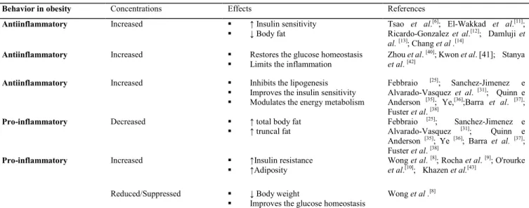
Table 1. Adipokines involved in the modulation of the energy metabolism
Behavior in obesity Concentrations Effects References
Antiinflammatory Increased ↑ Insulin sensitivity
↓ Body fat Tsao et al.
[6]; El-Wakkad et al.[11];
Ricardo-Gonzalez et al.[12]; Damluji et al. [13]; Chang et al .[14]
Antiinflammatory Increased Restores the glucose homeostasis
Limits the inflammation
Zhou et al. [40]; Kwon et al. [41]; Stanya et al. [42]
Antiinflammatory Increased Inhibits the lipogenesis
Improves the insulin sensitivity
Modulates the energy metabolism
Febbraio [25]; Sanchez-Jimenez e
Alvarado-Vasquez et al. [31]; Quinn e
Anderson [35]; Ye,[36];Barra et al. [37]; Fuster et al. [38]
Pro-inflammatory Decreased ↑ total body fat
↑ truncal fat Febbraio
[25]; Sanchez-Jimenez e
Alvarado-Vasquez [31]; Quinn e
Anderson [35]; Ye [36]; Barra et al. [37];
Fuster et al. [38]
Pro-inflammatory Increased ↑Insulin resistance
↑Adiposity Wong et al.
[8]; Rocha et al. [9]; O'rourke et al.[10]; Khazen et al.[43]
Reduced/Suppressed ↓ Body weight
Improves the glucose homeostasis
Wong et al .[8]
IL: Interleukin; IFN-γ: Interferon-gamma; ↑: increase; ↓decrease.
showed that depletion of IL-13 gene led to hyperglycemia accompanied by a high expression of genes required for synthesis of glucose in the liver, insulin resistance, decreased oxygen consumption, weight gain and increased triglyceride levels in blood and liver, contributing to the development of metabolic syndrome. However, its presence suppressed the postprandial hyperglycemia. IL-13 is probably necessary when the insulin signaling is compromised or suppressed, as in the cases of overfeeding, which leads to obesity. It acts as a physiological signal that activates the STAT3 pathway to suppress hepatic glucose production (gluconeogenesis) by
inhibiting the genes expression of EPKC and
glucose-6-phosphatase enzymes.
In this way, it is concluded that the inflammation present in adipose tissue induces the IL-13 expression, which serves to maintain a low level of chronic inflammation and, thereby, limit the action of pro-inflammatory cytokines and, therefore, help to the minimization of the characteristic inflammatory and metabolic effects of obesity.
Finally, the chart below summarizes the action of adipokines on the parameters that modulate energy metabolism and are also associated with the prevention or promotion of obesity (Table 1).
Final considerations
Given the aforementioned, scientific evidence
demonstrates that activation of NF-kB STAT- JNK pathways promotes the expression of cytokines with pro and anti-inflammatory effects, such as cytokines, IL-4, IL-13,
IL-15 and IFN -γ, which are associated with altered
biochemical, anthropometric and body composition
parameters that modulate energy metabolism and,
consequently, may predispose or prevent obesity and its metabolic changes.
Although this pathway activation might modulate the energy metabolism through the secretion of cytokines with anti-inflammatory effect, its blockade would be more
appropriate since the same route also secretes
pro-inflammatory cytokines, such as IFN-γ that at high concentrations favors the body weight gain and hence predisposes obesity.
Conflicting interests
The authors have declared that no conflict of interests exists.
Abbreviations
AgRP: Agouti:related peptide; cAMP: Cyclic adenosine
monophosphate; AOX: Antioxidants; C/EBP:
CCAAT/enhancer binding proteins; CART: Cocaine: and
amphetamine:regulated transcript; CPT1: Carnitine
Palmitoyltransferase 1; CPT2: Carnitine Palmitoyltransferase 2; PEPCK: Phosphoenolpyruvate carboxykinase; ROS:
Reactive oxygen species; GSK3β: Glycogen synthase Kinase
Page 7 of 8 Proteins; LPL: Lipoprotein Lipase; NADPH: Nicotinamide adenine dinucleotide phosphate; NF-Kβ: Nuclear Factor kappa Beta; NK: Natural Killers; NPY: Neuropeptide Y; PEPCK: Phosphoenolpyruvate carboxykinase ; PKA: Protein
kinase A; PKB: Protein kinase B; POMC:
Pro:opiomelanocortin; Melanocyte: stimulating hormone; PPAR: Peroxisome proliferator:activated receptor; mRNA: Messenger Ribonucleic Acid; STAT: Signal transduction and activation of transcription; WAT: White adipose tissue; BAT:
Brown adipose tissue; TNF-α: Tumor Necrosis Factor-Alpha;
UCP1: Uncoupling protein 1; UCP3: Uncoupling protein 3; VLDL: Very low density lipoprotein.
Author contributions
SFG have been involved in drafting the manuscript or revising it critically for important intellectual content; and have given final approval of the version to be published in all stages, from designing the study to review the final version. FCS attended the article translation and final review and ACPV participated in the final review, guidance and critical analysis. All authors read and approved the final manuscript.
References
1. Leite LD, Rocha ÉDdM, Brandão-Neto J. Obesity: an inflammatory disease. Cien Saude Colet 2009; 2: 85-95.
2. Miranda VPN, Peluzio MdCG, Franceschini SdCC, Priore SE. Marcadores inflamatórios na avaliação nutricional: relação com parâmetros antropométricos, composição corporal e níveis de atividade física. Rasbran 2014; 6: 61-72.
3. Van Guilder GP, Hoetzer GL, Greiner JJ, Stauffer BL, Desouza CA. Influence of metabolic syndrome on biomarkers of oxidative stress and inflammation in obese adults. Obesity (Silver Spring) 2006;14:2127-2131.
4. Choudhury S, Ghosh S, Gupta P, Mukherjee S, Chattopadhyay S. Inflammation-induced ROS generation causes pancreatic cell death through modulation of Nrf2-NF-kappaB and SAPK/JNK pathway. Free Radic Res 2015;49:1-41.
5. Queiroz JCFd, USP SP, Brasil, Alonso-Vale MIC, USP SP, Brasil, Curi R, USP SP, Brasil, et al. Control of adipogenesis by fatty acids. Arq Bras Endocrinol Metab 2009;53:582-594.
6. Tsao CH, Shiau MY, Chuang PH, Chang YH, Hwang J. Interleukin-4 regulates lipid metabolism by inhibiting adipogenesis and promoting lipolysis. J Lipid Res 2014,55: 385-397.
7. Wensveen FM, Valentic S, Sestan M, Wensveen TT, Polic B. The "Big Bang" in obese fat: events initiating obesity-induced adipose tissue inflammation. Eur J Immunol 2015;45:2446-2456.
8. Wong N, Fam BC, Cempako GR, Steinberg GR, Walder K, Kay TW, et al. Deficiency in interferon-gamma results in reduced body weight and better glucose tolerance in mice. Endocrinology 2011;152:3690-3699.
9. Rocha VZ, Folco EJ, Sukhova G, Shimizu K, Gotsman I, Vernon AH, et al. Interferon-gamma, a Th1 cytokine, regulates fat
inflammation: a role for adaptive immunity in obesity. Circ Res 2008;103:467-76.
10. O'Rourke RW, Metcalf MD, White AE, Madala A, Winters BR, Maizlin, II, et al. Depot-specific differences in inflammatory mediators and a role for NK cells and IFN-gamma in inflammation in human adipose tissue. Int J Obes (Lond) 2009;33:978-990.
11. El-Wakkad A, Hassan Nel M, Sibaii H, El-Zayat SR. Proinflammatory, anti-inflammatory cytokines and adiponkines in students with central obesity. Cytokine 2013;61:682-687.
12. Damluji AA, Ramireddy A, Al-Damluji MS, Marzouka GR, Otalvaro L, Viles-Gonzalez JF, et al. Association between anti-human heat shock protein-60 and interleukin-2 with coronary artery calcium score. Heart 2015; 101:436-441.
13. Ricardo-Gonzalez RR, Red Eagle A, Odegaard JI, Jouihan H, Morel CR, Heredia JE, et al. IL-4/STAT6 immune axis regulates peripheral nutrient metabolism and insulin sensitivity. Proc Natl Acad Sci U S A 2010;107:22617-22622.
14. Chang YH, Ho KT, Lu SH, Huang CN, Shiau MY. Regulation of glucose/lipid metabolism and insulin sensitivity by interleukin-4. Int J Obes (Lond) 2012;36:993-998.
15. Guyenet SJ, Schwartz MW. Clinical review: Regulation of food intake, energy balance, and body fat mass: implications for the pathogenesis and treatment of obesity. J Clin Endocrinol Metab 2012;97:745-755.
16. Drougard A, Fournel A, Valet P, Knauf C. Impact of hypothalamic reactive oxygen species in the regulation of energy metabolism and food intake. Front Neurosci 2015;9:56.
17. Sohn JW. Network of hypothalamic neurons that control appetite. BMB Rep 2015;48:229-233.
18. Bastos DHM, Rogero MM, Arêas JAG. Effects of dietary bioactive compounds on obesity induced inflammation. Arq Bras Endocrinol Metab 2009;53:646-656.
19. Souza CLd, Oliveira MRMd. Fatores associados ao metabolismo energético na obesidade. Nutrire 2010;35:145-164.
20. Flachs P, Rossmeisl M, Kuda O, Kopecky J. Stimulation of mitochondrial oxidative capacity in white fat independent of UCP1: a key to lean phenotype. Biochim Biophys Acta 2013;1831:986-1003.
21. Priyanka A, Nisha VM, Anusree SS, Raghu KG. Bilobalide attenuates hypoxia induced oxidative stress, inflammation, and mitochondrial dysfunctions in 3T3-L1 adipocytes via its antioxidant potential. Free Radic Res 2014;48:1206-1217.
22. Petrangeli E, Coroniti G, Brini AT, de Girolamo L, Stanco D, Niada S, et al. Hypoxia Promotes the Inflammatory Response and Stemness Features in Visceral Fat Stem Cells from Obese Subjects. J Cell Physiol 2015; doi: 10.1002/jcp.25113.
23. Xu D, Yin C, Wang S, Xiao Y. JAK-STAT in lipid metabolism of adipocytes. Jakstat 2013; doi: 10.4161/jkst.27203.
24. Richard AJ, Stephens JM. Emerging roles of JAK-STAT signaling pathways in adipocytes. Trends Endocrinol Metab 2011; 22:325-332.
Page 8 of 8 visceral adipose tissue inflammation. PLoS One 2012; doi: 10.1371/journal.pone.0048155.
26. Febbraio MA. Role of interleukins in obesity: implications for metabolic disease. Trends Endocrinol Metab 2014;25:312-319.
27. Boschini RP, Garcia Júnior JR. UCP2 and UCP3 genic expression: regulation by food restriction, fasting and physical exercise. Rev Nutr 2005;18:753-764.
28. Vosselman MJ, van Marken Lichtenbelt WD, Schrauwen P. Energy dissipation in brown adipose tissue: from mice to men. Mol Cell Endocrinol 2013;379:43-50.
29. Rachid TL, Penna-de-Carvalho A, Bringhenti I, Aguila MB, Mandarim-de-Lacerda CA, Souza-Mello V. PPAR-alpha agonist elicits metabolically active brown adipocytes and weight loss in diet-induced obese mice. Cell Biochem Funct 2015;33:249-256.
30. Ye J. Beneficial metabolic activities of inflammatory cytokine interleukin 15 in obesity and type 2 diabetes. Front Med 2014;9:139-145.
31. Shabalina IG, Petrovic N, de Jong JM, Kalinovich AV, Cannon B, Nedergaard J. UCP1 in brite/beige adipose tissue mitochondria is functionally thermogenic. Cell Rep 2013;5:1196-1203.
32. Ebert EC. IL-15 converts human intestinal intraepithelial lymphocytes to CD94 producers of IFN-gamma and IL-10, the latter promoting Fas ligand-mediated cytotoxicity. Immunology 2005;115:118-126.
33. Khazen W, Distel E, Collinet M, Chaves VE, M'Bika JP, Chany C,
et al. Acute and selective inhibition of adipocyte glyceroneogenesis and cytosolic phosphoenolpyruvate carboxykinase by interferon gamma. Endocrinology 2007; 148:4007-4014.
34. Waite KJ, Floyd ZE, Arbour-Reily P, Stephens JM. Interferon-gamma-induced regulation of peroxisome proliferator-activated receptor gamma and STATs in adipocytes. J Biol Chem 2001;276: 7062-7068.
35. Sanchez-Jimenez R, Alvarado-Vasquez N. IL-15 that a regulator of TNF-alpha in patients with diabetes mellitus type 2. Med Hypotheses 2013;80:776-777.
36. Barra NG, Chew MV, Holloway AC, Ashkar AA. Interleukin-15 treatment improves glucose homeostasis and insulin sensitivity in obese mice. Diabetes Obes Metab 2012;14:190-193.
37. Argiles JM, Lopez-Soriano FJ, Busquets S. Therapeutic potential of interleukin-15: a myokine involved in muscle wasting and adiposity. Drug Discov Today 2009;14:208-213.
38. Fehniger TA, Caligiuri MA. Interleukin 15: biology and relevance to human disease. Blood 2001;97:14-32.
39. Quinn LS, Anderson BG. Interleukin-15, IL-15 Receptor-Alpha, and Obesity: Concordance of Laboratory Animal and Human Genetic Studies. J Obes 2011; doi: 10.1155/2011/456347.
40. Barra NG, Reid S, MacKenzie R, Werstuck G, Trigatti BL, Richards C, et al. Interleukin-15 contributes to the regulation of murine adipose tissue and human adipocytes. Obesity (Silver Spring) 2010;18:1601-1607.
41. Fuster G, Almendro V, Fontes-Oliveira CC, Toledo M, Costelli P, Busquets S, et al. Interleukin-15 affects differentiation and apoptosis in adipocytes: implications in obesity. Lipids 2011;46:1033-1042.
42. Barra NG, Reid S, MacKenzie R, Werstuck G, Trigatti BL, Richards C, et al. Interleukin-15 contributes to the regulation of murine adipose tissue and human adipocytes. Obesity (Silver Spring) 2010;18:1601-1607.
43. Nielsen AR, Hojman P, Erikstrup C, Fischer CP, Plomgaard P, Mounier R, et al. Association between interleukin-15 and obesity: interleukin-15 as a potential regulator of fat mass. J Clin Endocrinol Metab 2008;93:4486-4493.
44. Zhou R, Qian S, Gu X, Chen Z, Xiang J. Interleukin-13 and its receptors in colorectal cancer (Review). Biomed Rep 2013;1:687-690.
45. Kwon H, Laurent S, Tang Y, Zong H, Vemulapalli P, Pessin JE. Adipocyte-Specific IKKbeta Signaling Suppresses Adipose Tissue Inflammation through an IL-13-Dependent Paracrine Feedback Pathway. Cell Rep 2014;9:1574-1583.